The End-systolic Wall Stress/End-systolic Volume Index Ratio for
Systolic Function in Anatolian Shepherd Dogs with Stage
B2 Degenerative Mitral Valve Disease
[1]Kürşad TURGUT
1,a İsmail ŞEN
2,bMehmet Ege İNCE
1,cAmir NASERİ
3,dHavva SÜLEYMANOĞLU
1,eMerve ERTAN
1,fVedat SAĞMANLIGİL
4,g [1] Oral presentation at congress: 32nd World Veterinary Congress (WVC), 13-17 September 2015 - Istanbul, Turkey1 Near East University, Faculty of Veterinary Medicine, Department of Internal Medicine, 99280, Nicosia, NORTH CYPRUS 2 Kyrgyz Turkish Manas University, Faculty of Veterinary Medicine, Department of Internal Medicine, 720011 Bishkek -
KYRGYZSTAN
3 Selcuk University, Faculty of Veterinary Medicine, Department of Internal Medicine, TR-42003 Konya - TURKEY 4 Near East University, Faculty of Veterinary Medicine, Department of Physiology, 99280, Nicosia, NORTH CYPRUS a ORCID: 0000-0001-8725-8044; b ORCID: 0000-0002-2965-7183; c ORCID: 0000-0002-1859-1449; d ORCID: 0000-0001-9140-5879 e ORCID: 0000-0002-6681-0212; f ORCID: 0000-0003-3436-4817; g ORCID: 0000-0001-9335-7348
Article ID: KVFD-2019-22661 Received: 17.05.2019 Accepted: 17.09.2019 Published Online: 17.09.2019
How to Cite This Article
Turgut K, Şen İ, İnce ME, Naseri A, Süleymanoğlu H, Ertan M, Sağmanligi V: The end-systolic wall stress/end-systolic volume index ratio for
systolic function in Anatolian shepherd dogs with stage B2 degenerative mitral valve disease. Kafkas Univ Vet Fak Derg, 26 (2): 299-303, 2020. DOI: 10.9775/kvfd.2019.22661
Abstract
This study was performed to evaluate the end-systolic wall stress/end-systolic volume index ratio (ESWS/ESV-I) for LV systolic function in Anatolian shepherd dogs (ASHs) with stage B2 degenerative mitral valve disease (DMVD). Thirty-eight adult ASHs with DMVD (experimental group; 20 B1 dogs and 18 B2 dogs) and 30 weight-matched adult healthy ASHs (control group) were used as subjects. There was no overlap for the ESWS/ESV-I ratio (0.83±0.03, range: 0.61-1.09 for B2 dogs, 1.34±0.05, range: 1.11-1.89 for B1 dogs, 2.17±0.0 range: 1.91-3.02 for the control dogs) among the groups. ESWS/ESV-I ratio (≤1.09) in ASHs with stage B2 DMVD may play a developmental role in left ventricular remodelling and risk factor for development of systolic dysfunction.
Keywords: Anatolian shepherd dog, Degenerative mitral valve disease, Echocardiography, End-systolic wall stress/end-systolic volume index ratio, Systolic function indices
Evre B2 Dejeneratif Mitral Kapak Hastalığı Olan Anadolu Çoban
Köpeklerinde Sistol-sonu Duvar Stresi/Sistol-sonu Hacim İndeks
Oranı İle Sistolik Fonksiyonun Değerlendirilmesi
ÖzBu çalışma, evre B2 dejeneratif mitral kapak hastalığı (DMVD) olan Anadolu çoban köpeklerinde (ASHs) sistol-sonu duvar stresi/sistol-sonu hacim indeks oranı (ESWS/ESV-I) ile sistolik fonksiyonu değerlendirmek için yapıldı. Çalışmada, DMVD’li 38 yetişkin ASHs (deney grubu; 20 B1 köpek ve 18 B2 köpek) ve 30 yetişkin sağlıklı ASHs (kontrol grubu) kullanıldı. Gruplar arasında ESWS/ESV-I oranı için (B2 köpeklerinde: 0.83±0.03, aralık: 0.61-1.09, B1 köpeklerinde 1.34±0.05, aralık: 1.11-1.89, kontrol köpeklerinde 2.17±0.0 aralık: 1.91-3.02) bir örtüşme yoktu. Evre B2 DMVD’li ASH’lerde, ESWS/ESV-I oranı (≤1.09) sol ventrikül yeniden yapılanması ve sistolik disfonksiyon gelişiminde risk faktörü olarak rol oynayabilir.
Anahtar sözcükler: Anadolu çoban köpeği, Dejeneratif mitral kapak hastalığı, Ekokardiyografi, Sistol-sonu duvar stresi/Sistol-sonu hacim indeksi oranı, Sistolik fonksiyon endeksleri
INTRODUCTION
The most common heart diseases causing heart failure (HF) in dogs is degenerative mitral valve disease (DMVD) [1,2].
Dogs in stage B2 may be days away from developing congestive heart failure (CHF), or may never develop CHF in their life time [2]. Systolic function is not easy to evaluate in
DMVD because of decreased afterload, increased preload
İletişim (Correspondence)
+90 533 8594430and enhanced sympathetic tone [3,4]. Therefore, systolic
function indices, such as fractional shortening (FS), end-systolic volume index (ESV-I) and E point septal separation (EPSS) remain high or no change during the compensated state of chronic mitral regurgitation (MR), despite reduced myocardial functioning [5].
Many authors have advocated the use of different parameters in dealing with wall stress to better define left ventricular function in humans. Alter et al.[6] suggested that elevated
end-diastolic wall stress (EDVS) was a strong predictor of systolic and diastolic dysfunction. Clerfond et al.[7] informed
that end-systolic wall stress (ESWS) was an independent predictor of LV remodelling and post-discharge HF after myocardial infarction (MI). Carabello et al.[8] suggested that
the end-systolic wall stress/end-systolic volume index ratio (ESWS/ESV-I) might be helpful in assessing left ventricular systolic function and operative risk in human patients with chronic, symptomatic MR.
End systolic volume (ESV) is independent of preload and has been shown to vary linearly with afterload. Afterload has been approximated by measuring either end-systolic pressure or ESWS [3]. The importance of ESWS/ESV-I ratio
and its applicability are unknown in dogs with DMVD. Our hypothesis is that ESWS/ESV-I ratio is related with ventricular function and thus provides a new criterion in determining LV remodelling and systolic function. It seemed logical to us to determine if the ESWS/ESV-I ratio would be higher (indicating relatively greater left ventricular shortening) or would be lower (indicating less shortening and thus relatively poorer left ventricular function). For this reason, our study aimed to assess the ESWS/ESV-I ratio for LV systolic function in Anatolian shepherd dogs (ASHs) with asymptomatic DMVD.
MATERIAL and METHODS
Legal Conformity and Ethics Statement
Ethical approval was received from the Faculty of Veterinary Medicine of Selçuk University (permit number 2012/053).
Animals
This retrospective study used 38 adult (age: madian 5.87 (3-9) years; weight: 41±3.29 (28-55) kg gender: 31 males, 7 females) ASHs with DMVD (experimental group) and 30 weight-matched, adult (age: madian 5.17 (2-9) years; weight: 40±6.33 (27-52) kg gender: 25 males, 5 females), healthy ASHs (control group) as subjects. All the ASHs had been presented for a cardiology consultation both for the identification of a heart murmur and for the evaluation of the cardiovascular system or for the regular checkup purpose.
Study Design and Clinical Examination
A clinical examination, an electrocardiography (ECG) (Vet ECG Electrocardiograph VE-300, Vega Group) study, thoracic
radiography for VHS determination [3] and arterial blood
pressure (BP, systolic and diastolic) measurements using oscillometric technique (DynaPulse DP 5200A Pathway Pulse Dynamic NIBP Monitor, Pulse Metric Inc., USA) [3]
were done in each ASH for both the experimental and control groups. The intensity of cardiac murmurs was evaluated according to Levine’s classification [3]. NT-proBNP
concentrations (CardioPet, NT-proBNP, IDEXX, Westbrook, ME) were measured by enzyme immunoassay (EIA). Each dog was examined using a standard 2-D, M-mode, and colour-flow Doppler imager.
The presence of ticked valve leaflets and/or prolapsed and the identification of MR was inclusion criteria in the experimental group. The control dogs had no symptoms of heart disease showing normal blood pressure, chest radiography, ECG, and echocardiographic findings. Dogs with any medical treatment for mitral valve disease or that had extra-cardiac disease based on history, physical examination, and a chemistry profile was excluded. The degree of heart disease was classified according to the ACVIM consensus statement [9], according to radiographic
and echocardiographic heart size. In order to differentiate class B animals as B1 and B2, the vertebral heart scale (VHS) and echocardiographic left atrial/aortic root (LA/ Ao) ratio were performed. From the experimental group, 18 dogs had both VHS scores >10.5 vertebrae and LA/Ao ratios >1.7 (stage B2), whereas 20 had both VHS scores ≤10.5 vertebrae and LA/Ao ratios ≤1.7 (stage B1).
Echocardiography
Transthoracic echocardiographic examinations (2-D, M-mode and Doppler) were performed in all dogs according to the techniques described by Turgut [3]. Transducer arrays of 4-7
MHz were used (SUIU, CZXL-43C).
The regurgitant jet area signal to the LA area (ARJ/LAA) and the LA/Ao ratio were classified as described by Turgut [3]. M-mode measurements including EPSS, LV
end-diastolic diameter (Ded), end-systolic diameter (Des), IVSed
and LV posterior wall thickness in end-systole (PWhes) were
obtained according to the leading-edge-to-leading-edge method. The FS, end-diastolic volume (EDV), ESV and stroke volume were calculated using the Teichholz method using the software program of the echocardiogram. The EDV, ESV and cardiac output values were indexed for body surface area. This was in order to obtain the EDV-I, ESV-I and the cardiac index (CI).
IVSd/LVd ratio; The IVSd/LVd ratio was determined by M-mode measurements from right parasternal long-axis 5-chamber view.
ESWS/ESV-I ratio; The ESWS is determined by the ratio of left ventricular internal dimension (Des) to the posterior
ESWS = (PxDes) × 1.35/PWhes; where ESWS is left ventricular
wall stress (g/cm2) at end-systole, P is the systolic BP (mmHg),
Des and PWhes are the left ventricular internal dimension and
posterior wall thickness (cm) at end-systole, respectively, and 1.35 is a conversion factor (mmHg to g/cm2). Thus, the
determination of ESWS and ESV-I allowed for calculation of the ESWS/ESV-I ratio.
Statistical Methods
All data (control, B1, and B2 groups) were evaluated by a homogenity test (Kolmogrov-Smirnov Z) to determine the data distributions. The values for FS, EPSS, NT-proBNP and LA/Ao were evaluated using Mann-Whitney U test, because of the data’s nonparametric distribution. Values for FS, EPSS, NT-proBNP and LA/Ao are reported as the median. The values for VHS, ESV-I, CI, EDV-I, the IVSd/ LVd ratio and the ESWS/ESV-I ratio were evaluated using a One-way ANOVA and Tukey post hoc test (SPSS 19.0). The values for VHS, ESV-I, CI, EDV-I, the IVSd/LVd ratio and the ESWS/ESV-I ratio are reported as the mean ± SE. The statistical significance level was set at P<0.05.
RESULTS
On the physical examination, 16 (80%) of the B1 dogs had left-apical holosystolic murmurs (13 with grade I-II/VI; 3 with grade III-IV/VI) and 4 (20%) had no an auscultable murmur. 17 (94.4%) of the B2 dogs had left-apical holosystolic murmurs (10 with grade I-II/VI; 7 with grade III-IV/VI) and 1 (5.6%) had no an auscultable murmur.
The left ventricular systolic function parameters (FS, ESV-I, EPSS and ESWS/ESV-I ratio), EDV-I, NT- proBNP, VHS, LA/Ao, IVSd/LVd and ESWS/ESV-I values in the control and the experimental (B1 and B2) groups of dogs are summarised in Table 1.
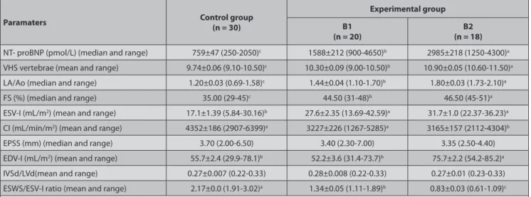
On the colour-flow Doppler examination, all 20 B1 dogs (100%) had mild MR (<20%), whereas among 18 B2 dogs, 6 (33%) had mild, 11 (61%) had moderate (20-40%) (Fig. 1) and 1 (6%) had severe (>50%) MR.
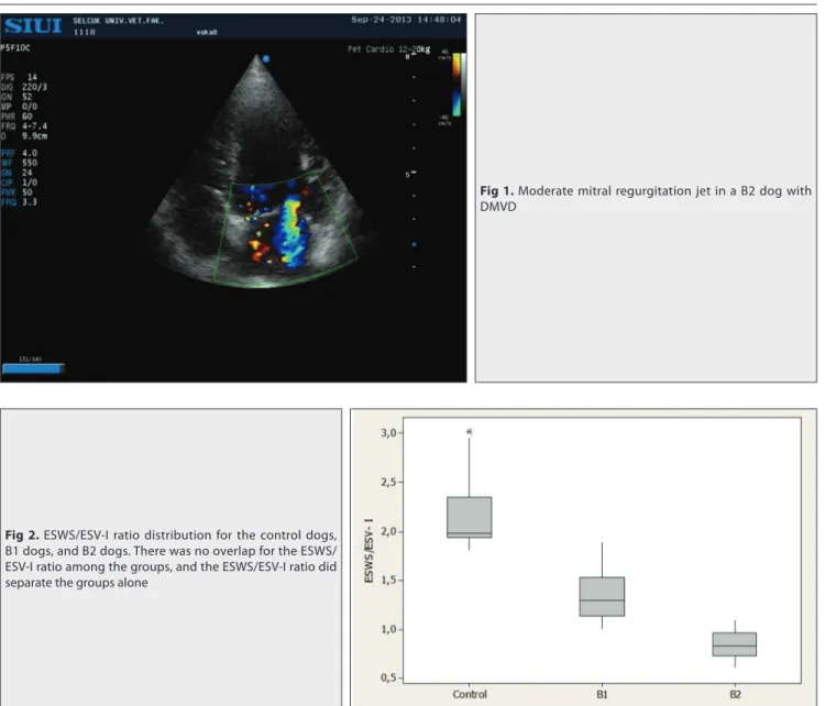
The mean ESWS/ESV-I ratio was (P<0.05) lower in the B2 dogs (0.83±0.03, range: 0.61-1.09) when compared with the B1 dogs (1.34±0.05, range: 1.11-1.89) and control dogs (2.17±0.0, range: 1.91-3.02). The difference between the B2 dogs and the B1 dogs (Table 1, Fig. 2) was also significant (P<0.05).
DISCUSSION
Many studies have provided important information on the natural progression of stage B2 DMVD and reported factors that can be used to identify which stage B2 dogs have higher versus lower risks of developing CHF [10,11]. The
results of studies on the effect of volume overload caused by DMVD on left ventricular function are controversial [2,4,8].
In our study, FS, ESV-I, CI showed a significant difference, especially in the B2 dogs (Table 1). However, there was some overlap for FS (29-45% for the control dogs, 31-48% for theB1 dogs, 45-51% for the B2 dogs) (Table 1), for the ESV-I (5.84-30.16 mL/m2 for the control dogs, 13.69-42.59
mL/m2 for the B1 dogs, 22.37-36.23 mL/m2 for the B2 dogs
(Table 1), for the CI (2907-6399 ml/min/m2 for the control
dogs, 1267-5285 mL/min/m2 for the B1 dogs, 2112-4304
mL/min/m2 for the B2 dogs) among the groups, and these
indexes did not manage to separate the groups (Table 1). This might be the result of decreased afterload, increased preload and sympathetic tone. For these reasons, we think that FS, ESV-I and CI are thought to be less sensitive indicators of systolic function in asymptomatic ASHs with DMVD, which is in agreement with previous studies [4,12]. All
these results may demonstrate that there seemed to be
Table 1. NT- proBNP, VHS, LA/Ao, LV, FS, ESV-I, CI, EPSS, EDV-I, IVSd/LVd and ESWS/ESV-I ratio in the control and experimental groups of dogs
Paramaters Control group
(n = 30)
Experimental group B1
(n = 20) (n = 18)B2
NT- proBNP (pmol/L) (median and range) 759±47 (250-2050)c 1588±212 (900-4650)b 2985±218 (1250-4300)a VHS vertebrae (mean and range) 9.74±0.06 (9.10-10.50)c 10.30±0.09 (9.00-10.50)b 10.90±0.05 (10.60-11.50)a
LA/Ao (median and range) 1.20±0.03 (0.69-1.58)c 1.44±0.04 (1.10-1.70)b 1.80±0.03 (1.73-2.10)a
FS (%) (median and range) 35.00 (29-45)c 44.50 (31-48)b 46.50 (45-51)a
ESV-I (mL/m2) (mean and range) 17.1±1.39 (5.84-30.16)b 27.6±2.35 (13.69-42.59)a 31.7±1.0 (22.37-36.23)a CI (mL/min/m2) (mean and range) 4352±186 (2907-6399)a 3227±226 (1267-5285)a 3165±157 (2112-4304)b
EPSS (mm) (median and range) 3.70 (2.00-6.50) 3.40 (2.30-7.00) 3.35 (2.50-4.40)
EDV-I (mL/m2) (mean and range) 55.7±2.4 (29.9-78.1)b 52.2±3.6 (31.4-73.7)b 75.7±2.2 (54.2-85.2)a
IVSd/LVd(mean and range) 0.27±0.007 (0.22-0.33) 0.28±0.008 (0.22-0.33) 0.27±0.01 (0.23-0.33)
ESWS/ESV-I ratio (mean and range) 2.17±0.0 (1.91-3.02)a 1.34±0.05 (1.11-1.89)b 0.83±0.03 (0.61-1.09)c
FS: fractional shortening; EF: ejection fraction; ESV-I: end-systolic volume index; EPSS: E point septal separation; EDV-I: end-diastolic volume index; CI: cardiac index; ESWS/ESV-I: end-systolic wall stress/end-systolic volume ratio; IVSd/LVd: interventricular septum thickness/left ventricular internal dimension ratio; a,b,c Values with different character in each row are statistically different with P<0.05
preserved systolic function and a hyper-dynamic phase of the heart in the B1 and B2 dogs.
Chamber enlargement may lead to increased peak systolic wall stress (by the Law of La Place), which causes wall thickening of a sufficient magnitude to normalise the systolic stress [3]. Suzuki et al.[13], have also reported that
relative wall thickness was not a factor in determining asymptomatic dogs with Class I DMVD and healthy dogs. Increased ESWS in LV volume overload causes mainly chamber enlargement, and eccentric hypertrophy. ESWS probably gives more accurate prediction when determining afterload than using pressure alone, because it deals with filling conditions, BP, wall thickness and curvature radius [7].
Clerfond et al.[7] informed that ESWS was an independent
estimator of LV remodelling and post-discharge HF after MI. When we consider ESV-I, dogs with ESV-I values>30 mL/m2 indicate impaired systolic function [3]. In our study,
ESV-I was high in the B2dogs (>30 mL/m2) (Table 1). For
this reason we thought ESWS/ESV-I would be useful in
evaluating systolic function. Carabello et al.[8] studied patients
with symptomatic, chronic, severe MR to determine which hemodynamic and angiographic factors might be prognostic of surgical outcome. They found that the ESWS/ESV-I ratio was more sensitive than EF or other hemodynamic variables in predicting surgical outcome in symptomatic patients with chronic MR. In our study, the mean ESWS/ESV-I ratio was lower (P<0.05) in the B2 dogs (0.83±0.03, range: 0.61-1.09) when compared with B1 dogs (1.34±0.05, range: 1.11-1.89) and control dogs (2.17±0.0 range: 1.91-3.02). The diff erence between the B2 dogs and the B1 dogs was also significant (P<0.05). There was no overlap for the ESWS/ ESV-I ratio among the groups, and the ESWS/ESV-I ratio did separate the groups alone (Table 1, Fig. 2). All the ESWS/ ESV-I ratios were ≤1.09 for the B2 group. This may indicate that the ESWS/ESV-I ratio is more accurate in assessing left ventricular function. Thus, the B2 dogs, who had a median FS of 46.50% (range: 45-51%), had a significantly lower ESWS/ESV-I ratio (≤1.09) than both control dogs and B1 dogs, indicating left ventricular function. The level of
Fig 2. ESWS/ESV-I ratio distribution for the control dogs,
B1 dogs, and B2 dogs. There was no overlap for the ESWS/ ESV-I ratio among the groups, and the ESWS/ESV-I ratio did separate the groups alone
Fig 1. Moderate mitral regurgitation jet in a B2 dog with
NT-proBNP secreted predominantly from the ventricle is elevated in response to cardiac remodelling [14,15]. In our
study, NT-proBNP levels (the B2 dogs: 2985±218 pmol/L (1250-4300 pmol/L), the B1 dogs: 1588±212 pmol/L (900-4650 pmol/L) and the control dogs: 759±47 pmol/L (250-2050 pmol/L) were irrespectively correlated with ESWS/ ESV-I ratio (Table 1). These results could be the reason of synthesis and secretion of NT-proBNP induced by the elevation of left ventricular ESWS in the B1 and the B2 dogs. It may suggest that the echocardiography-based approach could be improved through the determination of BNP.
Our results highlight that ESWS/ESV-I ratio (≤1.09) in ASHs with stage B2 DMVD may result in left ventricular remodelling and a risk factor in the development of systolic dysfunction; in other words, it may be to foresee the transition from asymptomatic to a symptomatic DMVD in dogs.
A
cknowledgmentsThis study was financed by the Scientific and Technological Research Council of Turkey (TÜBİTAK). Project No: 1120908 REFERENCES
1. Menciotti G, Borgarelli M: Review of diagnostic and therapeutic
approach to canine myxomatous mitral valve disease. Vet Sci, 4:47, 2017. DOI: 10.3390/vetsci4040047
2. Gordon SG, Saunders AB, Wesselowski SR: A symptomatic canine
degenerative valve disease: Current and future therapies. Vet Clin North
Am Small Anim Pract, 47, 955-975, 2017. DOI: 10.1016/j.cvsm.2017.04.003
3. Turgut K: Klinik Kedi ve Köpek Kardiyolojisi. Nobel Tıp Kitapevleri.
İstanbul, Türkiye, 2017.
4. Petric AD: Myxomatous mitral valve disease in dogs - An update
and perspectives. Mac Vet Rev, 38 (1): 13-20, 2015. DOI: 10.14432/j. macvetrev.2014.11.026
5. Chetboul V, Tissier R: Echocardiographic assessment of canine
degenerative mitral valve disease. J Vet Cardiol, 14, 127-148, 2012. DOI:
10.1016/j.jvc.2011.11.005.
6. Alter P, Koczulla AR, Nell C, Figiel JH, Vogelmeier CF, Rominger MB:
Wall stress determines systolic and diastolic function - Characteristics of heart failure. Int J Cardiol, 202, 685-693, 2016. DOI: 10.1016/j.ijcard. 2015.09.032
7. Clerfond G, Bière L, Mateus V, Grall S, Willoteaux S, Prunier F, Furber A: End-systolic wall stress predicts post-discharge heart failure
after acute myocardial infarction. Arch Cardiovasc Dis, 108, 310-320, 2015. DOI: 10.1016/j.acvd.2015.01.008
8. Carabello BA, Stanton P, Nolan MD, Lockhart B, Mcguire MD:
Assessment of preoperative left ventricular function in patients with mitral regurgitation: Value of the end-systolic wall stress-end-systolic volume ratio. Circulation, 64, 1212-1217, 1981.
9. Matos JM, Glaus TM: Medical treatment of canine heart failure. EJCAP,
20, 171-176, 2010.
10. Reynolds CA, Brown DC, Rush JE, Fox PR, Nguyenba TP, Lehmkuhl LB, Gordon SG, Kellihan HB, Stepien RL, Lefbom BK, Meier CK, Oyama MA: Prediction of first onset of congestive heart failure in dogs with
degenerative mitral valve disease: the PREDICT cohort study. J Vet Cardiol, 14 (1): 193-202, 2012. DOI: 10.1016/j.jvc.2012.01.008
11. Mattin MJ, Boswood A, Church DB, Lopez-Alvarez J, McGreevy PD, O’Neill DG, Thomson PC, Brodbelt DC: Prevalence of and risk
factors for degenerative mitral valve disease in dogs attending primary-care veterinary practices in England. J Vet Intern Med, 29, 847-854, 2015. DOI: 10.1111/jvim.12591
12. Chetboul V, Carlos Sampedrano C, Gouni V, Nicolle AP, Pouchelon JL, Tissier R: Ultrasonographic assessment of regional radial and
longitudinal systolic function in healthy awake dogs. J Vet Intern Med, 20, 885-893, 2006. DOI: 10.1111/j.1939-1676.2006.tb01802.x
13. Suzuki R, Matsumoto H, Teshima T, Mochizuki Y, Koyama H:
Left ventricular geometrical differences in dogs with various stages of myxomatous mitral valve disease. J Small Anim Pract, 54, 234-239, 2013. DOI: 10.1111/jsap.12058
14. Fox PR, Oyama MA, Hezzell, MJ, Rush JE, Nguyenba TP, DeFrancesco TC, Lehmkuhl LB, Kellihan HB, Bulmer B, Gordon SG, Cunningham SM, MacGregor J, Stepien RL, Lefbom B, Adin D, Lamb K: Relationship of plasma N-terminal pro-brain natriuretic peptide
concentrations to heart failure classification and cause of respiratory distress in dogs using a 2nd generation ELISA assay. J Vet Intern Med, 29, 171-179, 2014. DOI: 10.1111/jvim.12472
15. Strunz CMC, Marcondes-Santos M, Takada JY, Fragata FS, de Padua Mansur A: Quality of life score as a predictor of death in dogs with
degenerative mitral valve disease. Arq Bras Cardiol, 108 (4): 347-353, 2017. DOI: 10.5935/abc.20170032