Original Paper
Cells Tissues OrgansThe Effects of Hypericum perforatum L. on the
Proliferation, Osteogenic Differentiation, and
Inflammatory Response of Mesenchymal Stem
Cells from Different Niches
Ayşegül Mendi
aBeyza Gökçınar Yağcı
bNurdan Saraç
cMustafa Kızıloğlu
dAysel Uğur
aDuygu Uçkan
bDerviş Yılmaz
daDepartment of Medical Microbiology, Faculty of Dentistry, Gazi University, Ankara, Turkey; bPEDI-STEM Center for Stem Cell Research and Development, Hacettepe University, Ankara, Turkey; cDepartment of Biology, Faculty of Sciences, Muğla Sıtkı Koçman University, Muğla, Turkey; dDepartment of Oral and Maxillofacial Surgery, Faculty of Dentistry, Gazi University, Ankara, Turkey
Accepted after revision: June 26, 2018 Published online: August 17, 2018
DOI: 10.1159/000491633
Keywords
Mesenchymal stem cell · Osteogenic differentiation · Migration · Niche · Hypericum perforatum L. · Bone marrow · Dental pulp
Abstract
The aim of this study is to demonstrate and compare the dif-ferentiation, proliferation, migration and inflammatory be-havior of dental pulp- and bone marrow-derived mesenchy-mal stem cells (DP-MSCs and BM-MSCs) in response to a
Hy-pericum perforatum ethanol extract. Using xCELLigence, a
real-time monitoring system, a dose of 10 µg/mL was found to be the most efficient concentration for vitality. The IC50 values and doubling time were calculated. The results showed that H. perforatum L. was able to accelerate osteo-genic differentiation in DP-MSCs, but calcium granulation was impaired in BM-MSCs. H. perforatum L.-induced tion increased when compared to the TNF-α-induced migra-tion in a Transwell migramigra-tion assay, and the IL-6 cytokine
lev-els between cells also differed. It can be suggested that tis-sue memory is an important factor in MSCs, and that they differ in their response to external factors. In conclusion, H.
perforatum L. can be considered an excellent osteoinductive
agent for DP-MSCs but should not be used for BM-MSCs. Tis-sue-specific osteoinductive agents should be discussed in
future studies. © 2018 S. Karger AG, Basel
Introduction
Mesenchymal stem cells (MSCs) can be considered a promising tool in regenerative medicine due to their high therapeutic potential in the treatment of degenerative and metabolic diseases [Kornicka et al., 2017]. MSCs are ca-pable of self-renewal and differentiation into different lineages, including bone, cartilage, fat, tendon, muscle, and hematopoietic stroma [Deans and Moseley, 2000]. Cellular therapies involving MSCs require their isolation
C
ells
T
issues
mainly from bone marrow (BM), adipose tissue, the um-bilical cord, or dental pulp (DP), and in vitro expansion for further autologous or allogenic transplantations, and these have the potential to differentiate into a wide variety of cell lineages (i.e., osteoblasts, adipocytes, chondro-cytes, tenochondro-cytes, neurons and myocytes [Gronthos et al., 2000; Ding et al., 2011; Inoue et al., 2013]. Based on these advantageous properties, techniques to increase MSC proliferation and differentiation are under constant scru-tiny. Extensive use of synthetic and semi-synthetic sub-stances, i.e., the recombinant cytokines and growth fac-tors currently used as proliferative and differentiation factors in stem cell therapy, particularly for bone regen-eration, may lead to side effects and toxicity while also being exorbitantly expensive. Osteoinductive agents aim to stimulate seeded cell migration, proliferation, and mi-gration, and modulate immune responses, involving the stimulation of MSCs and/or osteoprogenitors to differen-tiate into osteoblasts. There are many osteogenic proteins that stimulate the proliferation and differentiation of MSCs and/or progenitors in vitro and in vivo [Govender et al., 2002; Giannoudis et al., 2005; Ko et al., 2013]. How-ever, they have a short half-life, and so require either high concentrations or sustained delivery for bone tissue engi-neering [Itoh et al., 2001]. That said, higher concentra-tions could lead to increased osteoclastic activity and bone resorption [Kaneko et al., 2000], and so it is appar-ent that alternative and natural ostoeinductive agappar-ents need to be identified.
Recent studies have shown that bioactive compounds, which occur naturally in seaweed, herbs, fruits, and veg-etables, have the ability to modulate self-renewal and the differential potential of adult stem cells, targeting a broad range of intracellular signal transduction pathways [Kor-nicka et al., 2017]. There are a number of ongoing trials, aiming to find a herbal extract that is less toxic and more affordable as a natural therapy [Udalamaththa et al., 2016]. Promoting endogenous stem cell multipotency and differentiation potential may also support
regenera-tive processes after MSC transplantation [Kornicka et al., 2017]. We suggest that natural ostoeinductive agents should be identified from MSCs of different origins if suc-cessful outcomes in bone regeneration are to be achieved.
Hypericum perforatum L., also known as St. John’s
wort [Oztürk et al., 2007], is known to have remarkable wound-healing and anti-inflammatory properties [Knüp-pel and Linde, 2004; Dole et al., 2015]. It is used to treat anxiety, depression, lacerations, burns, cancer, and bacte-rial and viral diseases, and is an antioxidant, analgesic, and neuroprotective agent [Rota et al., 2004]. Based on these data, we chose H. perforatum L. as a promising nat-ural osteoinductive agent, and compared its effects on proliferation, differentiation, migration, and immune re-sponse in MSCs from different niches.
Materials and Methods Extraction of Plant Samples
H. perforatum L., as naturally growing plants belonging to the Hypericaceae family, were purchased from a local market in Muğla, Turkey, and a voucher specimen (herbarium No: MUH 2796) was deposited in the Herbarium of the Faculty of Science, University of Muğla, Turkey. Air-dried plant samples were ex-tracted with ethanol (Merck, Taufkirchen, Germany) using a Soxhlet apparatus, and the extracts were evaporated and stored in sterile opaque glass bottles under refrigerated conditions until use. The total hypericin amount in the ethanol extract was determined as 0.2 mg (Dr. Yılmaz Klinik, Kayseri).
Isolation and Culture of MSCs
Human DP tissue was obtained from patients (aged 15–20 years) who were undergoing extraction of their third molars for orthodontic reasons at the Department of Oral and Maxillofacial Surgery, Gazi University, Ankara, Turkey. All patients signed an informed consent form. After the tooth surfaces were disinfected (75% ethanol), the tooth was drilled, and the DP extracted gently with forceps. The extracted pulp tissue was rinsed in α-MEM sup-plemented with 2 nM L-glutamine, 100 U/mL penicillin, 100 μg/ mL streptomycin, and 10% FBS (Invitrogen/GIBCO, Grand Is-land, NY, USA) (hereafter referred to as the MSC culture medi-um), after which it was minced into fragments 1–2 mm3 in size.
The tissue fragments were cultured on T75 plates (Nunc) in MSC culture medium at 37 ° C in a humidified atmosphere containing 5% CO2.
The human BM-MSCs were a kind gift from the Hacettepe University Center for Pediatric Stem Cell Research and Develop-ment. They were suspended in a concentration of 1 × 10⁶ cells/mL
in MSC culture medium, and the cultures were monitored regu-larly with an inverted microscope (Olympus CKX41, Tokyo, Ja-pan). The MSC culture medium was changed every 3 days. After reaching 70–80% confluence, the cells were harvested with 0.05% Trypsin/EDTA (Sigma Aldrich, St. Louis, MO, USA) and subcul-tured for further experiments. The experiments were done on pas-sage 2–3 cells.
Abbreviations used in this paper
BM-MSCs bone marrow-derived mesenchymal stem cells DP-MSCs dental pulp-derived mesenchymal stem cells
DT doubling time
MSCs mesenchymal stem cells
OCN osteocalcin
ON osteonectin
Immunophenotypic Analysis
The culture-expanded adherent cells were analyzed on flow cy-tometry (BD FACSAria, USA), and the antibody panel included CD105-PE (eBioscience, USA); CD44-PE (eBioscience); CD90-PE (BD, USA); CD166-PE (BD); CD146-PE (BD); CD73-PE (BD); and CD29-FITC (eBioscience) as mesenchymal stromal markers, as well as their isotype controls. CD45-FITC (BD); CD14-PE (BD); HLA DR-FITC (Chemicon, USA); and CD34-FITC (BD) were used as hematopoietic markers to exclude any cells of hematopoi-etic origin. The relative frequencies of the cells that expressed the respective surface markers were analyzed using FACS Diva soft-ware v6.0.0 (BD-Biosciences, San José, CA, USA) by acquiring 10,000 events for each sample.
Effect of H. perforatum L. on MSC Proliferation Using the xCELLigence System
Initially, the proliferation of DP-MSCs in a 24-well culture mi-croplate seeded at a density of 5,000 cell/cm2 was examined.
DP-MSCs were cultured in different concentrations (1, 3, 5, 10, 25, 50, 75, and 100 µg/mL) of H. perforatum L. up to the control group of 90% confluency. The cells were counted using the Trypan blue method (data not shown), and the 3 concentrations (i.e., 5, 10, and 25 μg/mL) that induced the cell number were selected for xCELLigence analysis. The xCELLigence system, made up of an impedance-based real-time cell analyzer (RTCA), an RTCA single plate (E-plate 96), an RTCA computer and a tissue-culture incuba-tor, was used according to the manufacturer’s instructions (Roche
Applied Science, Mannheim, Germany) [Roche Diagnostics, 2008]. The E-plate 96 was connected to the xCELLigence system and verified in a cell culture incubator to ensure that proper elec-trical contacts were established. The background impedance was measured. Subsequently, 100 μL of MSC culture medium contain-ing different concentrations of H. perforatum L. were added into each well of the E-plate 96, and the cells were resuspended (5,000 cells/cm2) in MSC culture medium containing the appropriate
concentration of H. perforatum L. Cell growth and proliferation were monitored every 30 min for up to 314 h for DP-MSCs, and 290 h for BM-MSCs.
Effect of H. perforatum L. on MSC Differentiation
The concentration that decreased the doubling time (DT) and increased the proliferation was selected based on the results of the xCELLigence system analysis. The selected concentration was add-ed to the osteogenic and adipogenic differentiation madd-edia [Pit-tenger et al., 1999], and the secreted osteocalcin (OCN) and osteo-nectin (ON) levels in the supernatants were assessed using an ELISA kit, in line with the manufacturer’s instructions (R&D Systems, Inc. Minneapolis, USA). The limits of detection for the ELISA were 1.2–75 ng/mL for OCN and 1.56–50 ng/mL for ON. The calcium ion concentration in the differentiation medium was measured using a QuantiChrom calcium assay kit according to the manufacturer’s instructions (DICA 500, BioAssay Systems, Hay-ward, CA, USA).
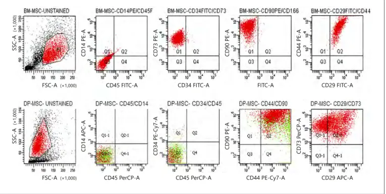
SSC-A CD14 PE-A (×1,000) (×1,000) SSC-A FSC-A (×1,000)
FSC-A CD45 PerCP-A CD45 PerCP-A CD44 PE-Cy7-A CD29 APC-A
CD29 FITC-A FITC-A CD34 FITC-A CD45 FITC-A CD14 APC-A CD73 PE-A CD34 PE-Cy7-A CD90 PE-A CD90 PE-A CD44 PE-A CD73 P erCP-A (×1,000)
Fig. 1. DP-MSCs and MSCs were identified as MSCs according to their expression of surface receptors.
BM-MSCs and DP-BM-MSCs were found positive for MSC markers (CD90, CD44, CD73, and CD29) and negative for hematopoietic stem cell markers (CD14, CD45, and CD34). Also, a subpopulation in DP-MSCs was found posi-tive for MSCs and negaposi-tive for hematopoietic markers.
BM-MSC-UNSTAINED BM•MSC-CD14PE/CD45F BM-MSC-CD34FITCICD73 BM-h1SC-CD90PEICDI 68 BM·MSC-CD29FITCICD4 4
g "' "'e " "' " " " S' 0 0 " . "
..
,,2 '< 0 e S' S' :!; 0 "e " " "e e e S' N N N.
.
e S' S' S' ro 100 l!-0 100 2lllDP-MSC-UNSTAINED DP-MSC-CD45/CD14 OP-MSC- CD34ICD45 DP-1,ISC-CD44/CD90 DP-MSC-C0291C073
0
~2
N
"'e "'e "'e
" ., "e •s-
..
; ., Ql-1 Q2'1 QI Q2 !l M$? "2 "..
,.. C :a "2 "e "!? '"'e ~ 100 1:0 lOD 1:0 10'Effect of H. perforatum L. on MSC Migration
To investigate the migration of DP-MSCs and BM-MSCs in response to TNF-α and the H. perforatum L. extract, we used a Transwell chamber assay in a 6-well microchemotaxis chamber with 8-μm pores (Corning Costar, USA). The upper chambers were loaded with 5 × 10⁴ MSCs in 500 μL of MSC culture medium,
and the MSC culture medium containing 10 μg/mL H. perforatum L. extract was placed in the upper chamber. After 48 h of incuba-tion at 37 ° C in 5% CO2, the migrated cells were counted using a Trypan blue cell viability assay. Each experiment was performed in duplicate, and the means of the garnered data were recorded for statistical analysis. We used 10 ng/mL TNF-α (Invitrogen/GIBCO) as the control [Lopez-Ponte et al., 2007].
Effect of H. perforatum L. on MSC Immunomodulatory Activities
DP-MSCs and BM-MSCs were plated at a density of 5,000 cell/ cm2 on 96-well culture plates and allowed to attach overnight. The
cells were pretreated with 10 μg/mL H. perforatum L. extract for 1 h, after which 10 ng/mL TNF-α (Invitrogen/GIBCO) was added. After 24 h, the cell culture supernatants were collected and stored at –80 ° C for use in the IL-6 and IL-10 ELISAs, according to the
manufacturer’s instructions, and medium alone, with TNF-α and H. perforatum L. were included as controls.
Statistical Analysis
All calculations were carried out using the RTCA integrated software of the xCELLigence system, which fits the curve of the selected sigmoidal dose response equations to the experimental data points. Data are presented as mean (μg/mL) ± SD (n = 4). For the proliferation experiments, a statistical analysis was performed using ANOVA (p < 0.05).
Results
Identification of MSCs
The common MSC markers (CD90, CD44, CD29, and CD73) were constitutively positive (>92%) and the hema-topoietic markers (CD14, CD45, and CD34) were nega-tive (>97.9) in the tested BM-MSC samples, indicating a mesenchymal origin of the cells (Fig. 1). Interestingly, 2
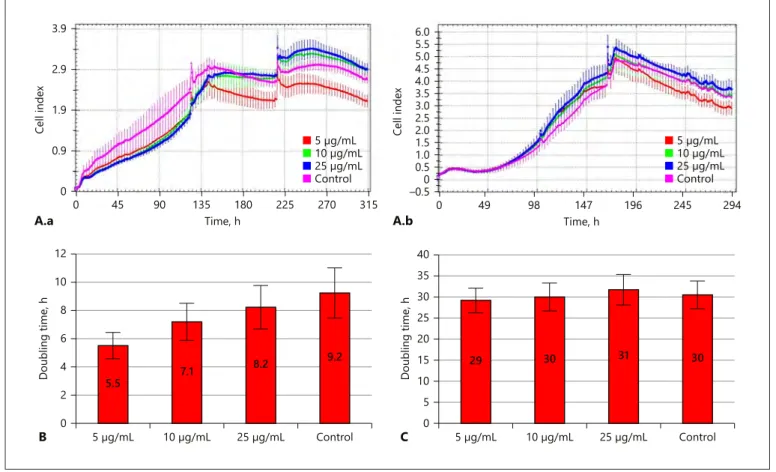
12 3.9 2.9 1.9 0.9 0 0 45 90 135 180 Time, h 225 270 315 10 8 Doubling time, h 6 4 2 5.5 5 µg/mL 10 µg/mL 25 µg/mL Control 7.1 8.2 9.2 0 40 35 30 25 Doubling time, h Cell index Cell index 20 15 10 5 29 5 µg/mL 10 µg/mL 25 µg/mL Control 30 31 30 0 6.0 5.5 5.0 4.5 4.0 3.5 3.0 2.5 2.0 1.5 1.0 0.5 0 –0.5 0 49 98 147 196 Time, h 245 294 ■ 5 µg/mL ■ 10 µg/mL ■ 25 µg/mL ■ Control ■ 5 µg/mL ■ 10 µg/mL ■ 25 µg/mL ■ Control B A.a A.b C
Fig. 2. Growth curve (A) and doubling time (B, C) graphs for DP-MSCs and BM-MSCs. A.a DP-MSCs showed a
rapid logarithmic phase and H. perforatum L. increased proliferation. A.b BM-MSCs showed a slow proliferation rate. B Population doubling time was decreased with H. perforatum in DP-MSCs. C H. perforatum L. showed no effect on the population doubling time of the BM-MSCs.
subpopulations were identified in the flow cytometer analysis, suggesting that the DP has multiple stem cell niches.
xCELLigence Assays
The Trypan blue assay showed that concentrations of 1–25 µg/mL increased cell viability, while 50–100 µg/ mL reduced viability, and 10 µg/mL was found to be the effective concentration (data not shown). Based on these findings, 5, 10, and 25 µg/mL were selected for the xCELLigence analysis system, and a cell proliferation graph, cell index, and DT values for DP-MSCs and BM-MSCs were produced (Fig. 2). The IC50 value was 250 µg/mL for DP-MSCs and 1,000 µg/mL for BM-MSCs (Table 1).
Differentiation Assay
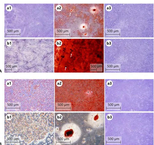
The DP-MSCs did not undergo an adipogenic differ-entiation (Fig. 3A.a1, b1). Approximately 20% of the cells became rounder, but no lipid droplets were observed. In contrast to the adipogenic differentiation, the DP-MSCs
underwent rapid osteogenic differentiation, with small, dark deposits becoming visible after 7–10 days, and in-creasing over the next several days of culture. Calcium mineralization was confirmed from positive Alizarin Red S staining on day 21 of the culture. The DP-MSCs that were treated with 10 μg/mL H. perforatum L. extract for
21 days exhibited calcium mineralization (Fig. 3B.a2, b2). Although ON and OCN levels were found in H.
perfora-tum L.-treated BM-MSCs, the calcium mineralization was
found to be reduced (Table 1). The levels of the ON and OCN osteogenic markers were also determined (Fig. 3B), and the data showed that the level of the early osteogenic marker (ON) decreased in the H. perforatum L.-treated group, whilst the levels of the late osteogenic marker (OCN) increased when compared to the control group.
Migration Assay
The spontaneous migration capacity of DP-MSCs and BM-MSCs in the presence of medium alone (without TNF-α or H. perforatum L.) was low, but it increased in the presence of TNF-α (Table 1), and H. perforatum L.
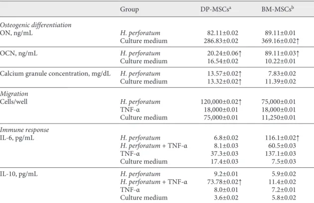
Table 1. H. perforatum L. effect on DP-MSCs and BM-MSCs
Group DP-MSCsa BM-MSCsb Osteogenic differentiation ON, ng/mL H. perforatum 82.11±0.02 89.11±0.01 Culture medium 286.83±0.02 369.16±0.02↑ OCN, ng/mL H. perforatum 20.24±0.06↑ 89.11±0.03↑ Culture medium 16.54±0.02 10.22±0.01
Calcium granule concentration, mg/dL H. perforatum 13.57±0.02↑ 7.83±0.02
Culture medium 13.32±0.02↑ 11.39±0.02 Migration Cells/well H. perforatum 120,000±0.02↑ 75,000±0.01 TNF-α 18,000±0.01 18,000±0.01 Culture medium 75,000±0.01 11,250±0.01 Immune response IL-6, pg/mL H. perforatum 6.8±0.02 116.1±0.02↑ H. perforatum + TNF-α 8.1±0.03 60.5±0.03 TNF-α 37.3±0.03 137.1±0.03 Culture medium 17.4±0.03 7.5±0.03 IL-10, pg/mL H. perforatum 9.2±0.01 5.9±0.02 H. perforatum + TNF-α 73.78±0.02↑ 11.4±0.02 TNF-α 8.0±0.01 7.2±0.01 Culture medium 3.6±0.02 5.8±0.02
DM-MSCs, dental pulp-derived mesenchymal stem cells; BM-MSCs, bone marrow-derived MSCs; ON, os-teonectin; OCN, osteocalcin; ↑, increased.
increased the migration of DP-MSCs 2-fold when com-pared to the control and the BM-MSCs.
Determining the Preventive Effect of H. perforatum L. on the Inflammatory Response of MSCs
Both IL-6 and IL-10 were present in the DP-MSC and BM-MSC cell culture supernatants (Table 1). Our find-ings showed that when used alone, H. perforatum L. re-duced the IL-6 level in DP-MSCs, and, as expected, TNF-α increased the IL-6 level. H. perforatum L., was able to re-duce the IL-6 level in DP-MSCs; however, in BM-MSCs, it increased the IL-6 level both when used alone and with TNF-α stimulation.
Discussion
Research into osteoinductive agents for bone tissue engineering is more likely focused on promoting cell pro-liferation, differentiation, and migration, and determin-ing immune response. This study has shown that the functions of MSCs may vary according to their site of
or-igin, even when they are treated with the same osteoin-ductive agent.
We demonstrated that DP-MSCs and BM-MSCs showed different population characteristics when the BM-MSC surface markers were taken as a reference. In-terestingly, we identified 2 subpopulations of DP-MSCs in the flow cytometry analysis, which suggests the pres-ence of multiple stem cell niches in the DP. This finding concurs with that of Pisciota et al. [2015], showing that the heterogeneity of the stem cell population within hu-man DP, particularly its peculiar embryological origin, may explain the existence of 2 different subpopulations. Of course, further studies could better sort and analyze subpopulations to identify their differences and prolif-eration potencies.
To determine the H. perforatum L. effect, we first sought to define effective concentrations of H.
perfora-tum L., selecting 5, 10, and 25 µg/mL for the xCELLigence
analysis. This analysis allowed real-time cell proliferation monitoring, leading to a robust cell count, and we were also able to obtain and compare the growth curves of 2 cell types. Growth curves provide information about 3
B A 500 µm 500 µm 500 µm 500 µm 500 µm 500 µm 500 µm 500 µm 500 µm 500 µm 500 µm 500 µm a1 b1 a1 a2 a2 b1 b2 b2 a3 a3 b3 b3
Fig. 3. I. Differentiation potential of
DP-MSCs (A) and BM-MSCs (B). A.a1, b1 Ad-ipogenic differentiation was not seen in either the controls or in H. perforatum L.-treated DP-MSCs. The osteogenic dif-ferentiation of the DP-MSCs increased in the H. perforatum L.-treated group (A.b2) when compared to the control group (A. a2). ×4 (Olympus CKX41). White arrows show the calcium granules in the culture. B.a1 BM-MSCs were well-differentiated into adipocytes in the control group. B.b1 However, this was found to be decreased by H. perforatum L. B.b2 Osteogenic differen-tiation was also decreased by H. perforatum L. B.a1 However, large calcium granules were observed in the control group.
parameters: the lag phase before cell proliferation is initi-ated after subculture, the DT in the middle of the expo-nential growth phase, and the terminal density. It was clear from the curves that the log phase of DP-MSCs was shorter than that of BM-MSCs. Cell indexes and prolif-eration graphs revealed that DP-MSCs entered the log phase earlier than BM-MSCs, while BM-MSCs showed increased proliferation at 180 h when compared with DP-MSCs, although the DP-MSCs were still alive in station-ary phase at 314 h. We also calculated the DT at the mid-log phase of the cells. A shortened DT with H. perforatum L. (10 µg/mL) in DP-MSCs suggests a more rapid increase in cell proliferation, and the healing fracture or implant region could be shortened.
Oriental medicine practices are based primarily on personal experience, but often rely on unknown mecha-nisms, leading to difficulties in dose specification. The xCELLigence system is a sophisticated cell-based assay. It offers an alternative to commercialized conventional cell analyses, in which expensive agents are used, by simulta-neous monitoring of cell proliferation and viability as well as label-free cytotoxicity [Hensten-Pettersen, 1988]. A lack of determination of concentrations can lead to con-flicting reports. Demiroğlu et al. [2005] reported a rare case of BM necrosis in a patient who had been taking H.
perforatum L. for the treatment of depression at a dose of
approximately 1,000 mg/day for 3 weeks; this could be at-tributed to the usage of high concentrations of H.
perfo-ratum L. Since BM-MSCs line the marrow and are
in-volved in hematopoiesis, high doses of extracts could lead to tissue necrosis. On the other hand, Knüppel and Linde [2004] reported that the available evidence suggests that
H. perforatum L. is well-tolerated and safe if taken under
the guidance of a physician who is aware of the potential risks under specific circumstances. In addition, Bray et al. [2002] administered extract of St John’s wort at a dose of 140 mg/kg in their animal study. There is no recommend-ed concentration of H. perforatum L., but we suggest that there should be a unique concentration if a clear conclu-sion is to be reached. We can conclude with confidence, based on the data obtained, that 10 µg/mL of H.
perfora-tum L. protects cell vitality; accordingly, this
concentra-tion has been used in subsequent studies.
To demonstrate whether the extracts induce a differ-entiation of MSCs, we constructed an in vitro model in which lipid-laden adipocytes, calcium granules, and early and late markers of osteogenesis were examined after 21 days. No adipogenic differentiation was seen in DP-MSCs, while BM-MSCs were well-differentiated. Our findings concur with those of Gronthos et al. [2000], who
expanded DP-MSCs from single-cell clones and demon-strated that these cells exhibit osteogenic differentiation, but do not form lipid-laden adipocytes. A reduced adipo-genic differentiation was seen in a microscopic analysis of
H. perforatum L.-treated BM-MSCs. A few clones of
DP-MSCs with H. perforatum L. tried to differentiate into ad-ipocytes, but they could not be determined. The osteo-genic differentiation potentials of DP-MSCs in vitro and in vivo have been well-documented in several studies [D’aquino et al., 2007]. During osteogenic differentiation, markers of undifferentiated cells are gradually turned off, and differentiation markers are then expressed sequen-tially [Huang et al., 2007]. Levels of ON (an early osteo-genic marker) and OCN (a late osteoosteo-genic marker) levels were determined, and H. perforatum L.-treated DP- and BM-MSCs showed reduced ON secretion compared to controls. ON is synthesized by preosteoblasts and has less affinity to collagen. The ON transcript is quite stable, with a half-life of >24 h under conditions of transcription ar-rest [Dole et al., 2015], and it can thus be concluded that the earlier synthesized ON, via the H. perforatum L.-treat-ed DP-MSCs and BM-MSCs, acceleratL.-treat-ed osteogenic dif-ferentiation. The preosteoblasts matured on approxi-mately day 14, and OCN was synthesized by these mature osteoblasts and marked the late phase of osteogenic dif-ferentiation. OCN synthesis leads to calcium deposits in the bone, has an affinity for collagen, and plays an impor-tant role in the mineralization and formation of bone [Ram et al., 2015]. The H. perforatum L.-treated DP-MSCs and BM-DP-MSCs exhibited higher OCN levels than the untreated cells; however, it is notable that calcium concentrations were found to be low in H. perforatum L.-treated BM-MSCs, despite the reduced ON and increased OCN levels. We suggest H. perforatum L. may disturb the calcium deposit development of the BM-MSCs.
Similarly, but using a different plant extract, Anpo et al. [2011] and our previous study [Mendi et al., 2017] pro-vide epro-vidence that eugenol/Syzygium aromaticum reduc-es the synthreduc-esis of collagen, which plays a critical role in osteogenesis. This is an interesting result, since the H.
per-foratum L. effect changes according to the origin of the
cells. H. perforatum L. could be used safely to induce and accelerate osteogenic differentiation for DP-MSCs, but not for BM-MSCs.
Evidence suggests that MSCs can target injured or ischemic tissues, which involves migration across layers of endothelial cells, and Lopez-Ponte et al. [2007] showed increased migration in response to TNF-α preincubation. In this study, we demonstrated that H. perforatum L. can stimulate greater DP-MSC migration than cells
stimulat-ed with TNF-α and BM-MSCs in vitro. Recruitment is important for tissue regeneration, and this is the first study to show the migration effect of H. perforatum L. on MSCs.
Next, we determined the response of DP-MSCs and BM-MSCs to H. perforatum L. and TNF-α-induced in-flammation. Both IL-6 and IL-10 were present in the cell culture supernatants, consistent with the findings of Egermann et al. [2005]. Our results show that when the extract alone is used, the IL-6 levels increased in BM-MSCs but decreased in DP-BM-MSCs. In contrast with this result, in the cells that were pretreated with H. perforatum L., before TNF-α stimulation, the IL-6 level was found to be decreased at a ratio of 20 and 60% in DP-MSCs and BM-MSCs, respectively. Pricola et al. [2009] showed that IL-6 is both necessary and sufficient for enhanced MSC proliferation, in that it protects MSCs from apoptosis, in-hibits the adipogenic and chondrogenic differentiation of MSCs, and increases the rate of in vitro wound-healing of MSCs. Based on these findings, we suggest that H.
perfo-ratum L. may lead to a direct increase of IL-6 and a
re-duced adipogenic differentiation indirectly in BM-MSCs. The need for bone regeneration in cranial, oral, maxil-lofacial, and orthopedic surgery is one of the central clin-ical issues in regenerative and rehabilitation medicine. We conclude that H. perforatum L. differentially affected DP-MSCs and BM-MSCs, especially with regard to IC50 values, osteogenic differentiation, and IL-6 secretion. There is emerging evidence that MSCs and their niches are critically important for tissue regeneration, based on
the secretion of interleukins in response to different stim-ulants, such as H. perforatum L., as shown in this study. We propose that therapeutic efforts to regenerate the stem cell niche are important for tissue engineering. In this regard, H. perforatum L. was found to be more effi-cient in DP-MSCs than BM-MSCs, suggesting that it can be used for topical and oromaxillofacial regions. Tissue-specific osteoinductive agents should be examined in fu-ture studies.
Acknowledgements
We give special thanks to Sevil Köse and Prof. Dr. Petek Korku-suz for their assistance with the xCELLigence assays and analysis.
Statement of Ethics
The research was approved by the Gazi University Ethics Com-mittee of Clinical Research.
Disclosure Statement
The authors declare no conflict of interests related to this study. Funding Sources
This study was supported by the Scientific and Technologi- cal Research Council of Turkey (TUBITAK, project No. SBAG 113S448).
References
Anpo, M., K. Shirayama, T. Tsutsui (2011) Cyto-toxic effect of eugenol on the expression of molecular markers related to the osteogenic differentiation of human dental pulp cells. Odontol 99: 188.
Bray, B.J., N.B. Perry, D.B. Menkes, R.J. Rosen-gren (2002) St. John’s wort extract induces CYP3A and CYP2E1 in the Swiss Webster mouse. Toxicol Sci 66: 27–33.
D’aquino, R., A. Graziano, M. Sampaolesi, G. Lai-no, G. Pirozzi, A. De Rosa, et al. (2007) Hu-man postnatal dental pulp cells co-differenti-ate into osteoblasts and endotheliocytes: a pivotal synergy leading to adult bone tissue formation. Cell Death Differ 14: 1162–1171. Deans, R.J., A.B. Moseley (2000) Mesenchymal
stem cells: biology and potential clinical uses. Exp Hematol 28: 875–884.
Demiroğlu, Y.Z., T.T. Yeter, C. Boğa, H. Özdoğu, E. Kızılkılıç, N. Bal, et al. (2005) Bone marrow necrosis: a rare complication of herbal
treat-ment with Hypericum perforatum (St John’s wort). Acta Medica 48: 91–94.
Ding, D.C., W.C. Shyu, S.Z. Lin (2011) Mesen-chymal stem cells. Cell Transplant 20: 5–14. Dole, N.S., K. Kapinas, C.B. Kessler, S.P. Yee, D.J.
Adams, R.C. Pereira, (2015) A single nucleo-tide polymorphism in osteonectin 3′ untrans-lated region regulates bone volume and is tar-geted by miR-433. J Bone Min Res 30: 723– 732.
Egermann, M., J. Goldhahn, E. Schneider (2005) Animal models for fracture treatment in os-teoporosis. Osteoporos Int 16: 129–138. Giannoudis, P.V., H. Dinopoulos, E. Tsiridis
(2005) Bone substitutes: an update. Injury
36(suppl 3): 20–27.
Govender, S., C. Csimma, H.K. Genant, A. Valen-tin-Opran, Y. Amit, R. Arbel, et al.; BMP-2 Evaluation in Surgery for Tibial Trauma (BESTT) Study Group (2002) Recombinant human bone morphogenetic protein-2 for
treatment of open tibial fractures: a prospec-tive, controlled, randomized study of four hundred and fifty patients. J Bone Joint Surg Am 84: 2123–2134.
Gronthos, S., M. Mankani, J. Brahim, P.G. Robey, S. Shi (2000) Postnatal human dental pulp stem cells (DPSCS) in vitro and in vivo. Pro Nat Acad Sci 97: 13625–13630.
Hensten-Pettersen, A. (1988) Comparison of the methods available for assessing cytotoxicity. Int Endod J 21: 89–99.
Huang, W., S. Yang, J. Shao, Y.P. Li (2007) Signal-ing and transcriptional regulation in osteo-blast commitment and differentiation. Front Biosci 12: 3068–3092.
Inoue, T., M. Sugiyama, H. Hattori, H. Wakita, T. Wakabayashi, M. Ueda (2013) Stem cells from human exfoliated deciduous tooth-de-rived conditioned medium enhance recovery of focal cerebral ischemia in rats. Tissue Eng A 19: 24–29.
Itoh, K., N. Udagawa, T. Katagiri, S. Iemura, N. Yasuda, K. Higashio, et al. (2001) Bone mor-phogenetic protein 2 stimulates osteoclast dif-ferentiation and survival supported by recep-tor activarecep-tor of nuclear facrecep-tor-κB ligand. En-docrinology 142: 3656–3662.
Kaneko, H., T. Arakawa, H. Mano, T. Kaneda, A. Ogasawara, M. Nakagawa, et al. (2000) Direct stimulation of osteoclastic bone resorption by bone morphogenetic protein (BMP)-2 and expression of BMP receptors in mature osteo-clasts. Bone 27: 479–486.
Knüppel, L., K. Linde (2004) Adverse effects of St. John’s wort: a systematic review. J Clin Psychiatry 65: 1470–1479.
Ko, E., K. Yang, J. Shin, S.W. Cho (2013) Biomac-romolecules 14: 3202−3213.
Kornicka, K., I. Kocherova, K. Marycz (2017) The effects of chosen plant extracts and com-pounds on mesenchymal stem cells – a bridge between molecular nutrition and regenerative medicine – concise review. Phytother Res 31: 947–958.
Lopez-Ponte, A., E. Marais, N. Gallay, A. Lan-gonne, B. Delorme, O. Herault, et al. (2007) The in vitro migration capacity of human bone marrow mesenchymal stem cells: com-parison of chemokine and growth factor che-motactic activities. Stem Cells 25: 1737–1745. Mendi, A., B. Gökçınar Yağcı, M. Kızıloğlu, N. Sa-raç, D. Yılmaz, A. Uğur, D. Uçkan (2017) Ef-fects of Syzygium aromaticum, Cinnamomum
zeylanicum, and Salvia triloba extracts on
proliferation and differentiation of dental pulp stem cells. J Appl Oral Sci 25: 515–522. Oztürk, N., S. Korkmaz, Y. Oztürk (2007)
Wound-healing activity of St. John’s wort (Hypericum perforatum L.) on chicken em-bryonic fibroblasts. J Ethnopharmacol 111: 33–39.
Pisciota, A., G. Carnevale, S. Meloni, M. Ricio, S.D. Biasi, L. Gibellini, et al. (2015) Human dental pulp stem cells (hDPSCs): isolation, enrichment and comparative differentiation of two sub-populations. BMC Develop Biol
15: 14–16.
Pittenger, M.F., A.M. Mackay, S.C. Beck, R.K. Jaiswal, R. Douglas, J.D. Mosca, et al. (1999) Multilineage potential of adult human mes-enchymal stem cells. Sci 284: 143–147.
Pricola, K.L., N.Z. Kuhn, H. Haleem-Smith, Y. Song, R.S. Tuan (2009) Interleukin-6 main-tains bone marrow derived mesenchymal stem cells stemness by an ERK1/2-dependent mechanism. J Cell Biochem 108: 577–588. Ram, V.S., S. Parthiban, U. Sudhakar, N.
Mithra-das, R. Prabhakar (2015) Bone biomarkers in periodontal disease: a review article. J Clin Diagn Res 9: 7–10.
Roche Diagnostics GmbH (2008) Introduction of the RTCA SP instrument. RTCA SP Instru-ment Operator’s Manual A. San Diego, Acea Biosciences, Inc., pp 14–16.
Rota, C., J.J. Carraminana, J. Burillo, A. Herrera (2004) In vitro antimicrobial activity of essen-tial oils from aromatic plants against selected foodborne pathogens. J Food Prot 67: 1252– 1256.
Udalamaththa, V.L., C.D. Jayasinghe, P.V. Udagama (2016) Potential role of herbal rem-edies in stem cell therapy: proliferation and differentiation of human mesenchymal stro-mal cells. Stem Cell Res Ther 7: 110–118.