Ankara ÜnivVet Fak Derg, 50,203-207, 2003
The effects of follicle diameter on the in vitro fertilization capacity of
bovine oocytes aspirated from the slaugthered ovaries
Mustafa ÜN, Şükrü KÜPLÜLÜ
Department of Gynaecology and Obstetrics, Faculty of Veterinary Medicine, Ankara University, Ankara
Summary: The aim of this study was to determine the maturation and fertilization capacities of bovine oocytes aspirated from slaugthered ovaries in relation with the follicle diameter. A number of 115 ovaries collected from the slaugthered cows at a local slaughterhouse were used as the materiaL. Peripheral follicles were counted according to the their diameters (2-6 mm-group i and 6-10 mm-group II). All follicles were punctured with an 18 G needIe hold on a 5 ml syringe and aspirated cumulus-oocyte complexes were classified in regard to their morphological apperance. Only Grade I, II and III oocytes were then placed in maturation medium (TCM-199+%20 (v/v) ECS+BSA) and incubated under an atmosphere of 5% C02 at 39°C for 22-24 hr. Af ter IVM, maturated oocytes were fertilized by adding 1-2 LLL (iX106), swim-up separated sperm to the fertilization media (Tyrode's albumin lactate pyruvate medium-TALP) for in vitro fertilization under an atmosphere of 5% C02 at 39°C for 18-19 hr. Heparine (LO Ilg/ml) was used as the capacitating agent. The datas obtained at the all stages were recorded and statistical evaIuation was done with the Student's T test. A total of 549 (4.77:t2.09) oocytes were aspirated from 588 (5.11:t2.39) follicles with an aspiration rate of 93.3% in group i and 275 (2.5:t1.87) COC were aspirated from 300 (2.72:t1.58) follicles with an aspiration rate of 91.7% in group II. After the maturation period 401 of 549 oocytes in group I and 217 of 275 oocytes in group II were found as mature with amaturation rate of 73% and 78.9%, respectively (p>0.05). In 165 of 401 in group I (41.1 %) and 107 of 217 incubated oocytes in group II (49.3%), both male and female pronuclei were detected. As a conclusion, it was evident that the ovaries collected from the slaugtherhouse are suffİcient potentials for in vitro embryo production, although a great variation between the maturation and fertilization capacities of oocytes aspirated from the peripheral follicles could be observed. It was also obvious that there is a significiant relation between the follicle diameter and maturation and fertilization capacities of oocytes since the fertilization rates increases as the follicle diameter rises.
Key words: Bovine, fertilization, in vitro, maturation, oocyte
Mezbahadan toplanan ovaryumlardan
aspire edilen sığır oositlerinin in vitro fertilizasyonu üzerine
fol-likül büyüklüklerinin
etkisi
Özet: Bu çalışmada, mezbahadan toplanan ovaryumlardan elde edilen sığır oositlerinin maturasyon ve fertilizasyon oranlarının folliklil çapı ile ilişkilendirilerek ortaya konması amaçlanmıştır. Çalışma materyali okdk bölge mezbahalarında kesilen hay-vanlardan toplanan 115 ovaryum kullanıldı. Ovaryumların üzerlerindeki yüzeysel folliküller çaplarına göre (2-6 mm-Grup i ve 6-10 mm-Grup II) sayıldı. Tüm folliküllerin 18 G'lik iğne ile punksiyonları yapılarak oosider aspire edildi. Aspire edilen kumulus-oosit kompleksIeri morfolojilerine göre sınıflandırıldı. Yanlızca
ı.
ve II. kalite oosider in vitro maturasyon vasatına (TCM-199+%20 (v/v) ECS+BSA) aktarılarak, 39°C sıcaklıkta %5 C02 atmosferinde 22-24 saat inkübe edildi. lnkubasyon sonrası perivitellin boşlukta Iç kutup hücresi ve kumulus ekspansiyonu görülen oositler mature olarak kabul edilerek in vitro fertilizasyon vasatına (Tyrode'nin al-bumin laktat piruvat vasatı-TALP) aktarıldı. Fertilizasyon swim-up testi ile immotil spermatozoon populasyonundan aynştırılmış, final konsantrasyonu 50xl06 spermatozoon/ml olan spermadan 1-2 Ill'nin (lx106) fertilizasyon vasatına aktrarılması ve vasatahe-parin (10 Ilg/ml) eklenerek kapasitasyonun sağlanması ile 3Ç)°C sıcaklıkta %5 C02atmosferinde i8-19 saatte gerçekleştirildi. Ça-lışmanın her basamağına ait veriler kaydedilerek Student's K testi ile istatistiki değerlendirmesi yapıldı. Çalışma sonucunda Grup .. I'de yer alan 588 (5. Jl:t2.39) ve Grup IL'de yeralan 300 (2.72:t1.58) follikülden aspire edilen toplam ve ovaryum.başına.ortalama.
oosit sayıları sırasıyla 549 (4.77:t2.09) ve 275 (2.5 :t 1.87) olarak kaydedildi. Aspirasyon başarıları ise Grup I'de %93.3 ve Grup II'de %91.7 olarak hesaplandı. Grup I'de aspirasyondan sonra I ve II. kalite oldukları belirlenen toplam 549 ve Gıup II'de 275 cu-mulus-oosit kompleksinin maturasyon kültürü sonrası Grup I'de 401 ve Grup II'de ise 217'sinde maturasyonun şekillendiği saptandı. Grup I ve II'de elde edilen maturasyon yüzdeleri ise sırasıyla %73 ve %78.9 olarak belirlendi (p>0.05). Grup I ve II'ye ait mature 00-sitier in vitro fertilizasyon işlemi için kullanıldı. Bu oositlerden Grup I'de 165 (%4J.1)'inde ve Grup II'de 107 (%49.3)'sinde, fer-tilizasyon kültürü sonrası mikroskobik incelemede, hem erkek hem de dişi pronukleus görülerek fertilize oldukları kabul edildi Sonu,>, olarak, mezbahadan toplanan sığır ovaryumları in vitro embriyo üretiminde iyi bir kaynak olduğu, ancak toplanan ovaryumların yü-zeyindeki folliküllerden aspire edilen oositlerin maturasyon ve fertilizasyon kapasiteleri old'i;;ça değişkenlik gösterdiği ve in vitro ça-lışmalarda kullanılabilecek oositlerin elde edildikleri folliküllerin çapları ile oositlerin maturasyon ve fertilizasyon başarıları arasında kuvvetli bir ilişki olduğu, follikül ölçüsü arttıkça fertilizasyon oranının yükseldiği sonucuna varıldı.
Ovary collection
A total of 482 ovaries were collected from the local slaughterhouse immedietaly after the culling of the cow and placed in a 500 ml-thermos filled with saline ineluding peniciline (100 IU/ml) and amphoteıicine-B (50 /lg/ml). Transportation duration was aimed to be minimized and ranged between 2-4 h.
Oocyte aspiration
Cumulus-oocyte complexes (COe) were obtained by aspiration of 2 to 7 mm (elassified with compass) foIlieles (group I) and 7- 1O riım foIlieles (grouo II) with a 10 cc
Introduction
In vitro boyine embryo production has improved
tremendously during the past decade and numerous research and practical applications have been developed for the production of boyine embryos in vitro (1,2). The primary techniques essential for embryo production are matmation of oocytes in vitro (IVM), in vitro fertilization (IVF) and in vitro embryo culture (3,4,9). Combining these techniques enables the large scale production of mature and fertilized oocytes for the introduction of genes, embryos for the production of embryonic stern cells and embryos after transfer, and oocytes and embryos for embryo transfer (12,14). The available statistics published by International Embryo Transfer Society show that the use of embryo transfer technology has increased rapidly during 1980's and the early 1990's (6). However, embryo production has stabilized over the past 5 years. Part of the reason for this plateau in the use of embryo transfer is the difficulty to harmonize this technology wİth the production goals of every herd(14).
The efficiency of embryo production to morula or blastocyst stages is stilI low (10,11). Although in vivo conditions ~ay never be duplicated in vitro, oocyte
matmation and also fertilization can be improved by experimentation with mbdium and supplements- especially the sera (18). The maturation of the oocyte is a complex phenomenon involving both the nueleus and the cytoplasm. Thompson (17) has stated the fact that there is a positive relation between the folliele diameter and the maturational capacity. Similarly, Gordon (6) explained this manner with the oocytes grow-up. Contrary, some authors (16,18,19) stated a disagreement to the sİted phenomen.
The objective of this study was to demonstrate that IVF technology can be an effective solution for upgrading the cattle population and to determine the effectiveness of the mostly used IVF techniques by the means of oocyte matmation and fertilization in relation with the folliele dian1eter.
Mustafa Ün - Şükrü Küp!ü!ü 204
Materİals and Methods
i i i i i i i i syringe fitted with an 18 gauge neddle a'nd the as~ıirated foIlicular fluid was pooled in a SO-ml
i
flask. Cumwus-oocyte complexes having a sandy cytoplasm and at least 2-4 layers of cumulus cells were chosen forAı
vitro.
i
maturatlon.
i
In vitro maturation
i
Selected oocytes were placed in culture ııtedium (TCM-199) (cal. no.M 2520, Sigma GmbH, Europe) added with ECS (20% v/v) and 0.6% BSA with a populaı~ion of LO oocytes in 50 /ll microdroplets, covered with laliquot volume of sterile mineral oi!. Cultures in study grou~s were done at 39°C and 5% CO, in humidified air for 24-
ılı.
i Mter the incubation period, oocytes showing the first polar bodyi
in perivitelline space and expanded cumulus cells were recorded and considered as matured. :
In vitro fertilization :
Before fertihzation, cumulus cells were rem(oved by pipetting with a large bore pipette (350 /lm i.d.). FerıPization medium was TALP modified by reducing Ca++ tCa-free TALP). Oocytes w~re fertilized using methods sibilar to those described by Gordon (6). Briefly, sperm bat had
i been frozen and then thawed were layered underi2 ml of sperm-TALP and incubated withan 45°-standinglposition for 1 hr at 39°C and 5% C02 in air. The upper lay/er of the tube was then collected (0.8 ml) and suspended wıith equal volume of sperm-TALP t~ a final concentration
9~
50x106motile sperm/ml. Fertilization was achieved by adding 1-2 /ll of sperm suspention directly to the fertilization! medium (fert-TALP) with the capacitating effect of hep!:ırİne (10
i
/lg/ml). At 24th hr postinsemination, eleavage ırate was i
determined according to the observation of l)oth two
pronuelei. i
i
Statistical analyses :
Statistical analyses of all datas obtained friom study group s were done withthe StudenCs T-test in ISPSS for Windows@ programıne and significiant differences were managed as 2 digits after comma.
i
i i
Results
iEvery 2-7 mm (group i) and 7-10 mm /egroup II) follieles on the same ovaries (A total of 1115 were collected) were used to have the maximum uniformity. The time sequence betweeiı the first and the last ovary put
i
into the thermos was 35-180 min. (mean 80 min) according i
to the culling que and density. The altera90n of the temperature of transport medium were recorded between 25-35°C and the transport time (i.e. from slaug~terhouse to laboratory) was 135-225 min.
i
i
Oocyte aspiration i
A total of 549 (4.77:t2.09) oocytes weı'e aspirated from 588 (5.11:t2.39) foilides with an aspir~tion rate of
i i
i i
Ankara Üniv Yet Fak Derg, 50, 2003 205
93.3% in group i and 275 (2.5:t:1.87)
eGe
were aspirated from 300 (2.72:t:1.58) foIlides with an aspiration rate of 91.7% in group II (Figure 1, 2 and 3). The follide numbers and population of ooeytes aspirated were found greatest in group i (p<O.Ol). However, no signifieiant differenee were deteeted in aspiration rates (Table 1).Table ]. Overall and mean folliele numbers and' total and mean numbers of aspirated oocytes with aspiration rates (%).
In vitrd maturation
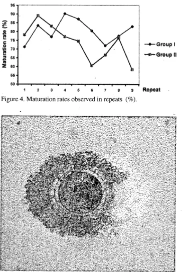
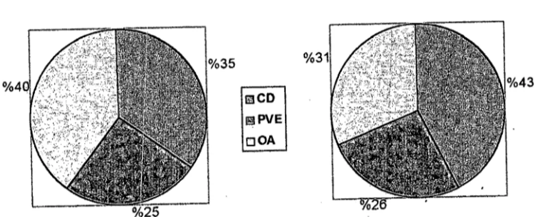
- Af ter the maturation period, 401 of 549 seleeted (good qua1ity) ooeytes in group i and 217 of 275 eultured ooeytes in group II were matured with amaturation of 73% and 78.9%, respeetively (Table 2, Figure 4 and 5), however, aberrant maturation findings were observed in 8% of ooeytes in group i and 5.1 % of ooeytes in group II (Figure 6).
--Group i
--Group ii
Table 2. In vitro maturation results recorded in study groups.
Parameter Group i Group II
(2-7 mm) (7-10 mm) Cultured oocyte (n) 549" 275b
Matured oocyte(n) 401 217
Maturation rate (%) 73% 78.9%
Aberrant maturated oocyte (n) 44:!:L.3g ]4:!:l.l 7h Aberrant maturation (%) 8%i 5.] O/J Means within a co1umn followed by different superscript letters differ (a,b,c,d p<O.oı and e,f,g,h,ı,j p<O.05).
9 Repeat Figure 4. Maturation rates observed in repeats (%).
A signifieiant differenee eould not be deteeted between study groups by the means of maturational eapaeity (p>0.05), but maturation rates differ between the
Figure 5. Maturated oocyte.
-+-Group i ...•.. Groupii Repeat -+-Group i ...•.. Groupii -+--Groupl ----Group ii Group II (7-10 mm) 300b 2.72:!: l.58d 275f 2.5:!:1.87h 9 1.7İ Group i (2-7 mm) 588" 5.11 :!:2.39c 54ge 4.77:!:2.09g 93.3İ 23456789 Parameter Follicle (n) Failiele (mean) Aspirated oocyte (n) Oocyte (mean) Aspiration rate (%) o 2 3 4 6 6 7 8 9 Repeat
Figure 3. Distribution of good quality COC's into repeats (p<O.Ol).
Repeat
1 2 4 6 6 7 8 9
Figure 2. Distribution of mean oocyte counts into repeats in group 1 and ll.
Means within a column followed by different superscript letters differ (a,b,c,d p<O.oı and e,f,g,h,ı p<O.05).
6
Figure i. Distribution of failiele counts into repeats in group i and ll. 100 90 80
i
70 ::J 60 o u 50ı
~
40'o
30 U. 20 10 O III ~4 U o .0 3~
iii5-
2 'oo o 1cl
206 Mustafa Ün - Şükrü Küplülü 8eD I!!ilPVE oOA i i i i i i i i i
i
iCD: Cumulus degeneration, PVE: Periviteııine space expantion, OA: Oolemma
Figure 6. Distribution of morphologiea!apperaneesobserved in aberrant maturation.
Figure 8.In vitrofertilized (pronuclear)ooeytes, Means within a eolumn followed by different superseript letters
differ (a,b,e,d p<O.05and e,f,g,h,l,j p<O.(5).
7 4 .2 20 26 60 66 i i i i i i -4-Groupl 1e-orouplı i i
i
i kepeat iFigure 7. Fertilizationrates in study groups and repeats
i
Fertilization rates were significantly huperior in group II to fertilizatiort rates observed in groud I (p<0.05), however fertilization rates were differ statisti(lalIy within
i the repeats of the same groups (p<0.05). :
i i
Discussion and Conclusion
i
The importance of ovary collection techniques and transfer expressions were discussed by diff~rent authors (6,7,10,11). Leibfried,Rutledge (l2) bundled the efficiency
i
of IVF system directIy to oocyte recovel)! conditions-mainly transfer temperature and speed. Gordoh (6) stated a _ 66
e
60 c .246 10 ~ 40i
36 LL 30 Group II (7,10 mm) 217b 107 49.3% 6(2.7j) 10 (4.6) 2(0.9) Groupi(2-7 mm) 40J a 165 41.1 % 1l(2.7i) 13(3.2) 3 (0.7) Parameter Cultured ooeyte (n) Fertilized ooeyte (n) Fertilizationrate(%) Parthenogenesis(n)(%) PoJyspermia(n)(%) Chromatine degenerationTable 3. Fertilization results in study groups.
In vitro fertilization
In vitro fertilization of 401 (group I) and 217 matured
oocytes (group II) has resulted 165 fertiIized in group I (41.1%) and 107 fertilized oocytes in group II (49.3%). In group I, II were (2.7%) parthenogenetic, polyspermia in 13 oocytes (3.2%) and 3 (0.7%) oocytes showed chromatine degeneration. Same parameters were found as, 6 (2.7%), 10 (4.6%) and 2 (0.9%) in
gioup
II (TabIe 3, Figure 7 and 8).repeats in the same study group (p<0.05) (Figure 9). In addition, aberrant maturation rates were greater in group I compared with group II (p<0.05).
Ankara Üniv Yet Fak Derg, 50, 2003 207
posıtıve correlation between ovary transport time and temperature and maturation capacity (60% at <15°C, 85% at >25°C). In the present study, the transport interval and temperature were carefuIly stabled (app 3 hour and 25-35°C). In order to achieve the satisfactory results suggested by workers above.
FoIlide diameter which the oocytes are aspirated from is an important varient and a good sign of the further maturation and fertilization capacities (6). Greve et al. (7) have suggested maturation rates of 85% and 95% for oocyles of foIlides <6 and >7 in dianıeter. Same suggestions were done by Küplülü and Ün (11) (70%-80%) and Telfer (16). All authors discussed this relation with the hormone receptors gained during the oogenesis. Similarly the fact that the lack of oocytes of smaIler «2 mm) foIlides achieve the second metaphase was highlighted by Gordon (6). The study of Gordon (6) has shown that there were no presence of LH receptors in such oocytes and the critical point of oogenesis for LH receptors is the reaching a diameter of 3-4 mm. Nevertheless, IVM of bovine oocytes depends upon the synthesis of several distinct and stilI almost defined protein s culminates the maturational capacities (14). This protein synthesis sequeııce alters during the foIlide development. Gordon (6) suggested a higher protein synthesis in oocytes aspirated from larger foIlides (>7 mm) where low at small foIlides (3-6 mm).
in the present study, in contrast with the studies above the maturational capacities of both oocytes from smaIl (2-6 mm) and larger (6-10 mm) foIlides were not differed (73%-78.9%) significantly. We considered the depud with the maluration tecnique as Jegwnow el aL. (8) stated the every similarity of tecniques when making comparision. However the main demonstration of the maturation is fertilization (6,7). Since the fertilization rates of oocytes of large foIlides were significantly higher than of smaIler ones (49.3% vs 41.1 %). Although it was evident that fıırther studies should be done to evaluate the ultrastructural basis of this high differantiation.
References
1. A van Soom, A de Kruif (1996): Oocyte maturation,
sperm capacitation and preimplantation development in
ıhe boyine: Jmplicationsfor iııvitro production ofembryos.
Reprad Dom Anim, 31, 687-70 i.
2. A van Soom, A de Kruif (1998): Bavine embriyonic de-velopment af ter in vivo and in vitro fertilizatioll. Reprod Dom Anim, 33, 261-265.
3. BoIs PEJ, Van Soom A, Ysebaert MT, Vandenheede JMM, A de Kruif (1996): Effects of aspiration vacuum
aııd ııeedle diameter on cumulus oocyte complex
morp-hology aııd developmental capaciıy ol'boviııe oocyles.
The-riogenology, 45,1001-1014.
4. Carolan C, Monaghan P, Gallagher M, Gordon i
(1994): Effecı ofrecovery method 011yield ofbovine
oocy-tes per ovary and their developmenıal compeıence af ter
maturation, fertilizatioıı and culture in vitro.
The-riogenology, 41,1061-1068.
5. Farın CE, Hasler JF, Martus NS, Stokes JE (1997): A
comparison of Menezo's B2 and tissue culture Medium-199
for in vitro produclion of boyine blastocysls.
The-riogenology, 48, 699-709.
6. Gordon i (1994): Laboratory Production of Catıle Emh-ryos. Cab International Co, Wallingford.
7. Greve T, Avery B, Callesen H (1993): Viahility of in vivo
and in vitro produced hoviııe emhryos. Reprad Dom Anim,
28,164-169.
8. Jewgwnow K, Hcerdegen B, Müller K (1999): LLL vitro
development of individually maturated havine oocytes in
relation tofollicular wall atresia. Theriogenology, 51, 745-756.
9. Khurana NK, Niemann H (2000): Effecls ol' oocyte
qu-ality, oxygen tension, emhryo deıısity. cumulus cells and
energy suhstrates on cleavage and morula/hlastocyst
for-mation ol'hovine emhryos. Theriogenology, 54, 741-756. 10. Küplülü
ş,
Ün M (2000): Mezhahadan elde edilen sığırovaryumlarında yüzeysel foUikül potansiyelinin helirlenmesi
ve oosit aspirasyonu. Ankara Üniv Yet Fak Derg, 47,
247-254.
lL. Küplülü
ş,
Ün M (2001): Sığırlarda foUikül hüyüklüğününoositlerin in vitro maturasyonu üzerine eıkisi. Ankara Üniv
Yet Fak Derg, 48, 201-205.
12. Leibfried-Rutledge ML (1999): Facton determining
competence of in vitro produced catıle emhryos.
Therigenology, 51, 473-485.
13. Loos DF, Van VUet C, Van Maurik P, Kruip Th AM
(1989): Morphology of immature hovine oocyles. Gam Res,
27, 197-204.
14. MacCallum C, Salamone D, Palasz AT (1997): Effect ql'
maturalian medium supplements on hovine oocyte
fertilization and emhryo development. Theriogenology, 47,
193-196.
15. Sosnowski J, Switonski M, Lechniak D, MoUnski K
(1996): Cytogenetic evctiuatioıı ql' in vitro maturated hovine oocytes collected .from ovaries ql' individual donors.
The-riogenology, 45, 865-872.
16. Telfer EE (1998): In vitro models for oocyte development. Theriogenology, 49, 451-460.
17. Thompson A (1997): Comparison hetween in vivo derived
and in vitro produced preelongation emhryos from
do-mestic ruminants. Reprad Fertil Dev, 9, 341-354.
18. Twagiramungu li,Morin N, Bordignon V, Smith L C, Bousquet D (1997): Iniluence ql' serum in culture system on the production and cryopreservation of in vitro-derived
hovine emhryos. Theriogenology, 47, 356-361.
19. Yamashita S, Satoh T, Hoshi H (1996): Bovine hlastocyst
formatian from IVM/IVF produced zygoles in serum and
serumfree medium. Theriogenology, 45,197-201
20. Yang X, Jiang S, Foote RH (1993): Bavine oocyte
de-velopmentfollowing different oocyte maturation and sperm
capacitation procedures. Mol Reprad Dev, 34, 94-100.
Geliş tarihi: 25.09.2002/ Kahul tarihi: 09.12.2002
Corresponding address:
Prof Dr. Şükrü Küplülü
Ankara Üııiversitesi Veteriner Fakültesi Doğum ve Jinekolqji Aııahilim Dalı, Dışkapı, 06110 Ankara.