Management of femoral fractures in dogs with unilateral
semicircular external skeletal fixator-intramedullary pin tie-in
configurations
Cenk YARDIMCI, Ahmet ÖZAK, Taylan ÖNYAY, Kamil Serdar İNAL, Birsen Deniz ÖZBAKIR
Department of Surgery, Faculty of Veterinary Medicine, Ondokuz Mayıs University, 55139, Kurupelit, Samsun, Turkey.Summary: The aim of the present study is to report the use of semicircular external skeletal fixator-intramedullary pin tie-in
configurations for management of femoral fractures in dogs. Twenty dogs (21 femoral fractures) with complete clinical and radiographic records for at least 24 weeks were included in the study. A unilateral, multiplanar, semicircular external skeletal fixator system was used in combination with intramedullary pin. Fracture description, history, frame configuration, complications, limb use, concomitant injury, fixator removal time, and functional outcome were evaluated. All of the fractures with sufficient follow up were healed. Eight dogs started to use the operated limb immediately after recovery from anaesthesia whereas initial limb use was 1 to 7 days in other dogs. Time for removal of the fixator ranged from 36 to 67 days (mean, 50 days). Functional outcome was excellent in 15 cases, good in 4 cases, fair in 1 case and poor in 1 case. As a result, it was concluded that semicircular external skeletal fixator - intramedullary pin tie-in configurations can be used as an alternative for the management of the femoral fractures in dogs.
Keywords: Dog, external skeletal fixation, femur, fracture, intramedullary pin.
Köpeklerde femur kırıklarının unilateral semisirküler eksternal fiksasyon intramedullar pin tie-in
kombinasyonu ile sağaltımı
Özet: Bu çalışmanın amacı köpek femur kırıklarının sağaltımında semisirküler eksternal fiksatör intramedullar pin tie-in
kombinasyonu ile ilgili sonuçların sunulmasıdır. Çalışmada en az 24 haftalık klinik kaydı ve radyografik verisi bulunan 20 köpek (21 femur) yer aldı. Unilateral, multiplanar, semisirküler eksternal fiksasyon sistemi ile intramedullar pin kombinasyonu kullanıldı. Kırığın tipi, anamnez, çerçevenin konfigürasyonu, komplikasyonlar, bacağın kullanımı, eşlik eden lezyon, fiksatörün çıkarılma zamanı ve sonuçlar değerlendirildi. Bütün kırıklarda kaynama şekillendi. Sekiz olgu operasyondan hemen sonra ilgili bacağını kullanmaya başlarken diğer olgularda bu sürenin 1 ile 7 gün arasında değiştiği gözlendi. Fiksatörün uzaklaştırılma süresi 36 ila 67 gün (ortalama, 50 gün) arasında değişim gösterdi. Fonksiyonel değerlendirme, olguların 15’inde mükemmel, 4’ünde iyi, 1’inde orta ve 1’inde ise zayıf olarak değerlendirildi. Sonuç olarak, semisirküler eksternal fiksasyon intramedullar pin tie-in konfigürasyonunun, köpeklerin femur kırıklarında bir alternatif sağaltım yöntemi olarak kullanılabileceği kanısına varılmıştır.
Anahtar sözcükler: Eksternal fiksasyon, femur, intramedullar pin, kırık, köpek.
Introduction
Dog femur fractures usually occur from motor vehicle accident and comprise more than 40% of long bone fractures (7, 15, 19, 20). The thigh muscles of the area obstructs closed reduction (2, 9, 13). Since proximity to the abdominal wall prevents the use of full-pins proximally, half pin configurations are generally used for the management of femoral fractures with external skeletal fixation (ESF). Such configurations do not provide adequate stabilization by themselves. Thus they are generally used together with an intramedullary pin which provides axial alignment and resistance to bending. Insertion of ESF pins to the femoral diaphysis in closed fashion may result muscle damage, significant pain, and decrease in limb activity. As a result of these
complications, muscle atrophy, reduced joint motion, and periarticular fibrosis may occur (13, 14, 20, 21).
A unilateral semicircular ESF system has been developed to enable the multi-plane half pin insertion. The aim of the present study is to share experiences regarding the use of semicircular external skeletal fixator-intramedullary pin tie-in configurations for management of femur fractures of 20 dogs.
Materials and Methods
Inclusion criteria and medications: Dogs (>14 kg)
with detailed clinical and radiographic data for their femoral fractures (20 dogs, 21 fractures) for at least 6 months were included. Detailed data about the dogs were summarized in Table 1. Ages of the dogs ranged from 5
T ab le 1 . P re o p era ti v e an d p o sto p era ti v e ca se d etails o f 2 0 d o g s m an ag ed w it h S ES F -IM p in ti e-in c o n fig u ra ti o n s T ab lo 1 . S EF S -I M p in t ie -in k on fig üra sy on u uy gu lan an 2 0 kö pe ğe a it p re op era ti f ve p osto pe ra ti f bu lg ular Ca se no S ig n a lm e n t H isto ry De sc ri p tio n o f th e fr a ct u re Co n fig u ra ti o n Co m p li ca ti o n s Fi rst u se o f th e li m b / IM p in -Fi x a to r re m o v a l (Da y s fo ll o w in g su rg er y ) Co n co m it a n t in ju ry /tre a tm e n t Fu n ctio n a l o u tc o m e 1 6 -m a-o ld , 2 8 k g , F c, A n ato li an sh ep h er d VT e Ob li q u e, m id -d iap h y se al, R i 4 m m kIM p in 4 a rc h ed , 2P l: 4 m m Ø n 2 ET HP o 2 mD: 4m m Ø 2 ET HP M il d DBIM P p 2 /2 1 -52 No n e Ex ce ll en t 2 5 -m -o ld , 2 6 k g , M d, T u rk ish Ka n g al VT Ob li q u e, m id -d iap h y se al, L j 4 m m IM p in 4 a rc h ed , 2P : 4 m m Ø 2 ET HP 2D: 4 m m Ø 2 ET HP M o d era te DBIM P M in o r p in trac t d isc h arg e r/2 1 -44 No n e Ex ce ll en t 3 4 -y b-o ld , 1 5 k g , M , G o rd o n se tt er VT Co m m in u ted , m id -d iap h y se al, L 4 m m IM p in 4 a rc h ed , 2P : 4 m m Ø 2 ET HP 2D: 4m m Ø 2 ET HP M il d DBI M P 4 /2 8 -62 Co n trala tera l h ip j o in t lu x ati o n / Kn o w les to g g le p in tec h n iq u e G o o d 4 6 -m -o ld , 1 6 k g , M , Bo x er HR f T ra n sv erse , d istal m etap h y se al, L 4 m m IM p in 3 a rc h ed , 2P : 4 m m Ø 2 ET HP 1D: 4m m Ø 2 ET HP M il d DBI M P M in o r p in trac t d isc h arg e r/2 1 -36 No n e Ex ce ll en t 5 14 -m -o ld , 3 3 k g , M , G er m an sh ep h erd G S I g Ob li q u e, d istal d iap h y se al, R 4 m m IM p in 4 a rc h ed , 3P : 4 m m Ø 3 ET HP 1 D: 4 m m Ø 2 ET HP M o d era te DBIM P M in o r p in trac t d isc h arg e r/2 1 -44 No n e Ex ce ll en t 6 9 -y -o ld , 2 6 k g , M , Hu n g arian h o u n d W B A h Ob li q u e, m id -d iap h y se al, R 4 m m IM p in 4 a rc h ed , 2P : 4 m m Ø 2 ET HP 2D: 4 m m Ø 2 ET HP M il d DBI M P r/2 8 -45 P n eu m o th o ra x / C h est tu b e ap p li ca ti o n Ex ce ll en t 7 11 -m -o ld , 2 2 k g , M , P itt b u ll terrie r HR Co m m in u ted , m id -d iap h y se al, L 4 m m IM p in 4 a rc h ed , 2P : 4 m m Ø 2 ET HP 2D: 4 m m Ø 2 ET HP M il d DBI M P M in o r p in trac t d isc h arg e 3 /2 8 -50 P n eu m o th o ra x / C h est tu b e ap p li ca io n Ex ce ll en t 8 8 -m -o ld , 1 4 k g , M , M ix b re ed VT T ra n sv erse , m id -d iap h y se al, L 4 m m IM p in 4 a rc h ed , 2P : 4 m m Ø 2 ET HP 2D: 4 m m Ø 2 ET HP M o d era te DBIM P P in trac t in fe cti o n 3 /2 1 -66 Ip silate ra l ti b ial fra ctu re /E S F G o o d 9 7 -m -o ld , 1 5 k g , M , G er m an p o in ter VT Ob li q u e, p ro x im al d iap h y se al, L 4 m m IM p in 3 a rc h ed , 1P : 4 m m Ø 2 ET HP 2D: 4 m m Ø 2 ET HP M il d DBI M P r/2 1 -40 No n e Ex ce ll en t 10 12 -m -o ld , 1 8 k g , F M ix b re ed VT T ra n sv erse , d istal m etap h y se al, L 4 m m IM p in 3 a rc h ed , 2P : 4 m m Ø 2 ET HP 1D: 4 m m Ø 2 ET HP M il d DBI M P 2 /2 1 -42 Ip silate ra l fe m o ra l n ec k fra ctu re /E x cisio n arth ro p las ty G o o d
11 4 -y -o ld , 3 3 k g , F , Do g o a rg en ti n o VT Ob li q u e, p ro x im al m etap h y se al, R 4 m m IM p in 3 a rc h ed , 1P : 4 m m Ø 2 ET HP 2D: 4 m m Ø 2 ET HP M o d era te DBIM P r/3 5 -60 Co n trala tera l d istal ra d iu s-u ln a fra ctu re /H y b rid ES F Ex ce ll en t 12 2 -y -o ld , 2 6 k g , F , G er m an sh ep h erd VT Co m m in u ted , m id -d iap h y se al, L 4 m m IM p in 4 a rc h ed , 2P : 4 m m Ø 2 ET HP 2D: 4 m m Ø 2 ET HP M il d DBI M P r/3 0 -48 No n e Ex ce ll en t 13 18 -m -o ld , 2 2 k g , M , G er m an P o in ter VT Ob li q u e, d istal d iap h y se al, L --- ---T ra n sv erse d istal d iap h y se al, R 4 m m IM p in 3 a rc h ed , 2P : 4 m m Ø 2 ET HP 1D: 4 m m Ø 2 ET HP --- ---4 m m IM p in 3 a rc h ed , 2P : 4 m m Ø 2 ET HP 1D: 4 m m Ø 2 ET HP M o d era te DBIM P M in o r p in trac t d isc h arg e M il d DBI M P M in o r p in trac t d isc h arg e 4 /3 3 -67 1 /2 1 -40 No n e F air --- Ex ce ll en t 14 14 -m -o ld , 2 9 k g , M ,G o ld en re tri ev er VT Co m m in u ted , m id -d iap h y se al, L 4 m m IM p in 4 a rc h ed , 2P : 4 m m Ø 2 ET HP 2D: 4 m m Ø 2 ET HP M il d DBI M P 3 /2 5 -55 Co n trala tera l ra d iu s-u ln a fra ctu re /E S F Ex ce ll en t 15 2 -y -o ld , 1 9 k g , M , Ku rz h aa r VT Co m m in u ted , d istal d ia p h y se al, R 4 m m IM p in 3 a rc h ed , 2P : 4 m m Ø 2 ET HP 1D: 2 m m Ø 2 ET HP M il d DBI M P 7 /2 8 -44 De g lo w in g in ju ry o f th e ip silate ra l m etata rsa ls / S ec o n d ary in ten ti o n h ea li n g G o o d 16 16 -m -o ld , 2 4 k g , M , M ix b re ed VT Ob li q u e, m id -d iap h y se al, R 4 m m IM p in 4 a rc h ed , 2P : 4 m m Ø 2 ET HP 2D: 4 m m Ø 2 ET HP M il d DBIM P r/2 1 -48 No n e Ex ce ll en t 17 10 -m -o ld , 2 3 k g , F , S ib erian h u sk y VT Co m m in u ted , p ro x im al d iap h y se al, L 4 m m IM p in 3 a rc h ed , 1P : 4 m m Ø 2 ET HP 2D: 4 m m Ø 2 ET HP M il d DBI M P M in o r p in trac t d isc h arg e 2 /2 8 -45 No n e Ex ce ll en t 18 22 -m -o ld , 2 3 k g , M , Dra h th aa r HR T ra n sv erse , m id -d iap h y se al, R 4 m m IM p in 4 a rc h ed , 2P : 4 m m Ø 2 ET HP 2D: 4 m m Ø 2 ET HP M il d DBI M P r/2 1 -52 No n e Ex ce ll en t 19 8 -m -o ld , 1 9 k g , F , G o ld en re tri ev er VT Ob li q u e, m id -d iap h y se al, R 4 m m IM p in 4 a rc h ed , 2P : 4 m m Ø 2 ET HP 2D: 4 m m Ø 2 ET HP M il d DBI M P 2 /2 1 -44 No n e Ex ce ll en t 20 3 -y -o ld , 2 0 k g , M , M ix b re ed VT Co m m in u ted , m id -d iap h y se al, L 4 m m IM p in 4 a rc h ed , 2P : 4 m m Ø 2 ET HP 2D: 4 m m Ø 2 ET HP M o d era te DBIM P Os teo m y eli ti s, No n u n io n 3 /3 2 -76 L e ft 9 -10 th rib f ra ctu re , p n eu m o th o ra x / Ch est tu b e ap p li ca ti o n P o o r a m , m o n th ; b y , y ea r; c F , fe m ale ; d M , m ale; e V T , v eh icu lar trau m a; f HR, h ig h rise ; g G S I, g u n sh o t in ju ry ; h W B A , w il d b o ar a tt ac k ; i R, rig h t; j L , lef t; k IM , in tram ed u ll ary ; l P , p ro x im al; m D, d istal; n Ø, diam eter; o ET H P , en d -th re ad ed h alf p in ; p DBIM P , d ra in in g f ro m th e b ase o f th e in tram e d u ll ary p in ; r , im m ed iate ly a fter re co v er y f ro m a n esth esia . a m , a y ; b y, y ıl ; c F , d işi ; d M , erk ek ; e V T , traf ik k az ası; f HR, yü ks ek ten d üşm e; g G S I, a teş li silah y ara la m ası; h W B A , y ab an d om uz u sa ld ırı sı; i R, sa ğ; j L , so l; k IM , in tra m ed u ll ar; l P , p ro k sim al; m D, d istal; n Ø, ça p; o ET HP , u cu y iv li y arı m p in ; p DBIM P , in tram ed u ll ar p in d ib in d en d re n aj; r , o p era sy o n d an h em en so n ra .
months to 9 years (mean 22 months). Body weights ranged from 14 - 33 kg (mean, 22.5 kg). Fracture types were: oblique (n=9) (Figure 3), comminuted (n=7) (Figure 4), and transverse (n=5) (Figure 5). Fractures were caused by vehicular trauma (n=15), high rise (n=3), gunshot injury (n=1), and wild boar attack (n=1).
Frame design and features: The main part of
semicircular ESF consisted of 6 hole 45° carbon-fiber arches while the other components of the system were 6 mm threaded rods, half pin fixation bolts, 6 mm nuts, 4 mm diameter negative profile end-threaded half pins (Figure 1). Caudal rod was adjusted 3-4 cm longer than the cranial rod in order to attach the IM pin to the ESF frame in “tie-in” fashion. Another 45° carbon-fiber arch was used as a connector for the linkage of the two fixation systems (Figure 2).
Surgical technique: After cranio-lateral approach
(16), bone fragments exposed, and a 4 mm Steinmann pin was inserted to the medullary cavity by retrograde fashion. After achieving anatomic bone alignment, end-threaded half pins were bicortically and perpendicularly inserted. The proximal pin was inserted just below the trochanter major while the most distal pin was inserted to the lateral epicondyle. Once sufficient reduction achieved, the frame was reinforced by inserting more half pins (depending of the fracture type) to the femoral shaft by avoiding muscles. In order not to harm the strong muscle groups, half pins of the femoral shaft were applied in slight craniomedial direction (Figure 2c). Before skin closure, all components were firmly secured to the frame and then IM pin was
tied-in to the caudal rod. The half ptied-in fixation bolt on the IM pin was placed 4-5 cm proximal to the exit point of the pin from the skin.
Figure 1. The principal connecting elements of semicircular ESF are 6 hole 45° (180 mm inside diameter, 1/8 ring arch, 7x18x85 mm) carbon-fiber arches (a). Up to 4 half pins may be connected to each arch in above or below fashion (b) and the semicircular shape of the arches are particularly advantageous due to its multiplanar structure (c, d).
Şekil 1. Semisirküler eksternal fiksasyon sisteminin ana bileşeni 6 delikli 45°’lik (180 mm çaplı halkanın 1/8’lik yayı, 7x18x85 mm) karbon fiber ark (a). Her ark, alttan ya da üstten seçenekleriyle (b) 4 taneye kadar yarım pin uygulamaya olanak sağlar. Ayrıca arkların semisirküler yapısı multiplanar uygulamalara da izin verir (c,d).
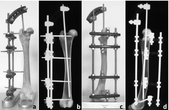
Figure 2. Dry bone sample and radiographic views of unilateral semicircular ESF-IM pin tie-in configuration in a dog femur model from caudal (a,b) and lateral (c,d) projections.
Şekil 2. Köpek femur modelinde unilateral semisirküler ESF-IM pin tie-in konfigürasyonunun kaudal (a,b) ve lateralden (c,d) görünümü
Figure 3. Dog 11. Preoperative (a,b) radiographs of a long oblique proximal metaphyseal fracture. Postoperative radiographs immediately after the operation (c,d). Note that two half pin application was carried out from the same arch.
Şekil 3. Olgu 11. Proksimal metafizer uzun oblik kırığın preoperatif (a,b) ve operasyondan hemen sonraki postoperatif (c,d) radyografileri. Aynı arktan iki farklı yarım pin uygulaması görülmekte.
Figure 4. Dog 14. Two days postoperative clinical view of a Golden retriever in which right transversal radius-ulna fracture was managed with semicircular ESF and left mid-diaphyseal comminuted femoral fracture was managed with semicircular ESF-IM pin tie-in configuration tie-in the same session (a). Craniocaudal radiographic projections of the operated femur immediately after the operation (b) and following fixator removal on postoperative 55th day (c).
Şekil 4. Olgu 14. Golden retriever ırkı bir köpeğin sağ transversal radius-ulna kırığına semisirküler ESF, sol orta diafizer parçalı femur kırığına ise semisirküler ESF-IM pin tie-in konfigürasyonu uygulandıktan 2 gün sonraki klinik görünümü (a). Femur’un operasyondan hemen sonra (b) ve postoperatif 55. günde fiksatörün çıkarılmasından sonra (c) çekilen kraniokaudal radyografileri.
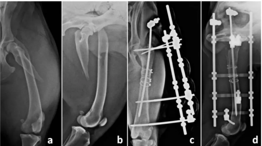
Figure 5. Dog 6. Preoperative mediolateral radiography of a Hungarian hound with an open mid-diaphyseal femur fracture 3 days after
a wild boar attack (a). Mediolateral radiographies immediately after the operation (b), following IM pin removal on 28th day (c) and 40
weeks after fixator removal (d). Clinical view of the dog on postoperative 8th month (e).
Şekil 5. Olgu 6. Yaban domuzu saldırısı sonucu açık orta diyafizer femur kırığı şekillenen Macar Barak ırkı köpeğin olaydan 3 gün sonra alınan preoperatif mediolateral radyografisi (a). Operasyondan hemen sonraki mediolateral (b), 28. günde IM pinin çıkarılmasından sonra (c) ve 40. haftada fiksatörün çıkarılmasından sonra (d) çekilen radyografiler. Olgunun postoperatif 8. aydaki klinik görünümü (e).
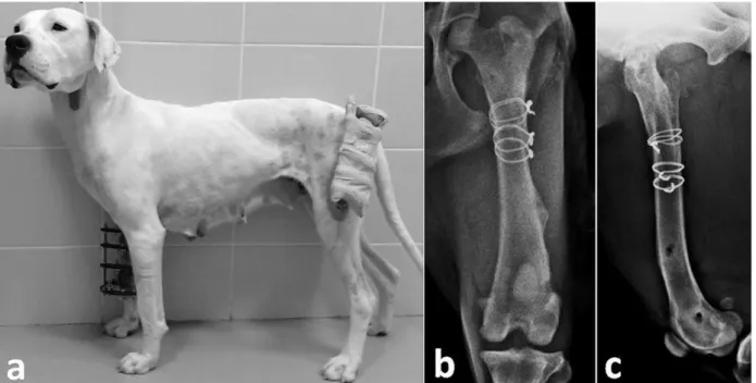
Figure 6. Dog 11. Clinical view of a multiple fracture case (a). The dog was able to bear weight on both the operated limbs immediately after recovery from the anaesthesia. Radiographic views of the same dog after removal of the fixator (b,c).
Şekil 6. Olgu 11. Çoklu kırık olgusunun klinik görünümü (a). Hasta operasyondan hemen sonra her iki bacağına da yüklenebiliyordu. Fiksatör çıkarıldıktan sonra çekilen radyografiler (b,c).
Postoperative period and evaluation of outcome:
Before discharge, clients were advised to keep the dogs indoor and clean the pin holes daily with povidone iodine (Batticon 10% sol). Follow-up examinations and radiographic assessments were performed every other week. The frame was removed in two stages; by removing of the IM pin and then the ESF. Time to remove the IM pins were determined depending on the radiographically visible woven bone image between the fragments. Bone healing was determined by observation of bridging new bone between the fragments, which was found to be sufficient for fixator removal.
Outcome was based on relative assessments including dogs willingness to use the operated limb, degree of weight loading, and presence of resistance to the flexion/extension of the hip and stifle joints. Final outcome scores were graded as: excellent (no obvious lameness, full weight bearing, functional use of the operated limb, no pain on palpation), good (obvious full weight bearing, no obvious lameness at a walk but slight lameness after extensive exercise, no pain on palpation), fair (obvious lameness but consistent weight-bearing, obvious resistance to flexion and extension) and poor (no limb use, non-weight-bearing lameness, resistance and pain on flexion and extension). Final clinical evaluations were made 4 to 6 months after removal of the fixator.
Results
In all dogs mild to moderate serosanguineous wound discharge was seen at the base of the IM pin over the
trochanteric fossa. Enlargement in the size of the IM pin hole was also seen in some active dogs due to the cyclic movement of the pin. This complication was minimized with daily wound care and activity restriction. Following removal of the IM pin, this problem was completely resolved. Mild discharge from the pin-skin contact area during consolidation period was the second most common problem and this complication resolved after cleaning of pin-skin contact areas and antibiotic therapy (Synulox 250 mg tablet, Pfizer, Italy). Poor wound hygiene was the major cause of the mild pin tract discharge.
In two dogs (dog no 8 & 20), purulent pin tract discharge was observed and staphylococcus epidermidis was cultured from both. In dog 8 infection was treated by regular use of oral amoxicillin-clavulonat and pin-skin interface cleaning. In dog 20 however, osteomyelitis was formed which resulted in nonunion and poor final clinical outcome.
In all dogs, semicircular ESF-IM pin combination provided a stable scaffold and all dogs except one tolerated the frame assembly well. In dog 13, because both femoral fractures were fixed, the dog had difficulty in a lateral recumbency position during the convalescence period. Radiographically, conical mild periosteal new bone formation was seen at the base of the ESF pins from where serosanguineous draining was apparent (Figure 6b).
Periosteal reaction was more significant around the pins closer to the metaphysis of the bone. All were
improved following removal of the pins during remodeling process.
All of the fractures except one (dog 20) healed. Eight of the dogs used the operated limb just after the operation (Figure 6a), while the other 12 dogs used their limbs between 1 to 7 days (mean 3 days). Intramedullary pin removal time ranged from 21 – 33 days (mean 25 days) while fixators were removed between 36 to 67 days (mean, 50 days; nonunion case -dog 20- is not included). Final clinical outcome was excellent in 15 femurs, good in 4, fair in 1, and poor in 1.
Discussion and Conclusion
A number of different external and internal fixation techniques were defined for stabilization of canine femoral fractures (6, 8, 15, 19-21). However, only few reports have been published that describe the fixation of femoral fractures with ESF-IM pin tie-in combination in dogs (1). In the present study, canine femoral fractures were managed with a novel custom designed semicircular ESF-IM pin tie-in system, which resulted in affirmative long-term results in dogs. This combined system provided sufficient stability and versatility that encouraged early and obvious painless limb use during daily activities in the postoperative period.
External skeletal fixation is usually applied in closed fashion but, reduction and realignment of the fragments are difficult in this technique. The femoral diaphysis is covered by strong muscle groups from lateral and cranial planes which makes reduction nearly impossible with closed reduction technique. Pins inserted to the femoral shaft with closed technique may result detrimental muscle lesions, reluctance and decrease in functional limb use that necessitates immediate frame removal to avoid muscle fibrosis, reduced joint motion and quadriceps tie-down (3, 13). In order to avoid this problem open reduction is essential and pins have to be inserted between these muscle bundles, not through them. The fascial plane between vastus lateralis and biceps femoris muscles over the lateral aspect of the femoral shaft is curvilinear which hinders safe pin application from a single linear connecting rod. In the present study, we could obtain proper pin applications through the safe intermuscular corridors by using an arch in semicircular shape and no complications related to muscular damage were encountered in any of the dog.
Fractures that involve the distal or proximal portion of the bone can be difficult to manage. The reason for that are tensile forces, higher shearing force due to the IM pin which poorly fills the medullary canal, and limited bone portion to secure implants. The shape of the arches used in this study provides a multiplanar pin insertion option and this system allows application of 1 to 4 pins from one arch which is very critical for the stabilization of fractures very close to joints (Figure 1b-d). Because the distal half of the dog femur is not aligned linear in the sagittal plane as in
feline femurs (12, 13, 15), the use of this system is more practical than the conventional ESF systems for treatment of the distal part fractures.
Although IM pin resists bending equally well in all directions due to the proximity of the neutral axis, resistance to disruptive forces of shearing, torsion and compression is inadequate if used alone (5, 11, 18). When used together, ESF and IM pin are more resistant against these forces. Under quasistatic uniaxial compressive loading, the mean yield strength, yield energy, and ultimate strength of the tied-in configuration were mentioned to be significantly larger than those not tied (13). Also, based on the results of a finite element analysis study (17), the use of the “tied-in” IM pin with the ESF is recommended in challenging fractures. Another study (18) demonstrated that if IM pin is embedded well in the metaphysis when used together with a unilateral frame, unilateral ESF frames reveal an equivalent or even higher stiffness modulus compared to a bilateral frame with an identical half pin arrangement.
The major purpose of all fracture treatment methods is to obtain the soonest possible recovery and early functional limb use (4). Early limb use promotes healing by allowing axial micromotion at the fracture site and also prevents inactivation atrophy in patients with bone fractures (11, 17).This is very critical in large and giant breed dogs with multiple fractures. In the present study 8 dogs used their fixed limb soon after the operation and also early ambulation was seen in 4 dogs with multiple major orthopaedic injuries.
This configuration had also disadvantages. Especially in young and vigorous dogs that used their operated limbs actively, increase in sanguineous secretion and enlargement of the pinhole wound, due to the cyclic activity was experienced. This complication occurred in all of the dogs in this case series but did not result in noticeable lameness. Interestingly, no mention of such complication was reported in previous studies in which “tie-in” configuration was used for the management of femoral fractures in dogs (1) and cats (10). However, since the wound healed and draining stopped after removing the IM pin, this was considered as a minor complication. Another possible complication the surgeon may encounter is the collision of the end-threaded half pins applied from the ESF frame with the IM pin inside the bone. We experienced this in “in vitro” laboratory trials in which dry bone samples were used. In some cases, it caused bone fissures especially in the isthmus which is the narrowest part of the femur, but no complication was seen in the proximal or distal regions. Neither resistance nor bone fissures were encountered during the operations of the present cases. The reason for that was thought to be the higher elasticity and lower brittleness of the live bone
tissue due to its greater water and collagen content compared with the dry bone.
According to the results of present study, in which most of the fractures represent a good to excellent bone healing and clinical outcome, it is believed that unilateral semicircular ESF–IM pin tie-in configurations can be used an alternative method for the management of canine femoral fractures.
References
1. Aron D, Foutz T, Keller W, et al. (1991): Experimental and clinical experience with an IM pin external skeletal fixator tie-in configuration. Vet Comp Orthop Traumatol, 4, 26-34.
2. Aron D, Toombs J, Hollingsworth S (1986): Primary treatment of severe fractures by external skeletal fixation: Threaded pins compared with smooth pins. J Am Anim Hosp Assoc, 22, 659-670.
3. Carmichael S (1991): The external fixator in small animal orthopaedics. J Small Anim Pract, 32, 486-493.
4. Egger EL (1992): Instrumentation for external fixation. Vet Clin North Am Small Anim Pract, 22, 19-43.
5. Egger EL, Histand M, Blass C, et al. (1986): Effect of Fixation Pin Insertion on the Bone‐Pin Interface. Vet Surg, 15, 246-252.
6. Farese JP, Lewis DD, Cross AR, et al. (2002): Use of IMEX SK-circular external fixator hybrid constructs for fracture stabilization in dogs and cats. J Am Anim Hosp Assoc, 38, 279-289.
7. Johnson JA, Austin C, Breur GJ (1994): Incidence of canine appendicular musculoskeletal disorders in 16 veterinary teaching hospitals from 1980 through 1989. Vet Comp Orthop Traumatol, 7, 5-18.
8. Kirkby KA, Lewis DD, Lafuente MP, et al. (2008): Management of humeral and femoral fractures in dogs and cats with linear-circular hybrid external skeletal fixators. J Am Anim Hosp Assoc, 44, 180-197.
9. Kraus KH, Toombs JP, Ness MG (2003): External fixation in small animal practice. Oxford: Blackwell Science, Oxford.
10. Langley‐Hobbs S, Carmichael S, McCartney W (1996): Use of external skeletal fixators in the repair of femoral fractures in cats. J Small Anim Pract, 37, 95-101.
11. Lincoln JD (1992): Treatment of open, delayed union, and nonunion fractures with external skeletal fixation. Vet Clin North Am Small Anim Pract, 22, 195-207.
12. Marcellin Little D (2003): Textbook of small animal surgery. 1818-1834. In: Slatter DH (Ed), External Skeletal Fixation. Saunders, Philadelphia.
13. Marti J, Miller A (1994): Delimitation of safe corridors for the insertion of external fixator pins in the dog 1: Hindlimb. J Small Anim Pract, 35, 16-23.
14. Özak A, Yardımcı C, Nisbet HÖ, et al. (2009): Treatment of long bone fractures with acrylic external fixation in dogs and cats: Retrospective study in 30 cases (2006-2008). Kafkas Univ Vet Fak Derg, 15, 615-622.
15. Piermattei DL, Flo GL, DeCamp CE (2006): Brinker, Piermattei and Flo’s Handbook of Small Animal Orthopedics and Fracture Repair. 512-561. In: Piermattei D, Flo G, and CE D (Ed), Fractures of the Femur and Patella. Saunders, St. Louis.
16. Piermattei DL, Johnson KA (2004): An atlas of surgical approaches to the bones and joints of the dog and cat. Saunders, Pennsylvania.
17. Radke H, Aron DN, Applewhite A, et al. (2006): Biomechanical Analysis of Unilateral External Skeletal Fixators Combined with IM‐Pin and Without IM‐Pin Using Finite‐Element Method. Vet Surg, 35, 15-23.
18. Shahar R, Shani Y (2004): Fracture stabilization with type II external fixator vs. type I external fixator with IM pin Finite element analysis. Vet Comp Orthop Traumatol, 17, 91-96.
19. Simpson DJ, Lewis DD (2003): Textbook of small animal surgery. 2059-2089. In: Slatter DH (Ed), Fractures of Femur. Saunders, Philedelphia.
20. Whitehair JG, Vasseur PB (1992): Fractures of the femur. Vet Clin North Am Small Anim Pract, 22, 149-159. 21. Yardımcı C, Özak A, Nisbet HO (2011): Management of
Femoral Fractures in Dogs with Unilateral Semicircular External Skeletal Fixators. Vet Surg, 40, 379-387. Geliş tarihi: 20.10.2016 / Kabul tarihi: 09.01.2017 Address for correspondence:
Doç. Dr. Cenk YARDIMCI Ondokuz Mayıs University, Faculty of Veterinary Medicine, Department of Surgery,
55139, Kurupelit, Samsun, Turkey. E-mail: [email protected] Tel:+90 362 3121919/1228 Fax: +90 362 4576922