Ankara Üniv Vet Fak Derg, 60, 151-153, 2013
Short Communication / Kısa Bilimsel Çalışma
A canine hemangiopericytoma case: Fine needle aspiration (FNA)
cytology and histopathological findings
Arda Selin COSKAN1, Binnur ONAL2, Mehmet Eray ALCIGIR1, Osman KUTSAL1
1 Department of Pathology, Faculty of Veterinary Medicine, Ankara University, Ankara; 2Department of Pathology&Cytology,
Ankara Dışkapı YB Training&Research Hospital, Ankara/Turkey.
Summary: Veterinary cytology is gaining importance in diagnostics of skin and subcutaneous masses of dogs. A case of
canine-hemangiopericytoma was described by cytopathological and histopathological findings on Fine needle aspiration (FNA) and excisional biopsy. A 13-year-old female terrier dog had a subcutaneous lesion on right leg. FNA was performed by the cytopathologist using 27-gauge needle and 10 ml. syringe attached to a syringe-holder. FNA and imprint slides from FNA aspirates and stained with May-Grunwald Giemsa,Papanicalau, Hematoxylin Eosin were prepared. Cyto-diagnosis was encountered with spindle cells. FNA can often provide information for a good diagnostic and management plan, especially for discrete, non-ulcerated lesions, allowing veterinarians to make more informed therapeutic choices.
Key words: Cytology, dog, fine needle aspiration, hemangiopericytoma.
Bir köpekte hemangioperisitom olgusu: İnce iğne aspirasyon (İİA) sitolojisi ve histopatolojik bulgular
Özet: Veteriner sitoloji ile köpeklerin deri-deri altı kitlelerinin tanı yöntemleri giderek önem kazanmaktadır. Bu makalede bir köpekte hemangioperistom olgusunda ince iğne aspirasyon (İİA) ve eksizyonel biyopside sitopatolojik ve histopatolojik bulgular tanımlanmıştır. 13 yaşlı terrier ırkı dişi bir köpeğin sağ ön ekstremitesinde saptanan deri altında kitle tespit edildi. İİA örneklemesi 27 G iğne ve CAMECO enjektör tutucuya ekli 10 ml.enjektör kullanılarak yapıldı. İİA örneklerinden yayma ve tuşe tekniklerine göre lamlar hazırlandı ve May-Grunwald Giemsa, Papanicolaou, Hematoksilen-Eozin ile boyandı. Sitopatolojik tanıda iğsi hücrelerle karşılaşıldı. İnce iğne aspirasyon sitolojisi özellikle ülserasyon göstermeyen lezyonlarda, yüksek tanı duyarlılığında ve hızlı bir yöntem olup, veteriner hekimlere daha bilinçli sağaltım seçimleri yapmasını sağlamaktadır.
Anahtar sözcükler: Hemangioperisitom, ince iğne aspirasyon, köpek, sitoloji.
Veterinary cytology is gaining importance in diagnostic procedures of cutaneous and subcutaneous
masses of dogs. A case of canine hemangiopericytoma
was described by cytopathological and histopathological findings on FNA and biopsy. Hemangiopericytoma is a mesencymal tumor originated from pericytes of vessels and developed in soft tissue. The tumor is generally localizated in hind and forelimbs (2, 4, 6).
In the case, a-13-year-old, female, terrier dog had a subcutaneous lesion on right leg. The animal was
operated under anesthesia at Department of Surgery of
Ankara University Veterinary Faculty. A FNA to the lesion was applied for both intraoperative consultation and increasing expertise on cytomorphological findings. The FNA was performed by the consultant cytopathologist using 27-gauge-needle and 10 ml.syringe attached to Cameco-syringe holder. Four times of needling were performed from different areas of the lesion and FNA slides were prepared. Some slides were
air-dried for May-Grunwald-Giemsa (MGG) staining and others were fixed in ethanol for Papanicolaou (PAP) and Hematoxylin-Eosine (HE) stainings. Operation material was evaluated at Pathology Department, tissue specimens were routinely processed; embedded in paraffine and stained by HE and Masson’s Trichrome for histopathological examinations.
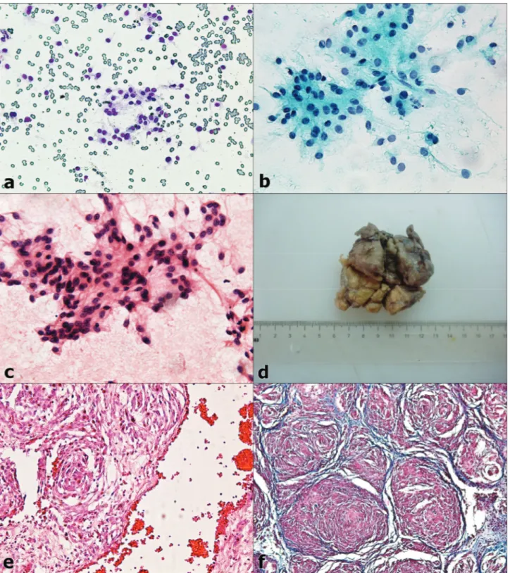
Cytopathological Findings: FNA material was
highly cellular with neoplastic cells, scattered individually or in clusters or whorles (Figure 1. a-b-c). The cytological architecture was relatively uniform with oval or spindle-shaped cells having cytoplasmic tails on both poles. Nuclei were also oval or elongated shaped with fine chromatine and rare prominent nucleoli. Cytoplasms were generally observed as light basophilic some of which contained few clear-vacuoles. No mitosis or necrosis was seen. Cytopathological diagnosis was ‘suspicious cytology of a spindle cell lesion’.
Arda Selin Coskan - Binnur Onal - Mehmet Eray Alcigir - Osman Kutsal 152
Macroscopical Findings: The excised-mass was
weighed of 42 g and diametered in 6×7×4 cm. The mass had firm in consistency. Cut surface had soft consistency and had excessive, lobular, grayish-white in appearance (Figure 1. d).
Histopathological Findings: The spindle-shaped
cells constituted whorls resembling fingerprint pattern. These cells, which interlacing collagen matrix, were having eosinophilic cytoplasm, ovoid nuclei and in some prominent nucleoli. In some areas, it was encountered to Figure 1: Cytological appearences of neoplastic pericytes; a:MGG x200, b: PAP x400, c: HE x400 ; d: Gross appearance of the mass ; e: Fingerprint appearance by neoplastic spindle- shaped- pericytes in histopathology, HE x 200 ; f: Hemangiopericytoma, Masson’s trichrome staining x100.
Şekil 1: Neoplazik perisitlerin sitolojik görüntüleri: a: MGG x200, b: PAP x400, c: HE x400 ; d: Kitlenin dış görünüşü ; e: Histopatolojideki neoplazik iğsi perisitlerin tarafından parmak izi görünümü, HE x 200 ; f: Hemangioperistoma, Masson’un trikrom boyaması x100.
Ankara Üniv Vet Fak Derg, 60, 2013 153 vessels containing few of eritrocytes in their lumina
(Figure 1. e-f).
The hemangiopericytoma is a tumor arising from
pericytes associated with blood vessels. Instead of
forming a useful tissue, these pericytes form tumors because improper cell division has left them with chromosomal damage (5,7). It was in the case, also described spindle shaped pericytes originated blood vessels and cellular architecture in routinly histopathology.
Hemangiopericytomas are classified as soft-tissue sarcomas and have uncommon metastasis however there is high incidence of recurrence. Even though the tumor show usually benign behaviour, they have potencially malignancy. Hemangiopericytomas are firm, small nodules that tend to occur singly, and vary in size from 0,5 to 25 cm. A slow growing tumor that develops in deep soft tissues (3,6,7). The most common sites of detection are hips, shoulders, limbs, abdomen, thorax and upper arms and upper legs of the dog. Hemangiopericytomas can appear bald, pigmented or even ulcerated skins (3,4). Macroscopically, the findings (size, shape, consistency, color and cut surface) were accorded to excepting out ulcerative skin in present case.
The tumor is usually seen in 8-14 years old dogs. Females appear to be affected more often than males. Hemangiopericytomas are more widespread in Springer Spaniel, Cocker Spaniel, German Shephards, Golden Retriever and Boxer dogs (3,5,7). In this case, the mass was localizated in hindlimb as mentioned literatures before. And also, old and gender of the dog was consisted with the knowledges. However, race was different from most affected races between dogs.
Histopathologically, hemangiopericytomas have a characteristic "fingerprint pattern" appearance that is composed of multiple layers of spindle cells arranged in a concentric whorls that frequently surrounds a central blood vessel, similar to our case. They may exhibit whorling patterns like fibrosarcomas, fibromatoses, cutaneous fibrous histiocytomas and schwannomas (3,4, 6,7). But, hemangiosarcoma is critical differentiation point of these kind of tumor encountering with eritrocytes as indicated in the case as regard histopathological features.
Despite the pitfalls inherent in interpreting aspiration material, the use of FNA has been rapidly increasing in diagnosing of soft tissue tumors; it is especially useful in documenting persistence, recurrence, or metastasis. The FNA material can be useful in the preoperative evaluation of neoplasia vs. inflammation as well as benign vs.malignant tumor. Cyto-diagnosis of soft tissue tumors could be difficult or impossible in some cases and core needle biopsy can help in tumor typing sometimes, however core needle biopsy has a higher unsatisfactory rate than FNA (2).
On the other hand, the exact typing of soft-tissue tumor may not be critical in the choice of therapy,
including surgery. Thus, even in cases with inconclusive cytodiagnosis, routine FNA may help the clinician to plan therapy by indicating the general cytological category and degree of malignancy.
Fine-needle aspirates of hemangiopericytomas usually display spindloid to polyhedral cells with light gray, wispy cytoplasm and a round to oval nucleus that may contain one or two nucleoli. Some neoplastic cells will have cytoplasm with a veil-like appearance. A few binucleated and multinucleated cells and scattered small lymphocytes also may be observed (1). Cytological features were generally accorded to these kind of knowledges, but last mentioned findings such as multinuclear structure and inflammatory cells were not noted in aspirates.
In conclusion, especially FNA cytology in prediagnosis and also histopathology can often provide information to formulate a good diagnostic and therapeutic plan, allowing veterinarians to make more informed choices (i.e limb-sparing surgery). Cytology tends to be of greatest value where lesions are discrete, free from ulceration and infection, such as the presented case.
Acknowledgement
Presented as poster at 36th European Congress of Cytology in Istanbul in 22-25 September 2011.
References
1. Chhieng D, Cohen JM, Waisman J, Fernandez G, Cangiarella J (1999): Fine-needle aspiration cytology of
hemangiopericytoma. Cancer Cytopathol. 87(4) 190–195.
2. DeMay R (1995): Soft tissue tumors. In: The Art & Science of Cytopathology. First ed. Vol. 2, ASCP Press, Chicago. pp.564.
3. Erer H, Kıran MM (2009): Veteriner Onkoloji. 4. ed. Damla Ofset, Konya.; pp.99-100.
4. Goldschmidt MH, Hendrick MJ (2002): Tumors of the
skin and soft tissues. In: Meuten, D.J. Ed. Tumors in
Domestic Animals. 4th. ed. Iowa State Press. Iowa, USA. Pp.94-95.
5. Mazzei M, Millanta F, Citi S, Lorenzi D, Poli A (2002):
Haemangiopericytoma: Histological spectrum, immunohistochemical characterization and prognosis. Vet
Dermatol, 13: 15-21.
6. McEntee K, Nielsen SW (1976): Tumors of the soft
tissues. In: Beveridge, W.I.B., Sobin, L.H. Ed. Bulletin of
The World Health Organization. Geneva. pp.102.
7. Schlafer DH, Miller RB (2007): Skin and appendages. In: M.Grant Maxie. Ed. Jubb Kennedy and Palmer’s Pathology of Domestic Animals. 5th. ed. Vol. 3, China.pp.762-763.
Geliş tarihi: 09.11.2012 / Kabul tarihi: 30.11.2012
Address for correspondence;
Res.Assist. Arda Selin Coskan
Ankara University, Faculty of Veterinary Medicine, Department of Pathology,06110, Diskapi-Ankara e-mail: [email protected]