R E S E A R C H
Open Access
GnRH agonist triggering affects the kinetics
of embryo development: a comparative
study
Ali Sami Gurbuz
1*, Funda Gode
2, Mehmet Sukru Uzman
1, Betul Ince
1, Melek Kaya
1, Necati Ozcimen
1,
Emel Ebru Ozcimen
3and Ali Acar
4Abstract
Background: To evaluate the effects of an ovulation triggering agent, human chorionic gonadotropin (hCG), versus
a gonadotropin-releasing hormone agonist (GnRHa) on early embryo developmentin vitro using a time-lapse
system.
Methods: Retrospective analysis of a prospectively collected database. A total of 739 embryos from 152 infertile couples undergoing intracytoplasmic sperm injection cycles.
Interventions : Embryo culture in a time-lapse incubator (EmbryoScope, Vitrolife, Göteborg, Sweden). Main Outcome Measures: Embryo morphokinetic parameters.
Results: In the 152 women, 252 embryos were derived from GnRHa-triggered cycles compared with 487 embryos derived from hCG-triggered cycles. Time-lapse analysis revealed that embryos from cycles triggered by a GnRHa cleaved faster than embryos derived from hCG-triggered cycles.
Conclusion: Triggering with a GnRHa inin vitro fertilization cycles affects embryo kinetics. Keywords: Agonist trigger, Oocyte, Embryo quality, Time lapse, Morphokinetic
Background
Embryo quality is one of the most important factors af-fecting the success ofin vitro fertilization (IVF). Currently, embryo quality is determined using morphological evalu-ation methods, and in most circumstances the embryolo-gist’s decision is the last step in choosing the embryo that is transferred to the patient. Although morphological evaluation has been the gold standard for many years, it is a subjective process with inter- and intra-observer vari-ability [1]. It is also a static evaluation method, and some abnormalities cannot be detected over the time interval involved in embryo evaluation. Time-lapse monitoring is a new technology that enables dynamic, more objective evaluation of embryos [2, 3].
The treatment protocol and duration and the type and dosage of drugs are clinician-dependent factors that might affect oocyte and embryo quality. Initially, IVF
treatment was performed in a natural cycle; however, over the last 20 years many different treatment protocols have been used [4]. Gonadotropin-releasing hormone agonists (GnRHa) have long been used to inhibit prema-ture luteinizing hormone (LH) release. In the last decade, however, a GnRH antagonist protocol has be-come preferred for pituitary desensitization worldwide, because it is a more patient friendly approach that also reduces the risk of ovarian hyperstimulation syndrome (OHSS) [5]. Another advantage of antagonist cycles is they enable the use of a GnRHa for triggering final oocyte maturation. There are some physiological differ-ences between human chorionic gonadotropin (hCG) and GnRHa triggers. Unlike hCG triggering of final oocyte maturation, GnRHa triggering is a more physio-logical approach, eliciting a surge of gonadotropins similar to that of the natural mid-cycle surge [6]. The serum LH and follicle-stimulating hormone (FSH) levels rise after 4 and 12 h, respectively, and are elevated for 24–36 h. The amplitudes of the surge are similar to
* Correspondence:[email protected]
1Novafertil IVF Centre, Yeni Meram yolu No:75, Meram, Konya, Turkey
Full list of author information is available at the end of the article
© 2016 Gurbuz et al. Open Access This article is distributed under the terms of the Creative Commons Attribution 4.0 International License (http://creativecommons.org/licenses/by/4.0/), which permits unrestricted use, distribution, and reproduction in any medium, provided you give appropriate credit to the original author(s) and the source, provide a link to the Creative Commons license, and indicate if changes were made. The Creative Commons Public Domain Dedication waiver (http://creativecommons.org/publicdomain/zero/1.0/) applies to the data made available in this article, unless otherwise stated.
those observed during the normal menstrual cycle [7]. However, hCG-mediated LH activity persists for several days into the luteal phase [8, 9].
Consequently, the two triggering agents affect oocyte maturation in different ways. Does this difference affect oo-cyte development and subsequent embryo quality? A recent study showed that GnRHa triggering results in the retrieval of more metaphase II (MII) oocytes compared with hCG triggering [8]. This was related to the endogenous FSH surge elicited along with the LH surge after GnRHa trigger-ing [8, 9]. Recently, Munoz et al. explored the effect of con-trolled ovarian stimulation and the ovulation triggering factor (GnRHa + hCG triggering versus GnRH antagonist + GnRHa triggering) on embryo development and kinetics [10]. They reported that embryos from cycles involving GnRH antagonist + GnRHa treatment cleaved faster than embryos derived from patients co-treated with a GnRHa + hCG. Their findings might be related to either the stimula-tion protocol or the triggering agent. Insufficient data have compared the steps following GnRHa- and hCG-triggered cycles using the same stimulation protocol, including fertilization and embryo developmental kinetics.
Therefore, this study compared the effects of hCG and GnRHa triggering on embryo developmental kinetics in antagonist cycles.
Methods
This retrospective cohort study analyzed the data on em-bryos from 152 couples undergoing intracytoplasmic sperm injection cycles from May 2014 to May 2015. The study was conducted at the Novafertil IVF Center in Konya, Turkey. The study protocol was approved by the Institu-tional Review Board. Exclusion criteria were endometriosis, poor ovarian reserve, azospermia, age > 36 years.
Ovarian stimulation
All patients followed a GnRH antagonist protocol. Ovarian stimulation was initiated with recombinant FSH (Puregon; MSD, Turkey or Gonal-F; Merck Serono, Turkey) on day 2 or 3 of the cycle and continued until the day of ovulation trigger. Cycles were monitored using ultrasound scanning. A GnRH antagonist, either ganirelix (Orgalutran; MSD, Turkey) or cetrorelix (Cetrotide; Merck Serono, Turkey), was administered when the leading follicle attained a max-imum diameter of 14 mm. When at least two follicles had reached diameters of 17 mm, final oocyte maturation was triggered by administering 0.2 mg of the GnRHa triptorelin (Gonapeptyl; Ferring, Turkey) in Group 1 or recombinant hCG (Ovitrelle; Merck Serono, Turkey) in Group 2.
Oocyte retrieval and intracytoplasmic sperm injection
Transvaginal oocyte retrieval was performed 35 h after triggering. Intracytoplasmic sperm injection was per-formed in all patients. Embryos were evaluated on third
day, and up to two embryos were transferred per patient on day 3 of development. All embryos were selected ac-cording to their morphological evaluation and embryo kinetics data were not used for embryo selection. For luteal support, all patients in Group 1 were given 90 mg progesteron gel (8 %) (Crinone gel, Merck Serono, Turkey), 50 mg/day of intramuscular progesterone (Progestan amp 50 mg, Koçak Farma, Turkey) and 4 mg/day estradiol hemihydrate (Estrofem 2 mg, Novo Nordis; Turkey). All patients in Group 2 were given 90 mg progesteron gel (8 %) (Crinone gel, Merck Serono, Turkey).
Time-lapse imaging
Images of each embryo were acquired every 20 min in seven focal planes, initiated after insemination. The im-ages were analyzed using Embryo Viewer software, which annotates embryonic developmental events with the corresponding time in hours after microinjection. The times from insemination to the following events were analyzed: when two pronuclei were visible (2PN); when second polar body was detected (PB2), pronuclear fading (PNF), when both pronuclei disappear; first cleav-age, when the zygote divides into two cells (t2); and when cleavage giving rise to 3 to 9 cells is observed for the first time (t3 to t9, respectively). The intervals be-tween two consecutive cleavages were also analyzed. The duration of the second cell cycle (cc2= t3 – t2) is the time from the division into a two-blastomere embryo until the time to the division into a three-blastomere embryo, and second synchrony (s2= t4– t3) is the time from this division into a four-blastomere embryo.
Morphokinetic categories
Recently, Meseguer et al. reported the optimal ranges of the morphokinetic parameters t5, s2, and cc2 [11]. The ranges of these parameters used in this current study were as follows: t5= 48.8–56.6 h, s2< 0.76 h, and cc2< 12 h. Embryos within these ranges were described as optimal embryos having the highest probability of implantation.
Statistical analysis
The Statistical Package for the Social Sciences 20 (SPSS, Chicago, USA) was used for the statistical analysis. The results were analyzed using Student’s t-test to compare timings and the chi-square test to compare proportions. Ap-value of less than 0.05 was considered to be statisti-cally significant.
Results
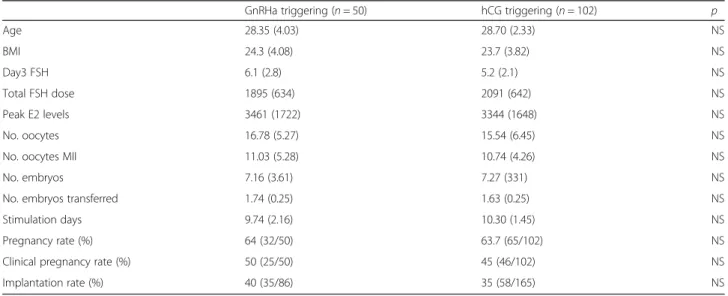
A total of 152 women were included in this study. The groups did not differ in terms of age, body mass index (BMI), day 3 FSH, total FSH dose, peak E2 levels, numbers of oocytes, MII oocytes, embryos, embryos transferred,
stimulation days, or implantation and pregnancy rates (Table 1).
When the embryo developmental kinetics were evalu-ated (Table 2), some early developmental events oc-curred significantly later in embryos derived from cycles triggered with hCG (n = 102 patients, n = 487 embryos) than in embryos derived from cycles triggered with a GnRHa (n = 50 patients, n = 252 embryos). The times from insemination to tPB2, tPNF, t2, t3, t5, and t6 were significantly shorter in the GnRHa triggered group (Group 1). The groups did not differ significantly in terms of t4, t7, t8, t9, cc2, and s2, but cc3was significantly shorter in Group 1 than in Group 2.
The percentage of optimal embryos based on the rele-vant morphokinetic variables were compared according to type of triggering agent. Variables analyzed were the time of cleavage to five cells (t5), second cell cycle (cc2), duration of the period as a two-cell blastomere embryo (t3– t2), and synchrony in division from two-cell blasto-mere embryos to four-cell blastoblasto-mere embryos (s2). There were no significant difference between groups ac-cording to t5. However percentage of optimal embryos according to s2 and cc2 were significantly higher in GnRH agonist group (Group1) than hCG triggering group (p < 0.05) (Table 3)
Discussion
Embryo competence depends on oocyte quality, which is affected by different factors, including the treatment mo-dality [12, 13]. We do not know the exact effect of the dosages and types of drugs on oocytes in IVF. Therefore, it was suggested that mild stimulation protocols and nat-ural cycles would reduce aneuploidy rates and increase embryo quality [12]. However, every step during
ovulation induction could affect the oocytes and subse-quent embryo development.
Triggering oocyte maturation is the last important step of ovulation induction. For a long time, hCG has been used as a triggering agent because of its homology with LH and extended life. Although the extended half-life of this molecule might be advantageous for luteal support, its effect on oocyte maturation is not clear and hCG-mediated LH activity differs from natural cycles. Recently, GnRH agonists have been used as the trigger-ing agent, especially in patients at a high risk of OHSS in GnRH antagonist cycles. The GnRHa displaces the GnRH antagonist from the GnRH receptor, inducing ini-tial activation (flare-up) of LH and FSH, similar to that of the natural cycle before receptor downregulation [14]. This seems to be a more physiological mode of oocyte maturation. We examined whether there is a difference in the development of these oocytes after GnRHa trig-gering. Limited data are available on this subject in the literature. To our knowledge, this is the first study to evaluate embryo kinetics after the use of different oocyte-triggering agents in patients undergoing the same treatment protocol. Our results confirmed that the oocyte-triggering agent affected the early developmental kinetics of oocytes and embryos.
Interestingly, we found that the time intervals during early embryo development were shorter in GnRHa-triggered cycles. A recent report evaluated the effect of different treatment protocols combined with different triggering agents and found that embryos cleaved faster, especially in the early developmental steps, following an antagonist protocol plus GnRHa triggering compared with a GnRHa protocol plus hCG triggering [10]. Our results were similar. However, the stimulation protocols
Table 1 Demographic characteristics of the two study groups
GnRHa triggering (n = 50) hCG triggering (n = 102) p
Age 28.35 (4.03) 28.70 (2.33) NS BMI 24.3 (4.08) 23.7 (3.82) NS Day3 FSH 6.1 (2.8) 5.2 (2.1) NS Total FSH dose 1895 (634) 2091 (642) NS Peak E2 levels 3461 (1722) 3344 (1648) NS No. oocytes 16.78 (5.27) 15.54 (6.45) NS
No. oocytes MII 11.03 (5.28) 10.74 (4.26) NS
No. embryos 7.16 (3.61) 7.27 (331) NS
No. embryos transferred 1.74 (0.25) 1.63 (0.25) NS
Stimulation days 9.74 (2.16) 10.30 (1.45) NS
Pregnancy rate (%) 64 (32/50) 63.7 (65/102) NS
Clinical pregnancy rate (%) 50 (25/50) 45 (46/102) NS
Implantation rate (%) 40 (35/86) 35 (58/165) NS
Results are presented as means (SD) when appropriate
also differed in the aforementioned study groups. There-fore, both stimulation and triggering factors affected the results. In comparison, in our study, both groups followed a GnRH antagonist protocol. Therefore, their result might also be related to triggering factors, rather than the stimulation protocol.
In this study, we found that oocytes were fertilized earlier and that the PNF, t2, t3, t5, and t6intervals were shorter, while we did not detect any difference in the later events. What is the role of early developmental kin-etics in subsequent embryo development? Previous stud-ies have compared early and late cleaving embryos and found that significantly more early cleaving embryos were good-quality embryos and the transfer of early cleavage embryos resulted in higher implantation and pregnancy rates [15–17]. Recently, Lemmen et al. re-ported that the disappearance of pronuclei and first div-ision occur earlier in embryos that implant and cell number is higher on day 2 of embryonic development [18]. Wong et al. found a correlation between reaching
the blastocyst stage and the first, second, and third cell divisions [19]. Meseguer et al. evaluated the use of mor-phokinetics as a predictor of embryo implantation and reported six discriminative morphokinetic parameters (t2, t3, t4, t5, cc2, and s2) that were correlated with im-plantation [11]. In our study, the durations of t2, t3, and t5 of GnRHa-triggered embryos were shorter than for hCG-triggered embryos. Unfortunately, we could not evaluate the relationship between these differences in early developmental kinetics and pregnancy rates be-cause we did not use embryo developmental kinetics for embryo selection. However, the proportion of optimal embryos based on the relevant morphokinetic variables (s2 and cc2) which are considered to have a strong pre-dictive potential of embryo competence were signifi-cantly higher in GnRH agonist triggered group. Thus the optimal time intervals stated above are related to higher implantation potential [17–19].
What causes the difference in the early embryologic developmental kinetics between GnRHa- and hCG-triggered cycles? One explanation might be the oocyte maturation process. Some recent studies focused on the oocyte maturation rate after hCG and GnRHa triggering, and found that the number of oocytes retrieved, percent-age of mature oocytes, and number of top-quality em-bryos were either comparable or in favor of the GnRHa trigger [8, 20]. These findings were related to different physiological events that happened after hCG and GnRHa triggering. Therefore, the effects of these agents differ in duration and receptor activation in some in-stances [20–22]. The duration of the LH surge after hCG triggering is longer than with GnRHa triggering,
Table 2 Embryo developmental kinetics according to the type of oocyte maturation triggering agent
GnRHa triggering (Group 1 n:252) hCG triggering (Group 2 n:487) P
tPB2 4.5 (1.7) 5.4 (1.8) 0.000 tPNf 24.8 (5.2) 26.7 (5.2) 0.000 t2(h) 32.2 (6.2) 34.3 (5.5) 0.009 t3(h) 38.5 (6.0) 39.9 (5.1) 0.027 t4(h) 40.6 (6.3) 41.7 (5.3) 0.101 t5(h) 47.0 (7.9) 50.8 (7.1) 0.000 t6(h) 52.1 (7.3) 54.6 (6.2) 0.002 t7(h) 57.5 (8.2) 57.8 (5.2) 0.871 t8(h) 63.2 (6.3) 62.5 (5.1) 0.589 t9+(h) 64.5 (5.9) 64.9 (4.8) 0.996 cc2(h) 6.9 (5.9) 6.3 (4.1) 0.321 s2(h) 2.6 (4.2) 2.0 (2.2) 0.151 cc3(h) 9.7 (6.1) 11.5 (4.5) 0.006 t4– t2(h) 9.5 (6.4) 8.6 (4.6) 0.071 t8– t4(h) 24.3 (6.3) 23.5 (5.1) 0.251
Results are presented as means(SD) when appropriate
t time, h hour, tPB2appearance of second polar bodytPNfboth pronuclei faded,cc cell cycle s, synchrony
Table 3 Percentages of optimal embryos whose cleavages are included in optimal timing ranges with a predicted higher implantation potential (Meseguer et al. 2011) according to type of triggering
Embryo category GnRHa triggering hCG triggering p
T5 (%) 15.4 17.4 NS
S2 (%) 42.0 24.8 0.000
CC2 (%) 52.3 43.1 0.005
Data are presented as % (n) for each category. The proportions of optimal embryos in each category were compared using theχ2
test. NS. no statistically significant differences were found
i.e., days versus 24 h, respectively [18]. LH has a greater impact on AKT and extracellular signal regulated kinase (ERK1/2) phosphorylation and is responsible for granu-losa cells proliferation, differentiation, and survival, while hCG generates more intracellular cAMP accumulation, which stimulates steroidogenesis [22]. HCG induces ele-vated follicular fluid progesterone levels, suggesting that there are differences in the oocyte microenvironment just before ovulation compared with the endogenous LH surge [23]. Recent studies also suggested a potential fa-vorable impact of FSH in the process of nuclear matur-ation by actively promoting the resumption of meiosis [19, 24, 25]. Erb et al. reported a significantly greater yield of high-quality embryos in the GnRHa-triggered group in donor cycles [26]. They suggested that the lon-ger half-life of hCG causes over-luteinization of the re-cruited follicles, affecting oocyte and embryo quality.
Although GnRHa triggering seems to favor oocyte maturation and embryo development, one major disad-vantage of GnRHa-triggered cycles is the luteolytic effect after GnRHa triggering, which may necessitate adding low-dose hCG for luteal support in fresh IVF cycles [8]. A dual or double trigger using both GnRHa and hCG might improve oocyte and embryo quality, while sup-porting the luteal phase. Lin et al. found that signifi-cantly more oocytes were retrieved, with more mature oocytes, and more embryos cryopreserved, with a signifi-cant increase in implantation, clinical pregnancy, and live birth rates, as compared with the hCG-triggered group [27]. Decleer et al. reported a greater number of excellent oocytes and cryopreserved embryos after dual triggering, compared with hCG only [28]. A recent paper recommended prolonging the time interval between ovulation triggering with a GnRHa and oocyte pick-up to overcome any existing impairment in granulosa cell function, oocyte meiotic maturation, or cumulus expan-sion for patients with abnormal follicular maturation [29]. We did not evaluate the effects of a dual trigger on oocyte development. Future studies evaluating the em-bryo developmental kinetics after a dual trigger will give more information on the subject.
Our findings suggested favorable embryo develop-mental kinetics after GnRHa triggering. This may be re-lated to the more physiological maturation process in response to GnRHa as previously reported by others [24–26]. Therefore, another patient-friendly option might be GnRHa triggering combined with transfer of these embryos in a natural thaw cycle with a natural endometrium, especially for patients at high risk of OHSS. Elective vitrification is an alternative embryo transfer strategy to achieve better perinatal outcomes following assisted reproduction technology treatment [30]. Future randomized controlled studies will provide more information.
Conclusions
In conclusion, the method used to trigger oocyte matur-ation seems to affect the dynamic parameters of early oocyte and embryo development. Larger randomized controlled studies are needed to evaluate the clinical ef-fects of these findings.
Competing interests
The authors declare that they have no competing interests. Authors’ contribution
ASG conception of study, acquisition of data, revising the manuscript for intellectual content, approval of the final version. FG conception of the study, data analysis and interpretation, drafting the manuscript, approval of final version. MSU revising the manuscript for intellectual content, BI approval of the final version acquisition of data, approval of the final version MK revising the manuscript for intellectual content, approval of the final version. NO acquisition of data, approval of the final version, EEO revising the manuscript for intellectual content, approval of the final version. AA acquisition of data, approval of the final version. All authors read and approved the final manuscript.
Acknowledgement
We thank all patients and staff of the study centre (AhmetŞalvarcı, Fuat Ali, Dilek Incesu).
The English in this document has been checked by at least two professional editors, both native speakers of English. For a certificate, please see: http:// www.textcheck.com/certificate/e3pxvx.
Funding
No funding was saught for this report. Author details
1Novafertil IVF Centre, Yeni Meram yolu No:75, Meram, Konya, Turkey. 2
Department of Obstetrics and Gynecology, Izmir University Hospital, Izmir, Turkey.3Department of Obstetrics and Gynecology, Baskent University
Hospital, Konya, Turkey.4Department of Obstetrics and Gynecology,
Necmettin Erbakan University, Konya, Turkey.
Received: 4 December 2015 Accepted: 18 March 2016
References
1. Baxter Bendus AE, Mayer JF, Shipley SK, Catherino WH. Interobserver and intraobserver variation in day 3 embryo grading. Fertil Steril. 2006;86:1608–15. 2. Montag M, Toth B, Strowitzki T. New approaches to embryo selection.
Reprod Biomed Online. 2013;27(5):539–46.
3. Armstrong S, Arroll N, Cree LM, Jordan V, Farquhar C. Time-lapse systems for embryo incubation and assessment in assisted reproduction. Cochrane Database Syst Rev. 2015;27(2):CD011320.
4. Steptoe PC, Edwards RG. Birth after reimplantation of a human embryo. Lancet. 1978;2(8085):366.
5. Al-Inany HG, Youssef MA, Aboulghar M, Broekmans F, Sterrenburg M, Smit J, Abou-Setta AM. Gonadotropin-releasing hormone antagonists for assisted reproductive technology. Cochrane Database Syst Rev. 2011;11(5):CD001750. 6. Kol S, Humaidan P, Itskovitz-Eldor J. GnRH agonist ovulation trigger and
hCG-based, progesterone-free luteal support: a proof of concept study. Hum Reprod. 2011;26:2874–7.
7. Youssef MAF, Abdelmoty HI, Ahmed MAS, Elmohamady M. GnRH agonist for final oocyte maturation in GnRH antagonist co-treated IVF/ICSI treatment cycles: Systematic review and meta-analysis. J Adv Res. 2015;6:341–9. 8. Humaiden P, Papanikolaou EG, Kyrou D, Alsbjerg B, Polyzos NP, Devroey P,
Fatemi HM. The luteal phase after GnRH-agonist triggering of ovulation: present and future perspectives. Reprod Biomed Online. 2012;24:134–41. 9. Kol S, Humaiden P. LH (as HCG) and FSH surges for final oocyte maturation:
sometimes it takes two to tango? Reprod Biomed Online. 2010;21:590–2. 10. Munoz M, Cruz M, Humaidan P, Garrido N, Perez-Cano I, Meseguer M. The
type of GnRH analogue used during controlled ovarian stimulation influences early embryo developmental kinetics: a time lapse study. Eur J Obstet Gynecol Reprod Biol. 2013;168:167–72.
11. Meseguer M, Herrero J, Tejera A, Hilligsoe KM, Ramsing NB, Remohi J. The use of morphokinetics as a predictor of embryo implantation. Hum Reprod. 2011;26:2658–71.
12. Rubio C, Mercader A, Alama P, Lizan C, Rodrigo L, Labarta E, Melo M, Pellicer A, Remohi J. Prospective cohort study in high responder oocyte donors using two hormonal stimulation protocols: impact on embryo aneuploidy and development. Hum Reproduc. 2010;25:2290–7.
13. Baart EB, Macklon NS, Fauser BJ. Ovarian stimulation and embryo quality. Reproduct Biomed Online. 2009;18 Suppl 2:45–50.
14. Humaidan P, Polyzos NP, Alsbjerg B, Erb K, Mikkelsen AL, Elbaek HO, Papanikolaou EG, Andersen CY. GnRHa trigger and individualized luteal phase hCG support according to ovarian response to stimulation: two prospective randomized controlled multi-centre studies in IVF patients. Hum Reprod. 2013;28(9):2511–21.
15. Orvieto R. Triggering final follicular maturation-hCG, GnRH-agonist or both, when and to whom? J Ovarian Res. 2015;8:60.
16. Lundin K, Bergh C, Hardarson T. Early embryo cleavage is a strong indicator of embryo quality in human IVF. Hum Reprod. 2001;16:2652–7.
17. Fenwick J, Platteau P, Murdoch AP, Herbert M. Time from insemination to first cleavage predicts developmental competence of human
preimplantation embryosin vitro. Hum Reprod. 2002;17:407–12.
18. Lemmen JG, Agerholm I, Ziebe S. Kinetic markers of human embryo quality using time-lapse recordings of IVF/ICSI fertilized oocytes. Reprod Biomed Online. 2008;17:385–91.
19. Wong CC, Loewke KE, Bossert NL, Behr B, De Jonge CJ, Baer TM, Reijo Pera RA. Non invasive imaging of human embryos before embryonic genome activation predicts development to the blastocyst stage. Nat Biotechnol. 2010;28:1115–21. 20. Van Montfoort AP, Dumuolin JC, Kester AD, Evers JL. Early cleavage is a
valuable addition to existing embryo selection parameters: a study using single embryo transfers. Hum Reprod. 2004;19:2103–8.
21. Fauser BC, De Jong D, Olivennes F, Wramsby H, Tay C, Itskovitz Eldor J, et al. Endocrine profiles after triggering of final oocyte maturation with GnRH agonist after cotreatment with the GnRH antagonist ganirelix during ovarian hyperstimulation for in vitro fertilization. J Clin Endocrinol Metab. 2002;87:709–15.
22. Casarini L, Lispi M, Longobardi S, Milosa F, La Marca A, Tagliassacchi D, et al. LH and HCG action on the same receptor results in quantitatively and qualitatively different intracellular signaling. PLoS One. 2012;7:e46682. 23. Yding Andersen C, Westergaard LG, Figenschau Y, Bertheussen K, Forsdahl
F. Endocrine composition of follicular fluid comparing human chorionic gonadotrophin to a gonadotrophin-releasing hormone agonist for ovulation induction. Hum Reprod. 1993;8:840–3.
24. Zelinski-Wooten MB, Hutchison JS, Hess DL, Wolf DP, Stouffer RL. Follicle stimulating hormone alone supports follicle growth and oocyte development in gonadotrophin-releasing hormone antagonist-treated monkey. Hum Reprod. 1995;10:1658–66.
25. Yding Andersen C, Leonardsen L, Ulloa-Aguirre A, Barrios-De Tomasi J, Moore L, Byskov AG. FSH-induced resumption of meiosis in mouse oocytes: effect of different isoforms. Mol Hum Reprod. 1999;5:726–31.
26. Erb TM, Vitek W, Wakim ANG. Gonadotropin realesing hormone agonist or human chorionic gonadotropin for final oocyte maturation in an oocyte donor program. Fertil Steril. 2010;93:374–8.
27. Lin MH, Wu FS, Lee RK, Li SH, Lin SY, Hwu YM. Dual trigger with combination of gonadotropin-releasing hormone agonist and human chorionic gonadotropin significantly improves the live-birth rate for normal responders in GnRH antagonist cycles. Fertil Steril. 2013;100(5):1296–302. 28. Decleer W, Osmanagaoglu K, Seynhave B, Kolibianakis S, Tarlatzis B, Devroey P.
Comparison of hCG triggering versus hCG in combination with a GnRH agonist: a prospective randomized controlled trial. Facts Views Vis Obygyn. 2014;6(4):203–9.
29. Uygur D, Alkan RN, Batuoglu S. Recurrent empty follicle syndrome. J Assist Reprod Genet. 2003;20:390–2.
30. Li Z, Wang Y, Ledger W, Edgar DH, Sullivan EA. Clinical outcomes following cryopreservation of blastocysts by vitrification or slow freezing: a population-based cohort study. Hum Reprod. 2014;29(12):2794–801.
• We accept pre-submission inquiries
• Our selector tool helps you to find the most relevant journal
• We provide round the clock customer support
• Convenient online submission
• Thorough peer review
• Inclusion in PubMed and all major indexing services
• Maximum visibility for your research Submit your manuscript at
www.biomedcentral.com/submit