Does topical bevacizumab prevent postoperative
recurrence after pterygium surgery with conjunctival
autografting?
窑Clinical Research窑
1Department of Ophthalmology, School of Medicine, Baskent University, Konya 42080 Turkey
2Department of Ophthalmology, School of Medicine, Izmir University, Izmir 35530, Turkey
3Department of Ophthalmology, Bay覦nd覦r Hospital, Ankara 06520 Turkey
4Department of Ophthalmology Sel觭uk University Faculty of Medicine, Konya 42050 Turkey
Correspondence to: Aylin Karalezli. Department of Ophthalmology, School of Medicine, Baskent University, Konya 42080, Turkey. [email protected]
Received: 2013-06-07 Accepted: 2013-09-02
Abstract
·
AIM: To assess the effect of topical bevacizumab use on postoperative pterygium recurrence in eyes who underwent pterygium excision with limbal -conjunctival autograft transplantation (LCAT).·
METHODS: eighty -eight eyes of 88 patients with primary pterygium were included. Pterygia were graded preoperatively from type 1 to type 3 (type 1 atrophic, type 3 inflamed) according to the inflammatory status. The eyes were preoperatively randomized to receive topical steroid and antibiotic treatment (group 1, 46 eyes) and additional topical bevacizumab (5 mg/mL; group 2, 42 eyes) in the postoperative period. All eyes underwent pterygium excision and LCAT. Medications were tapered and discontinued at one month. Postoperative complications and recurrence rates were recorded.·
RESULTS: The mean follow -up duration was 29.3 依 4.2mo (24-52mo) and 28.5依3.4 (24-48mo) in group 1 and 2, respectively ( 跃0.05). There were no statistically significant differences regarding the age or gender between groups ( 跃0.05). Also, the difference between groups with respect to pterygium type was not significant. During the follow -up period, recurrence developed in 2 eyes (4.3%) in group 1, whereas in one eye (2.4% ) in group 2. No statistically significant difference between groups was found in recurrence rates ( 跃0.05). No re-operation for recurrence was necessary during the follow-up period in both groups.·
CONCLUSION: Topical bevacizumab seems to have no additonal effect on pterygium recurrence after LCAT.·
KEYWORDS: limbal-conjuctival autograft; topical bevacizumab; pterygium; recurrence; topical bevacizumab DOI:10.3980/j.issn.2222-3959.2014.03.23Karalezli A, Kucukerdonmez C, Akova YA, Koktekir BE. Does topical bevacizumab prevent postoperative recurrence after pterygium surgery with conjunctival autografting? 2014;7(3):512-516
INTRODUCTION
P
terygium, histopathologically characterized by elastotic degeneration of collagen and fibrovascular proliferation, is a very common conjunctival degenerative condition. Current treatment for pterygium focuses on surgical excision and prevention of recurrence. Conjunctival autografting after pterygium excision is associated with very low rates of recurrence and complications when compared to other surgical techniques. The surgeon's skill and experience affect the recurrence rate, which varies between 2% and 39% with this technique[1,2].In the literature, some medical approaches were also used intraoperatively and postoperatively to prevent the recurrence including adjunctive therapy with beta radiation, thiotepa, mitomycin C, 5-fluorouracil, and corticosteroids ( triamcinolone)[3-7]. However, the value of such therapy is limited because of potential ocular side effects, such as increased intraocular pressure, secondary bacterial infection, scleral ulceration, poor epithelial healing, and superficial punctate keratitis.
Several studies suggest that vascular endothelial growth factor (VEGF) plays an important role in the development of pterygium. Furthermore, VEGF has been identified in the epithelium of pterygium [8-12]. The overexpression of VEGF in pterygium tissue led us to consider to evaluate the role of anti-VEGF therapy, which could induce regression of blood vessels and hence retard progression of pterygium. Bevacizumab (Avastin; Roche, USA) is a recombinant humanized monoclonal antibody against VEGF that neutralizes all isoforms of human VEGF and inhibits VEGF-induced proliferation of endothelial cells[13,14]. To date, several studies regarding the use of topical or subconjunctival bevacizumab as an off-label treatment for pterygium have been published [15-19]. Nonetheless, the results remain
栽藻造押8629原愿圆圆源缘员苑圆 8629-82210956 耘皂葬蚤造押ijopress岳员远猿援糟燥皂
inconclusive. Our study is the first randomized controlled trial that evaluated the effect of topical bevacizumab on postoperative recurrence after pterygium surgery with limbal-conjunctival autograft transplantation (LCAT). SUBJECTS AND METHODS
Eighty-eight consecutive patients (88 eyes) with primary nasal pterygium were enrolled in this prospective study. Patients with immune system, eyelid or ocular surface diseases ( blepharitis, Sj觟gren syndrome and dry eye), and a history of ocular surgeries within the previous 6mo were excluded from the study. All patients were informed about the design of the study and the procedure, and written informed consent was obtained from all patients. The study adhered to the tenets of the Declaration of Helsinki, and was approved by the Ethics Committee at Baskent University Faculty of Medicine (KA 11/34).
Patients were randomized into two groups. Randomization was accomplished by a list created by a random-number generator. One eye of each patient was included in the study. If the patient had bilateral pterygium, one eye was selected randomly and included in the study.
The pterygia were graded according to the grading system developed by Tan [20] as grade 1 (atrophic) with episcleral vessels under the body of the pterygium not obscured and clearly distinguishable; grade 3 (fleshy), episcleral vessels totally obscured; or grade 2 (intermediate), all other pterygia not falling into grades 1 or 3 (Figure 1). The pterygia extended at least 3 mm beyond the limbus in all cases.
To ensure consistency, all operations were performed under an operating microscope by the same surgeon (AK). After administration of topical 0.5% proparacaine hydrochloride ( HCL) ( Alcaine , Alcon , Switzerland ) , lidocaine HCL 40 mg/2 mL+epinephrine 0.025 mg/mL (Jetokain, Adeka, Turkey) was injected under the conjunctiva at the superotemporal bulbar region and into the body of the pterygium. The head of the pterygium was excised completely from the cornea, and the body was dissected and excised with conjunctival scissors. Abnormal scar tissue on the corneal surface was polished. Minimal cauterization was used to control bleeding. Graft transplantation was carried out after calculating the size of the defect (mm2) and determining the correct size (oversized by 1 mm horizontally and vertically compared with the bare sclera defect). Conjunctival autografts were taken from the superior temporal conjunctiva to have a large area for harvesting an appropriate-size graft and to achieve a more cosmetically acceptable appearance in the operated eye as a result of the coverage of the donor site by the upper lid. We were careful to place the limbal edge of the graft at the host limbal tissue. The graft was subsequently moved to the nasal area and attached to the conjunctival edges and episclera with fibrin
glue (Tisseel, Baxter, USA) in all cases. The eye was covered with an eye pad after administration of topical antibiotic ointment (Tobrex, Alcon).
Postoperatively, 46 patients (46 eyes, group 1) received dexamethasone 0.1% (Maxidex, Alcon) and tobramycin 0.3% (Tobrex, Alcon) eye drops. The medications were tapered over the course of four weeks. In group 2 (42 eyes), additional 5 mg/mL topical bevacizumab was used four times daily for one month postoperatively. These drops were prepared from the commercial bevacizumab solution (25 mg/mL Avastin, Genentech, USA) under sterile conditions and stored in sterile vials at +4℃ for 48h. Patients were examined on the first postoperative day, weekly until one month, and monthly thereafter. Postoperative complications and recurrence rates were recorded. Recurrence was defined as any fibrovascular growth that passed the corneal limbus by more than 1mm. Statistical Analysis To determine the sample size, 琢=0.05 and power=80% (1-茁=0.80) were accepted. The minimum sample size as calculated using the PASS statistical software program (NCSS, Kaysville, UT, USA). Assessment for normal distribution of the data was performed with the D'Agustino-Pearson test. The Student test for independent samples was used to compare parametric data and the Mann-Whitney U test was used for unpaired nonparametric data by using SPSS version 13.0 (SPSS Inc., Chicago, Illinois, USA). values less than 0.05 were considered significant.
RESULTS
The demographic data are summarized in Table 1. There were no statistically significant differences between the two groups with regard to age and sex ( =0.311, =0.534, respectively).
The mean follow-up period was 29.3依4.2mo (24-52mo) in
Figure 1 The preoperative grading of the pterygia Grade 1 (top) refers to episcleral vessels under the body of the pterygium not obscured and clearly distinguishable, grade 3 (bottom) to episcleral vessels totally obscured and grade 2 (middle) to all other pterygia not falling into grades 1 or 3.
group 1 and 28.5依3.4 (24-48mo) in group 2, and the difference was not statistically significant ( =0.621) (Table 1). Also, the difference between groups with respect to pterygium type was not significant. In both groups, type 2 pterygium was the most common type ( =0.826) (Table 2). Figures 2 and 3 show the postoperative appearances of the patients at the first postoperative day, the first week and 6th month in group 1 and group 2 additional bevacizumab therapy, respectively. Pterygium recurrence was observed in 2 eyes (4.3%) in group 1, and in one eye (2.4%) in group 2. No statistically significant difference between groups was found regarding to recurrence rates ( =0.092, Table 1). The recurrences occurred at the third and fourth month in group 1 and at the fourth month in group 2. The preoperative grades of these recurrent pterygia were grade 2 in both groups. The recurrent cases were closely followed-up for continuing growth of fibrovascular tissue onto the cornea. No reoperation was necessary during the follow-up period in both groups.
Topical bevacizumab was well tolerated in all patients. No irritation, burning, and/or any systemic side effects of bevacizumab were reported by the patients during the follow-up period.
DISCUSSION
Inflammation and fibrovascular proliferation are important factors in the pathogenesis of the pterygium which is a degenerative process. The extent and severity of the fibrovascular growth of pterygium seem to comprise a reliable morphologic index for predicting recurrence after surgery [20]. Several methods have been proposed to reduce the recurrence rate, but none has been shown to be superior to the other.
Several active angiogenic and epithelial growth factors including fibroblast growth factor, heparin-binding epidermal growth factor, and connective tissue growth factor contribute in the fibrovascular pathogenesis of pterygium. However, VEGF, which has been shown to be significantly increased in the excised pterygium tissues, plays the main role[11]. It is also
well-known that VEGF is the key factor in wound healing response. It is found to be elevated in active wounds and it plays a role in the fibroblast and inflammatory cell migration through acting as a signal transducer [21]. Thus, VEGF has been the primary target of many current antiangiogenic therapies and hence, by blocking VEGF, the remodeling and wound healing process the postoperative period of pterygium
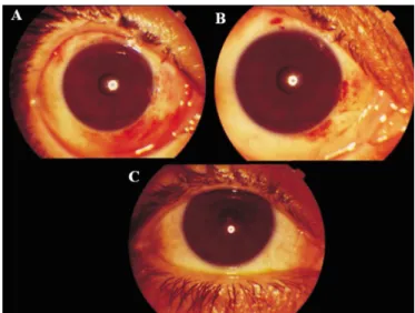
Figure 2 Postoperative photographs of a patient in group 1 (topical steroid) A: The graft is in place at postoperative 1d; B: There is no graft displacement and the vascularization is greatly reduced at postoperative 7d; C: A normal conjunctival healing without any recurrence at postoperative 6thmonth.
Figure 3 Postoperative photographs of a patient in group 2 (topical steroid and bevacizumab) A: The appearance at postoperative 1d showing subconjunctival hemorrhage and slight graft edema; B: The subconjunctival hemorrhage is reduced and the graft is healthy at 7d; C: There is no recurrence at postoperative 6th
month.
Table 1 Patients’ demographics, follow up periods and recurrence rates in two groups
Parameters Group 1 (steroid)
n=46 Group 2 (steroid+bevacizumab) n=42 P Gender(M/F) 21/25 22/20 0.534 Age (a) 53.04±11.81 58.82±12.02 0.311 Follow-up (mo) 29.3±4.2 28.5±3.4 0.621 Recurrence rate 2 (4.3 %) 1 (2.4 %) 0.092
Table 2 Pterygium types in two groups
Types Group 1 (steroid)
n=46 Group 2 (steroid+ bevacizumab) n=42 P Type 1 11 (24%) 7 (17%) 0.213 Type 2 22 (48%) 21 (50%) 0.826 Type 3 13 (28%) 14 (33%) 0.534
栽藻造押8629原愿圆圆源缘员苑圆 8629-82210956 耘皂葬蚤造押ijopress岳员远猿援糟燥皂
would be also changed and recurrence rate may be reduced as a result of reduction of fibrovascular tissue formation. There have been several studies on the use of bevacizumab in primary and recurrent pterygia. Main treatment approaches included subconjunctival and intralesional injection of the drug as well as topical administration to reduce the recurrence rate or to inhibit an impending recurrent pterygium. Also, brief reports and controlled trials were published about intralesional injections of the drug without any surgery [17,22]. Main outcome measures were the reduction in size in short term, but the effect of bevacizumab was not found to be clinically significant in these studies. Bahar and co-authors [19] reported that there were neither ocular or systemic adverse effects in patients treated with subconjunctival bevacizumab for recurrent pterygium, nor beneficiary effects seen on regression of corneal neovascularization. Razeghinejad [16] reported single dose of intraoperative subconjunctival bevacizumab injection in 30 eyes of 30 patients who underwent primary pterygium excision with rotational flap application and did not find any effect on the recurrence rate. Lekhanont [23] have conducted a randomized controlled study in patients with impending recurrent pterygia. They have applied three different dosages of bevacizumab with subconjunctival route, which showed no beneficial effects on the regression of the lesions or on the recurrence rate, but only a transient decrease in the conjunctival vascularization was observed. Ozgurhan [24] applied topical bevacizumab as adjunctive therapy 1mo after conjunctival autograft surgery for recurrent ptergia and found that the drug was safe and effective to prevent corneal neovascularization. But they did not find statistically significant effect on the recurrence rate .
Uy [25] has applied topical bevacizumab in 2 Steven Johnson cases with corneal neovascularization and they reported that the drug was well tolerated and induced regression of ocular surface neovascularization, conjunctival injection, and corneal opacification. In another study by Manzano 26], the effect of topical bevacizumab on experimental corneal neovascularization in rats was studied. They reported that 4 mg/mL topical administration of bevacizumab had decreased corneal neovascularization by 40% following chemical injury.
Wu [18] reported having success with topical bevacizumab in a patient suffering from impending recurrent pterygium. Fallah and coauthors [15] also reported that short term use of topical bevacizumab was effective in delaying of recurrence of impending pterygia in a study including 54 patients. However, the use of mitomycin C in their study might be an additive factor in the inhibition of recurrence. Conjunctival autografting is considered the most effective technique of preventing recurrence [20,28]. Although it is safe and effective, more surgical expertise, technical ability, and
surgical time are needed to secure the grafts with sutures[28]. Earlier studies with the use of fibrin-based adhesives for attaching conjunctival grafts in pterygium surgery showed that this technique reduces the surgery time, suture-related complications and patient discomfort [19,29-31]. In the current study, we performed pterygium excision and LCAT and used fibrin glue for fixation of the autograft in both groups. In our knowledge, this study is the first prospective, randomized clinical trial with long-term follow-up in the literature. Moreover, the lack of any significant difference between groups in patient age and preoperative pterygium type which may have an effect on the recurrence rates, strengthens the credibility of the results.
In conclusion, topical bevacizumab was well tolerated as an additional therapy after pterygium surgery with autografting. The drug did not have any side effects on wound healing or epithelization. However, the recurrence rate seems to be unaffected despite one month topical bevacizumab therapy. Bevacizumab seems to have no beneficial effect on lowering the recurrence of primary ptergia when used topically after operation.
ACKNOLEDGEMENTS
Conflicts of Interest: Karalezli A, None; Kucukerdonmez C, None; Akova YA, None; Koktekir BE, None.
REFERENCES
1 Hirst LW. The treatment of pterygium. 2003;48 (2):
145-180
2 Ang LP, Chua JL, Tan DT. Current concepts and techniques in pterygium
treatment. 2007;18(4):308-313
3 Yamada T, Mochizuki H, Ue T, Kiuchi Y, Takahashi Y, Oinaka M. Comparative study of different β-radiation doses for preventing pterygium
recurrence. 2011;81(5):1394-1398
4 D侏az L, Villegas VM, Emanuelli A, Izquierdo NJ. Efficacy and safety of intraoperative mitomycin C as adjunct therapy for pterygium surgery.
2008;27(10):1119-1121
5 Joselson GA, Muller P. Incidence of pterygium recurrence in patients
treated with thio-tepa. 1966;61(5 Pt 1):891-892
6 Zaky KS, Khalifa YM. Efficacy of preoperative injection versus intraoperative application of mitomycin in recurrent pterygium surgery.
2012;60(4):273-276
7 Salustiano Correa E Silva R, de Pereira Avila M, Rassi AR, Ximenes L, da Silva DS Jr, de Paula AC. Intra-operative use of 5-Fluorouracil in
pterygium surgery: a comparative study. 2013;28 (1):
34-36
8 Liang K, Jiang Z, Zhao B, Shen J, Huang D, Tao L. The expression of vascular endothelial growth factor in mast cells promotes the
neovascularisation of human pterygia. 2012;96 (9):
1246-1251
9 Fukuhara J, Kase S, Ohashi T, Ando R, Dong Z, Noda K, Ohguchi T, Kanda A, Ishida S. Expression of vascular endothelial growth factor C in
human pterygium. 2013;139(2):381-389
10 Jin J, Guan M, Sima J, Gao G, Zhang M, Liu Z, Fant J, Ma JX. Decreased pigment epithelium-derived factor and increased vascular
endothelial growth factor levels in ptergia. 2003;22(5):473-477
Al-Samir K, Geerling G, Paulsen FP Differential expression of vascular endothelial growth factor implies the limbal origin of ptergia.
2005;112(6):1023-1030
12 Aspiotis M, Tsanou E, Gorezis S, Ioachim E, Skyrlas A, Stefaniotou M, Malamou-Mitsi V. Angiogenesis in pterygium: study of microvessel density,
vascular endothelial growth factor, and thrombospondin-1. 2007;21
(8):1095-1101
13 Ranieri G, Patruno R, Ruggieri E, Montemurro S, Valerio P, Ribatti D. Vascular endothelial growth factor (VEGF) as a target of bevacizumab in
cancer: from the biology of the clinic. 2006;13 (16):
1845-1857
14 Ferrara N, Hillan KJ, Novotny W. Bevacizumab (Avastin), a humanized anti-VEGF monoclonal antibody for cancer therapy.
2005;333(2):328-335
15 Fallah MR, Khosravi K, Hashemian MN, Beheshtnezhad AH, Rajabi MT, Gohari M. Efficacy of topical bevacizumab for inhibiting growth of
impending recurrent pterygium. 2010;35(1):17-22
16 Razeghinejad MR, Hosseini H, Ahmadi F, Rahat F, Eghbal H. Preliminary results of subconjunctival bevacizumab in pterygium excision.
2010;43(3):134-138
17 Teng CC, Patel NN, Jacobson L. Effect of subconjunctival bevacizumab
on primary pterygium. 2009;28(4):413-419
18 Wu PC, Kuo HK, Tai MH, Shin SJ. Topical bevacizumab eyedrops for limbal-conjunctival neovascularization in impending recurrent pterygium.
2009;28(1):103-104
19 Bahar I, Kaiserman I, McAllum P, Rootman D, Slomovic A. Subconjunctival bevacizumab injection for corneal neovascularization in
recurrent pterygium. 2008;33(1):23-28
20 Tan DTH, Chee SP, Dear KBG, Lim AS. Effect of pterygium morphology on pterygium recurrence in a controlled trial comparing conjunctival
autografting with bare sclera excision. 1997;115 (10):
1235-1240
21 Wilgus TA, Ferreiara AM, Oberyszyn TM, Bergdall VK, Dipietro LA. Regulation of scar formation by vascular endothelial growth factor.
2008;88(6):579-590
22 Fallah Tafti MR, Khosravifard K, Mohammadpour M, Hashemian MN, Kiarudi MY. Efficacy of intralesional bevacizumab injection in decreasing
pterygium size. 2011;30(2):127-129
23 Lenkhanont K, Patarakittam T, Thongphiew P, Suwan-apichon O, Hanutsaha P. Randomized controlled trial of subconjunctival bevacizumab
injection in impending recurrent pterygium: A pilot study. 2012;31
(2):155-161
24 Ozgurhan EB, Agca A, Kara N, Yuksel K, Demircan A, Demirok A. Topical application of bevacizumab as an adjunct to recurrent pterygium
surgery. 2013;32(6):835-838
25 Uy HS, Chan PS, Ang RE. Topical bevacizumab and ocular surface neovascularization in patients with Steven-Johnson syndrome. 2008;27(1):70-73
26 Manzano RP, Peyman GA, Khan P, Carvounis PE, Kivilcim M, Ren M, Lake JC, Chévez-Barrios P. Inhibition of experimental corneal
neovascularization by bevacizumab (Avastin). 2007;91
(6):804-807
27 Zheng K, Cai J, Jhanji V, Chen H. Comparison of pterygium recurrence rates after limbal conjunctival autograft transplantation and other
techniques: meta-analysis. 2012;31(12):1422-1427
28 Han SB, Hyon JY, Hwang JM, Wee WR. Efficacy and safety of limbal-conjunctival autografting with limbal fixation sutures after
pterygium excision. 2012;227(4):210-214
29 Karalezli A, Kucukerdonmez C, Akova YA, Altan-Yaycioglu R, Borazan M. Fibrin glue versus sutures for conjunctival autografting in pterygium surgery: a prospective comparative study.
2008;92(9): 1206-1210
30 Koranyi G, Seregard S, Kopp ED. Cut and paste: a no suture, small
incision approach to pterygium surgery. 2004;88 (7):
911-914
31 Marticorena J, Rodriguez-Ares MT, Tourino R, Mera P, Valladares MJ, Martinez-de-la-Casa JM, Benitez-del-Castillo JM. Pterygium surgery;