Ankara Üniversitesi Tıp Fakültesi Mecmuası 2008, 61(3)
CERRAHİ BİLİMLER / SURGICAL SCIENCES Olgu Sunumu / Case Report
Recurrent Hemangiopericytoma With Multiple Extracranial
Metastates: A Case Report
Multiple Ekstrakranial Metastazı olan Rekürren Hemanjioperisitoma: Olgu Sunumu
Kutsal Devrim Seçinti, Gökmen Kahiloğulları, Hakan Tuna, Yusuf Şükrü Çağlar
Ankara Üniversitesi Tıp Fakültesi, Nöroşirürji Anabilim Dalı
162
Received: 08.12.2008 • Accepted: 12.01.2008 Corresponding author
Dr. Kutsal Devrim Seçinti
Ankara Üniversitesi Tıp Fakültesi, Nöroşirürji Anabilim Dalı Phone : + 90 (312) 508 25 98/2793
E-mail address : [email protected]
Hemangiopericytoma is an uncommon, highly vascular tumor of pericyte origin that may occur anywhere capillaries are found. These tumors are usually located in the musculoskeletal system, retroperitoneum and the skin. Their intracranial localization is rare. Hemangiopericytomas have a predilection for both local and distant central nervous system recurrences and exhibit an incre-ased tendency to metastasize compared with nonmalignant meningiomas. The biological beha-vior of hemangiopericytoma is sometimes malignant although it is considered grade II-III. Local tumor recurrence after many years is not uncommon, and late and widespread extracranial me-tastases may occur although this is rare in other intracranial tumors. A 46-year old male who had a recurrent intracranial hemangiopericytoma with multiple metastases such as cervical vertebrae, paravertebral soft tissue, lung, kidney and shoulder has been reported
.
Key Words: Hemangiopericytoma, Meningioma, Extracranial metas, Recurrence
Hemanjiyoperisitomalar nadir görülen, oldukça vasküler, kapiller damarların bulunduğu her yer-de görülebilen perisit orijinli tümörlerdir. Bu tümörler genelyer-de kas iskelet sisteminyer-de yerleşirler. Intrakranial yarleşimleri oldukça nadirdir. Hem local hem de santral sinir sisteminin uzak bölgele-rinde rekürrens yapmaya meyillidirler ve malign olmayan menenjiyomlara kıyasla uzak metastaz yapma ihtimalleri daha fazladır. Hemanjiyoperisitomların biyolojik davranışları bazen grade II-III olarak sınıfl andırılmalarına neden olacak kadar malign olabilmektedir. Uzun yıllar sonra lokal tü-mor rekürrensi çok sık değildir ancak yaygın ekstrakranial metastaza rastlanabilmektedir. Bu yazı-da, servikal vertebra, paravertebral yumuşak doku, akciğer, böbrek ve kas-iskelet sistemi metastazı olan, aynı zamanda da intracranial rekürrensi olan bir hemanjiyoperisitom olgusu sunulmuştur. Anahtar Kelimeler: Hemanjioperisitom, Menenjiom, Ekstrakranial metastaz, Rekürrens
Hemangiopericytoma (HPC) is an un-common, highly vascular tumor of pericyte origin that may occur any-where capillaries are found and it accounts for only 1.6-2.4 % of meningeal tumors and less than 1 % of all intracranial tumors (1-5). HPCs are usually located in the musculoskeletal system (lower ex-tremities, pelvis, vertebrae), retro-peritoneum and the skin, but they have been reported in the larynx, spleen, lung, kidney, pancreas, me-diastinium, orbita, adrenal gland, lymph nodes, liver, bone, breast, and pleura. Intracranial and to the other region metastasis of HPC is seen very rare together (6-8).
In this report, we describe a patient with hemangiopericytoma to the other side of brain and a large me-tastasis to the cervicothoracic spine and humerus which occurred with a delay of 4 years after the removal of right frontal meningeal HPC.
Case Report
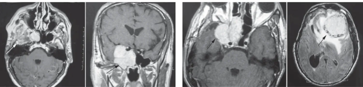
The patient, a 46-year old male, had undergone surgery for the removal of right sided anterior cranial fossa tumor with invasion of right cav-ernous sinus (Figure 1A, 1B). At that time, he had diplopia,
head-Journal of Ankara University Faculty of Medicine 2008, 61(3)
163 Kutsal Devrim Seçinti, Gökmen Kahiloğulları, Hakan Tuna, Yusuf Şükrü Çağlar
ache, visual deficit in his right eye and hypoesthesis on the right side of his face. Hemangiopericyto-ma was detected in his first histo-pathologic investigation and crani-al radiotherapy was performed. One year later, the patient started to
present clinical symptoms, such as headache and diplopia. MRI re-vealed a tumor recurrence at the same region of the first localiza-tion (Figure 2A). He underwent a second neurosurgical operation, with subtotal removal of the le-sion which was located in the fron-tal lobe, anterior cranial fossa, and the right cavernous sinus.
Three years later, he was referred to our department because of con-fusion and headache on left side, nubness and intractable pain at the left side of his body, with gait disability and dizziness. In his neurological examination; hypoes-thesia under the level of C5 , in-creased the deep tendon reflexes on the left, clonus and babinski signs were positive, truncal atax-ia, and also disability on pressure and heat discrimination on the left were found. Cranial MRI revealed a new mass in the left frontal lobe, but no recurrence on the right side (Figure 2B). For the purpose of explaining this clinical situation, servical MRI was performed and in-dicating a mass lesion (Figure 3A, 3B) wich was invading fourth cer-vical vertebrae corpus completely.
The lesion was extending to the left lamina vertebrae via left foramen intervertebrale and also invading left vertebral pedicle as same as other classical metastatic lesions of spinal column. Left vertebral ar-tery was encased by the mas but its lumen was still patent. Cranio-cervical angiograpy also showed a highly vascular lesion in his cer-vicothoracic region (Figure 3C). Left shoulder MRI was performed because of his left-sided arm and shoulder pain and detected a bone metastasis to his left humerus (Fig-ure 3D). Thorax CT revealed left-sided parenchymal multiple meta-static nodules in the lungs, while abdomi-nal CT showed multiple cystic lesions in the left kidney. The patient
un-derwent a third surgical opera-tion with sub-total removal of his cranial lesion, but not for the others. The lesion was diagnosed as hemangioperi-cytoma as ex-pected. The patient did not accept any ad-juvant or
surgi-cal therapy; however, radiotherapy was offered as a choice. Current-ly, the patient is followed-up with 6-month intervals of craniocervical MRI and whole body CT scan for recurrences, metastases, and the lesion progression.
Discussion
HPC is commonly affect adults in the fifth or sixth decade of life. The biological behavior of HPC is sometimes malignant although it is considered grade II-III. Local tu-Figure 1A - 1B. Tumor, involing the anterior skull bose and right
covernous sinus car be in A and B.
Figure 2A - 2B. Tumor recurrence can be seen at the same localization (A). And a new mass detected in his control WRI (B) at the left frontal lobe.
Figure 3A - 3B - 3C - 3D. Cervical metastasis can be seen in A and B. Cervical angiography revealed a highly vascular lesion at servical region (C). The left sholder MRI revealed a metastatic lesion in bony structures (D).
Ankara Üniversitesi Tıp Fakültesi Mecmuası 2008, 61(3)
164 Recurrent Hemangiopericytoma With Multiple Extracranial Metastates: A Case Report
mor recurrence after many years is not uncommon, and late and widespread extracranial metasta-ses may occur (7). Metastatic HPC to the spine is rare. Review of the literature identified only eight re-ported cases (9).
The preoperative differential diag-nosis between meningiomas and HPCs confers therapeutic advan-tages. May any radiologic inves-tigation help surgeons? In a re-cent study, Akiyama et al. reported some radiological features that may help preoperative differential diagnosis (3). Their analysis indi-cated that HPCs have multilobu-lar or irregumultilobu-lar margins, strong contrast enhancement, mainly pial-cortical blood supply, un-like meningiomas, which do not show hyperostosis or calcification. Compatible with their aggressive behavior, HPCs tend to show fea-tures such as irregular or lobulat-ed borders, apparent parenchy-mal invasion (mushrooming), and more heterogeneous contrast en-hancement than meningiomas. A recent study suggested that in vivo magnetic resonance spectroscopy could distinguish between menin-giomas and HPCs due to the higher levels of myo-inositol in the HPCs. Recurrent solitary fibrous tumor, schwannoma, meningioma should also be considered in differential
diagnosis (10).
Microneurosurgery and adjuvant ra-diotherapy constitute the main treatment. The tumor should be surgically removed completely, in-cluding the excision of involved dura mater and bone; when this is not possible, aggressive cauter-ization of the dura and bone is mandatory. Radiation therapy for treatment of this tumor has been reported to be ineffective, but the literature has increasingly docu-mented tumor response to great-er than 450 cGy of local thgreat-erapy. The response rates of HPCs to cur-rently available chemotherapy are poor (2,5). However, complete re-section of the tumor is sometimes impossible because of the risk of hemorrhage during the operation (1,4,5). Hemorrhage represents the most frequent cause of surgery related death, as well as greatest hindrance to tumor removal. Ear-ly reports noted operative mortal-ity rates that varied from 9 to 24 % (1,4). To reduce the risk, some au-thors prefer to use radiotherapy as neoadjuvant treatment (2). In our case; medulla spinalis was
un-der compression at the lesion level and this situation was able to ex-plain why the patient’s comex-plains and the neurological findings were on the same side. The nubness
and intractable pain , motor and sensorial deficitis on his left side were thought to be due to com-pression of fasciculus anterolatera-lis and tractus corticospinaanterolatera-lis. On the other hand, gait disability and dizziness, truncal ataxia, and also disability on pressure and heat discrimination on the left were thought to be due to compression of fasciculus cuneatus and gracilis.
Conclusion
There is a general consensus on the aggressive behavior of hemangio-pericytomas. Surgical resection is usually the initial treatment mo-dality. However, even after the re-section, their high recurrence rate and tendency to metastasize make HPCs very challenging to manage as our case. Radical surgery is the treatment of choice but must be supplemented with postoperative radiotherapy, which has proved to be the therapy most strongly relat-ed to the final prognosis. On the other side, in the case of discrep-ancy between the clinical signs and neurological findings, we decided that a hole spinal MRI should be performed for the aim of searching vertebral column metastasis and medulla spinalis compression.
REFERENCES
1- Spatola C, Privitera G. Recurrent intracranial hemangiopericytoma with extracranial and unusual mul-tiple metastases: case report and review of the literature. Tumori. 2004;90:265-8.
2- Sheehan J, Kondziolka D, Flickin-ger J, et al. RadiosurFlickin-gery for treat-ment of recurrent intracranial he-mangiopericytomas. J Neurosurg. 2002;51:905-1.
3- Akiyama M, Sakai H, Onoue H, et al. Imaging intracranial haemangiope-ricytomas: study of seven cases. Ne-uroradiology. 2004;46:194–7.
4- Hattingen E, Pilatus U, Good C, et al. An unusual intraventricular haeman-giopericytoma: MRI and spectroscopy. Neuroradiology. 2003;45:386–9. 5- Brunori A, Delitala A, Oddi G, et al.
Recent experience in the manage-ment of meningeal hemangiopericy-tomas. Tumori. 1997;83:856-61. 6- Koyama H, Harada A, Nakao A, et al.
Case report and review of literature. Intracranial hemangiopericytoma with metastasis to the pancreas. J Clin Gastroenterol. 1997;25:706-8. 7- Grunenberger F, Bachellier P,
Che-nard MP, et al. Hepatic and pulmo-nary metastases from a meningeal hemangiopericytoma and severe
hypoglicemia due to abnormal sec-retion of insulin-like growth factor: a case report. Cancer. 1999;85:2245-8. 8- Marsot-Dupuch K, Cauquil P, Munt-lak H, et al. Bone metastases of a hemangiopericytoma. J Radiol. 1984;65:41–5.
9- Woitzik J, Sommer C, Krauss JK. De-layed manifestations of spinal metas-tases: a special feature of hemangio-pericytoma. Clin Neurol Neurosurg. 2003;105:159-66.
10- Bikmaz K, Cosar M, Kurtkaya-Yapicier O, et al. Recurrent solitary fibrous in the cerebellopontine angle. J Clin Neurosci 2005;12:829-32.