Morphometric Study on the Digital Bones in the Domestic Cattle
[1]Ozan GÜNDEMİR
1,a
Ermiş ÖZKAN
1,bRıfat MUTUŞ
2,c[1] This study was funded by Scientific Research Projects Coordination Unit of Istanbul University-Cerrahpasa (Project Number: TSA-2018-29873)
1 Istanbul University-Cerrahpasa, Department of Anatomy, Faculty of Veterinary Medicine, TR-34320 Istanbul - TURKEY 2 Istanbul Gelisim University, School of Health Sciences, TR-34310 Istanbul - TURKEY
a ORCID:0000-0002-3637-8166; b ORCID:0000-0002-5000-5075; c ORCID:0000-0001-5140-2462
Article ID: KVFD-2019-22291 Received: 24.03.2019 Accepted: 22.07.2019 Published Online: 22.07.2019
How to Cite This Article
Gündemir O, Özkan E, Mutuş R: Morphometric study on the digital bones in the domestic cattle. Kafkas Univ Vet Fak Derg, 26 (1): 75-82, 2020.
DOI: 10.9775/kvfd.2019.22291
Abstract
In this study, the phalanges of the forelimb and hindlimb of 18 adult Holstein breed cattle were used. Morphometric measurements were taken from 144 digital bones. In contrast to classical references, it was concluded that the greatest lengths (GLpe) were longer in the hindlimb than the forelimb for the phalanx proximalis and phalanx media. In the phalanx proximalis and phalanx media, the SD*100/GLpe index value was high in the forelimb and low in the hindlimb. It was observed that the differences between the Bp (Breadth of the proximal end) values of phalanx proximalis and Bd (Breadth of the distal end) values in phalanx media were significant for the inner bones of the forelimb and their hindlimb counterparts, while the other values were statistically not significant. The presence of an asymmetry between the osteometric measurements of the internal and external bones of the digits could only be observed between the GL values of the phalanx media of forelimb (P<0.05). We concluded that the asymmetry seen in the forelimb in Holstein breed cattle maybe a result of being kept on concrete ground as dairy cows.
Keywords: Digital bones, Cattle, Morphometry
Evcil Sığırlarda Parmak Kemikleri Üzerine Morfometrik Çalışma
Öz
Bu çalışmada, 18 adet erişkin Holstein ırkı sığıra ait ön ve arka ayak parmak kemikleri (phalanx proximalis, media ve distalis) kullanıldı. Toplam 144 parmak kemiğinin her birinden morfometrik ölçümler alındı. En büyük uzunluk (GLpe) ölçümlerinin hem Ph1, hem de Ph2’lerde, klasik kaynakların aksine arka ayaklarda daha fazla olduğu sonucuna varıldı. Ph1 ve Ph2’lerde SD*100/GLpe indeksinin ön ayaklarda daha yüksek, arka ayaklarda ise düşük olduğu gözlendi. Ph1’de Bp, Ph2’de de Bd değerlerinin ön ayakların internal kemikleriyle arka karşılıkları arasındaki farklarının istatistiksel açıdan anlamlı olması dışında, diğer farklılıkların istatistiki açıdan anlam taşımadığı gözlendi. Parmakların internal ve external kemiklerinin osteometrik ölçümleri arasında bir asimetrinin varlığı sadece ön Ph2’nin GL değerleri arasında gözlenebildi (P<0.05). Ön ayak kemikleri arasındaki bu asimetrinin, Holstein ırkı sığırların süt amaçlı yetiştiriciliği ve bunların nispeten sert zeminde tutulmuş olmalarının etkin olabileceği sonucuna varıldı.
Anahtar sözcükler: Morfometri, Sığır, Parmak kemikleri
INTRODUCTION
Ruminants have 4 digital elements and have two digits on each foot [1,2]. The abaxial pairs of the digit-forming elements have been reduced and functionally joined as a single bone as evidence of their common origin [1]. Each metapodium forms the manus with the medial and lateral digital bones found in pairs. Although the phalanx bones in hindlimb are generally similar to the phalanx bones in forelimb, phalanx proximalis and phalanx media are reported to be shorter in the hindlimb
than forelimb [3,4]. This comparison in ruminants was
not made only for the forelimb and hindlimb, but also
for measurements in the inner and outer digits [5-7]. In
some studies, the results of these comparisons show no
statistical differences, although some present asymmetry [5-8].
In the comparison of phalanges forming the medial and lateral digits belonging to the same foot, it was reported that the average length of phalanx proximalis and phalanx
media of the 4th digit was longer than those of the 3rd
digit and the phalanx distalis of the 3rd digit had a larger
mean value [2].
İletişim (Correspondence)
+90 212 4737070/17331The main differences observed in the forelimb and hind-limb were both length and width of phalanx proximalis and phalanx media, which is reported as being shorter in hindlimb in comparison to forelimb in classical anatomy
resources [4], while Ocal et al.[8] reported that phalanx
proximalis and phalanx media are shorter in the forelimb. Nevertheless, it was stated that the most significant difference was in the width.
It has been suggested that the double digits of the
artiodactyls are not in equal length, indicating that they have different functions in posture and carrying
weight [2]. Asymmetry in the lower extremities in cattle
was observed especially in thoracic extremities [9]. It
was reported that there were no significant differences between the corresponding measurements of the right and left extremities of the buffalos and the total length of the digital bones of the medial and lateral digits of the
same extremities [5]. The movement of the animal on soft or
hard ground has an effect on the emergence of asymmetric
condition [2]. The anatomic position of the ruminant feet
causes them to be exposed to significant physiological
stresses that can result in pathological changes [10]. This may
lead to the emergence of common aseptic inflammations
expressed as laminitis [11]. Although the weight of the
animal is carried by the central digits in Bovidae, it is stated that the fact that the digits are not of equal length has an effect on the localization and incidence of the pathologies
observed in the digits [2,12]. It is stated that the digital bones
are symmetrical in the oxen resulting in the equal weight loading to foot during standing and walking, which is the reason why the biomechanical lesions of the foot are less
common than the cattle [5].
Approximately 90% of the clinical lameness cases in cattle are caused by digital lesions and 92% of these lesions affect
the hindlimb [13-15]. It has been reported that approximately
2/3 of the digital lesions also affect the lateral hindlimb
hooves [16,17]. The biomechanical properties of the feet of the
ruminants cause a difference in the balance distribution of the weight between the rear hooves. Chronic overloading of the lateral hind hooves is considered a predisposing condition for cows. A similar situation exists in the fore-limbs. While standing, the majority of the weight is carried
by the medial hoof. The 3rd inner digit of the hoof has a
greater length than the 4thdigit [6]. It is therefore more affected by diseases such as sole ulceration.
In addition to the difference in the distribution of the load on the legs due to the differences in the length of the digits in ruminants, the way the front and rear legs are connected to the body is also thought to affect the biomechanical properties of the foot [2,6]. The fact that the forelimbs are connected to the muscles while the hindlimb connected in the joint style can affect the biomechanics of digital
anatomy in these animals [3]. It is suggested that the effect of different hooves on the forelimb and hindlimb may be
related to the anatomical features of these digits [16].
In this study, we aimed to reveal the morphometric
differences between the digital bones of the forelimb
and hindlimb and between the right and left digits in Holstein breed cattle. We believe that the obtained data will contribute to the evaluation and identification of
artiodactyl digital bones in archaeozoological studies [18-21],
easier evaluation of digital pathologies of the foot and better understanding of biomechanical properties by
using morphometric data [22-24].
MATERIAL and METHODS
This study was accepted by the ethics committee of the Istanbul University (Decision number: 35980450-050.01.99).
In this study, digital bones of forelimb and hindlimb (phalanx proximalis, media and distalis) of 18 adult Holstein breed cattle were used obtained from the slaughterhouses of Istanbul region. For this purpose, each of the digital bones taken from the slaughterhouse was coded and recorded. Then these bones were boiled and subjected to
maceration [25].
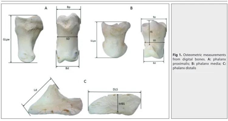
Morphometric measurements were obtained from each of 144 digital bones using digital calliper. The morphometric
measurements are (Fig. 1) [26];
Phalanx Proximalis
1. Greatest length of the abaxial half (GLpe) 2. Breadth of the proximal end (Bp)
3. Smallest breadth of the diaphysis (SD) 4. Breadth of the distal end (Bd)
Phalanx Media
1. Greatest length of the abaxial half (GLpe) 2. Breadth of the proximal end (Bp)
3. Smallest breadth of the diaphysis (SD) 4. Breadth of the distal end (Bd)
5. Greatest length (in dorsal direction) (GL)
Phalanx Distalis
1. Greatest diagonal length of the sole (DLS) 2. Length of the dorsal surface (Ld)
3. Middle breadth of the sole (MBS)
All measurements were based on von den Driesch [26]. SPSS
statistical package program (SPSS for Windows, version 21.0) was used for statistical analysis. Descriptive mean
values and SD values were calculated. One-way analysis of
variance (ANOVA) was used to evaluate all data. Tukey test was used to determine from which group the differences originated. Statistical differences between groups were presented as tables for each feature.
RESULTS
The osteometric measurements of the phalanx proximalis
of the forelimb and hindlimb are shown in Table 1.
There was no statistically significant diff erence in the GLpe
measurements between phalanx proximalis of the forelimb
and hindlimb, whereas the GLpe measurements showed a smaller value in the forelimb compared to the hindlimb. Statistically significant differences were found between the GLpe measurements of the phalanx proximalis of
forelimb and hindlimb except the left forelimb (P<0.05). The diff erence between the internal phalanx proximalis of the left forelimb and the GLpe measurements of the external phalanx proximalis of the right hindlimb was not statistically significant.
Although Bp measurement results were relatively small in the hindlimb phalanx proximalis compared to the fore-limbs, statistically significant differences were observed between the internal phalanx proximalis of the left fore-limb and the Bp measurements of both the internal and external phalanx proximalis of the left hindlimb (P<0.05). In addition, the diff erences between both the phalanx proximalis of forelimb and hindlimb were not significant. The SD value was relatively slightly higher in the phalanx proximalis of forelimb, but the differences between all phalanx proximalis of both the hindlimb and forelimb were not statistically significant. The same was true for Bd value.
Three diff erent index values were calculated for phalanx proximalis in Table 2. These were Bp*100/GLpe, SD*100/ GLpe and Bd*100/GLpe. While there were no statistically
significant diff erences between the right and left digital bones, it was observed that the diff erences between all three index values of the phalanx proximalis of forelimb and hindlimb were significant (P<0.05). All three index values of the forelimb phalanx proximalis were higher than the hindlimb and the diff erence was statistically significant. SD*100/GLpe index, which is considered as the fineness index, was higher than the hindlimb in the forelimb and the diff erences were statistically significant (P<0.05). Osteometric measurements of phalanx media were given in
Table 1. There were no statistically significant diff erences
between the phalanx media of forelimb and hindlimb in both of the anterior and posterior areas. While GLpe measurements were relatively small in the phalanx media of forelimb compared to the hindlimb, there was a significant difference between the external phalanx media of the forelimb and the internal phalanx media of the hindlimb (P<0.05). Although the diff erence between the left forelimb external phalanx media and left hindlimb internal phalanx media was not statistically significant, the observed diff erences were remarkable.
Phalanx media’s GL length in dorsal direction was diff erent from that in GLpe. There were no significant diff erences in the GL measurement of the phalanx media between both the right/left and external/internal parts of the hindlimb, but some diff erences were found in the forelimb itself. In addition, the GL values of the forelimb were smaller than the hindlimb and the diff erence was statistically significant. The GL value of the external phalanx media of the left and right forelimb was measured as the smallest value. No statistically significant diff erence was found between them.
Fig 1. Osteometric measurements
from digital bones. A: phalanx proximalis; B: phalanx media; C: phalanx distalis
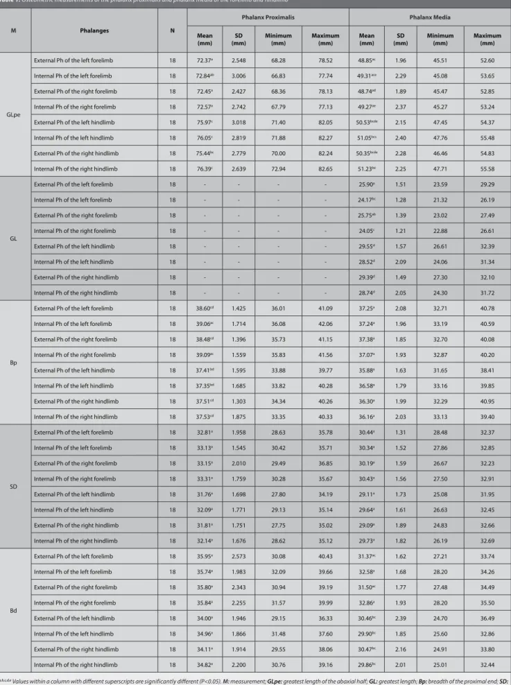
Table 1. Osteometric measurements of the phalanx proximalis and phalanx media of the forelimb and hindlimb
M Phalanges N
Phalanx Proximalis Phalanx Media
Mean (mm) SD (mm) Minimum (mm) Maximum (mm) Mean (mm) SD (mm) Minimum (mm) Maximum (mm) GLpe
External Ph of the left forelimb 18 72.37a 2.548 68.28 78.52 48.85ac 1.96 45.51 52.60
Internal Ph of the left forelimb 18 72.84ab 3.006 66.83 77.74 49.31ace 2.29 45.08 53.65
External Ph of the right forelimb 18 72.45a 2.427 68.36 78.13 48.74ad 1.89 45.47 52.85
Internal Ph of the right forelimb 18 72.57a 2.742 67.79 77.13 49.27ae 2.37 45.27 53.24
External Ph of the left hindlimb 18 75.97c 3.018 71.40 82.05 50.53bcde 2.15 47.45 54.37
Internal Ph of the left hindlimb 18 76.05c 2.819 71.88 82.27 51.05bcs 2.40 47.76 55.48
External Ph of the right hindlimb 18 75.44bc 2.779 70.00 82.24 50.35bcde 2.28 46.46 54.83
Internal Ph of the right hindlimb 18 76.39c 2.639 72.94 82.65 51.23be 2.25 47.71 55.58
GL
External Ph of the left forelimb 18 - - - - 25.90a 1.51 23.59 29.29
Internal Ph of the left forelimb 18 - - - - 24.17bc 1.28 21.32 26.19
External Ph of the right forelimb 18 - - - - 25.75ab 1.39 23.02 27.49
Internal Ph of the right forelimb 18 - - - - 24.05c 1.21 22.88 26.61
External Ph of the left hindlimb 18 - - - - 29.55d 1.57 26.61 32.39
Internal Ph of the left hindlimb 18 - - - - 28.52d 2.09 24.06 31.34
External Ph of the right hindlimb 18 - - - - 29.39d 1.49 27.30 32.10
Internal Ph of the right hindlimb 18 - - - - 28.74d 2.05 24.30 31.72
Bp
External Ph of the left forelimb 18 38.60cd 1.425 36.01 41.09 37.25a 2.08 32.71 40.78
Internal Ph of the left forelimb 18 39.06ac 1.714 36.08 42.06 37.24a 1.96 33.19 40.59
External Ph of the right forelimb 18 38.48cd 1.396 35.73 41.15 37.38a 1.85 32.70 40.08
Internal Ph of the right forelimb 18 39.09ac 1.559 35.83 41.56 37.07a 1.93 32.87 40.20
External Ph of the left hindlimb 18 37.41bd 1.595 33.88 39.77 35.88a 1.63 31.65 38.41
Internal Ph of the left hindlimb 18 37.35bd 1.685 33.82 40.28 36.58a 1.79 33.16 39.85
External Ph of the right hindlimb 18 37.51cd 1.303 34.34 40.26 36.30a 1.99 32.29 40.95
Internal Ph of the right hindlimb 18 37.53cd 1.875 33.35 40.33 36.16a 2.03 33.13 39.40
SD
External Ph of the left forelimb 18 32.81a 1.958 28.63 35.78 30.44a 1.31 28.48 32.37
Internal Ph of the left forelimb 18 33.13a 1.545 30.42 35.71 30.34a 1.52 27.86 32.85
External Ph of the right forelimb 18 33.15a 2.010 29.49 36.85 30.19a 1.59 26.67 32.23
Internal Ph of the right forelimb 18 33.31a 1.759 30.28 35.67 30.43a 1.56 27.50 32.91
External Ph of the left hindlimb 18 31.76a 1.698 27.80 34.19 29.11a 1.73 25.08 31.95
Internal Ph of the left hindlimb 18 32.09a 1.771 29.13 35.14 29.64a 1.61 26.63 32.45
External Ph of the right hindlimb 18 31.81a 1.751 27.75 35.02 29.09a 1.89 24.83 32.66
Internal Ph of the right hindlimb 18 32.14a 1.676 28.62 35.12 29.73a 1.82 26.19 32.69
Bd
External Ph of the left forelimb 18 35.95a 2.573 30.08 40.43 31.37ac 1.62 27.21 33.74
Internal Ph of the left forelimb 18 35.74a 1.983 32.09 39.66 32.58a 1.68 28.20 34.26
External Ph of the right forelimb 18 35.80a 2.343 30.94 39.19 31.50ac 1.77 27.48 34.49
Internal Ph of the right forelimb 18 35.84a 2.255 31.57 39.99 32.86a 1.93 28.20 35.50
External Ph of the left hindlimb 18 34.00a 1.946 29.15 36.33 30.46bc 2.39 24.70 36.49
Internal Ph of the left hindlimb 18 34.96a 1.866 31.48 37.60 29.90bc 1.85 25.60 32.86
External Ph of the right hindlimb 18 34.11a 1.914 29.55 38.06 30.47bc 2.16 24.91 33.80
Internal Ph of the right hindlimb 18 34.82a 2.200 30.76 39.16 29.86bc 2.01 25.01 32.44
a,b,c,d,e Values within a column with different superscripts are significantly different (P<0.05). M: measurement; GLpe: greatest length of the abaxial half; GL: greatest length; Bp: breadth of the proximal end; SD:
There was a difference between the GL measurements of the internal phalanx media of the external phalanx media. The GL value of internal phalanx media was relatively large compared to the external ones. There was no statistically significant difference between the GL measurements of internal phalanx media of the right and left feet.
No statistical differences were found between the forelimb and hindlimb of the Bp and SD values. However, there were differences in the Bd value of phalanx media were only between internal and external phalanx media GL values. These differences were statistically significant. The GL value of the internal phalanx media of the forelimbs had the highest value.
Phalanx media index (Bp*100/GLpe; SD*100/GLpe and Bd*100/GLpe) values are given in Table 2. In general, there were significant differences between the index values of the phalanx media (except the right external phalanx media) of forelimb and hindlimb. The index values of phalanx media in the forelimb had a higher value.
Differences between phalanx media index values of right and left feet were not statistically significant. There was no significant difference between Bp*100/GLpe value of the right rear external phalanx media and the index values of the front internal phalanx media. The same was also true for differences between the left front external and right rear external values of Bd*100/GLpe index value. For fineness index SD*100/GLpe, the forelimb had a higher value than the hindlimb and the differences were statistically significant (P<0.05).
The osteometric measurements of phalanx distalis and the index value of this bone are given in Table 3. The DLS value was higher in the forelimb than in the hindlimb. However, the highest value was in internal phalanx distalis. There was a statistically significant difference in DLS between the internal phalanx distalis in the forelimb and all phalanx distalis in the hindlimb (P<0.05). In the case of the external forelimb only significant difference was observed between the left phalanx distalis and right external hindlimb (P<0.05).
Table 2. The indices of the phalanx proximalis and phalanx media of the forelimb and hindlimb
Indices Phalanges N
Phalanx Proximalis Phalanx Media
Mean
(mm) (mm)SD Minimum(mm) Maximum(mm) Mean(mm) (mm)SD Minimum(mm) Maximum(mm)
Bp*100/GLpe
External Ph of the left forelimb 18 53.38a 2.04 48.10 56.79 76.24a 2.70 69.52 80.45 Internal Ph of the left forelimb 18 53.65a 2.01 50.35 56.60 75.59ac 3.74 70.12 82.17 External Ph of the right forelimb 18 53.15a 1.99 49.01 56.50 76.71a 3.25 69.52 84.06 Internal Ph of the right forelimb 18 53.90a 2.05 50.46 58.05 75.30ac 3.59 70.79 81.23 External Ph of the left hindlimb 18 49.27b 1.97 45.43 51.90 71.07b 3.01 65.68 75.59 Internal Ph of the left hindlimb 18 49.15b 2.46 45.47 54.04 71.70b 2.94 66.04 76.67 External Ph of the right hindlimb 18 49.74b 1.44 47.11 52.74 72.18bc 4.15 66.08 83.10 Internal Ph of the right hindlimb 18 49.15b 2.49 45.01 53.11 70.60b 3.10 65.31 76.32
SD*100/GLpe
External Ph of the left forelimb 18 45.35a 2.40 39.71 48.76 62.35a 2.20 58.91 66.61 Internal Ph of the left forelimb 18 45.50a 1.52 43.25 48.85 61.57a 2.93 57.60 70.36 External Ph of the right forelimb 18 45.76a 2.35 41.56 49.95 61.97a 3.15 56.70 68.55 Internal Ph of the right forelimb 18 45.91a 1.92 43.25 50.19 61.78a 2.43 59.42 69.87 External Ph of the left hindlimb 18 41.83b 2.23 37.84 45.49 57.64b 2.93 52.04 61.56 Internal Ph of the left hindlimb 18 42.20b 2.00 39.36 46.03 58.11b 2.88 53.41 63.28 External Ph of the right hindlimb 18 42.17b 2.14 38.19 45.87 57.82b 3.54 51.66 63.89 Internal Ph of the right hindlimb 18 42.07b 1.78 38.62 44.91 58.05b 2.85 53.20 62.88
Bd*100/GLpe
External Ph of the left forelimb 18 49.66a 2.87 42.11 55.40 64.25ac 3.32 57.83 71.62 Internal Ph of the left forelimb 18 49.06a 1.76 45.81 51.95 66.14a 3.29 59.63 73.16 External Ph of the right forelimb 18 49.39a 2.35 43.61 53.12 64.66a 3.56 58.42 71.65 Internal Ph of the right forelimb 18 49.38a 2.49 45.61 53.96 66.72a 3.23 60.76 71.85 External Ph of the left hindlimb 18 44.78b 2.37 39.68 48.00 60.34b 4.76 51.26 70.25 Internal Ph of the left hindlimb 18 45.98b 2.00 43.02 49.25 58.62b 3.53 52.53 66.56 External Ph of the right hindlimb 18 45.23b 2.30 40.67 49.86 60.60bc 4.71 51.83 67.81 Internal Ph of the right hindlimb 18 45.59b 2.64 41.51 52.71 58.31b 3.50 50.80 66.27
Although the Ld value was relatively similar between the phalanx distalis of forelimb and hindlimb, the differences between the values of the internal phalanx distalis in the right external phalanx distalis of forelimb and hindlimb
were significant (P<0.05). There were not any other statistically significant differences.
The MBS value was higher in the forelimb than in the hindlimb. The difference between them was statistically significant (P<0.05). There was no significant difference between the phalanx distalis value of the left external digit of forelimb and the others.
The MBS*100/DLS value, calculated using the length (DLS) and width (MBS) values of phalanx distalis, was slightly higher in the hindlimb but the differences were not statistically significant.
DISCUSSION
It is suggested that in ruminants which have an even number of digits in each foot, the pairs of digits are not of the equal length due to the asymmetry in these pair of digits, which indicates that they have different function in relation to stature and load bearing [2,9]. It was reported that
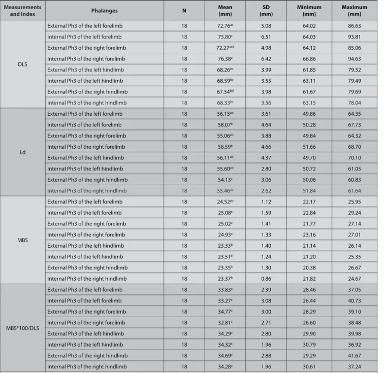
Table 3. The measurements and indices of the phalanx distalis of the forelimb and hindlimb Measurements
and Index Phalanges N
Mean (mm) SD (mm) Minimum (mm) Maximum (mm) DLS
External Ph3 of the left forelimb 18 72.76ac 5.08 64.02 86.63
Internal Ph3 of the left forelimb 18 75.80a 6.51 64.03 93.81
External Ph3 of the right forelimb 18 72.27acd 4.98 64.12 85.06
Internal Ph3 of the right forelimb 18 76.38a 6.42 66.86 94.63
External Ph3 of the left hindlimb 18 68.26bc 3.99 61.85 79.52
Internal Ph3 of the left hindlimb 18 68.59bc 3.55 63.11 79.49
External Ph3 of the right hindlimb 18 67.54bd 3.98 61.67 79.69
Internal Ph3 of the right hindlimb 18 68.33bc 3.56 63.15 78.04
Ld
External Ph3 of the left forelimb 18 56.15ab 3.61 49.86 64.35
Internal Ph3 of the left forelimb 18 58.07b 4.64 50.28 67.73
External Ph3 of the right forelimb 18 55.06ab 3.88 49.84 64.32
Internal Ph3 of the right forelimb 18 58.59b 4.66 51.66 68.70
External Ph3 of the left hindlimb 18 56.11ab 4.37 49.70 70.10
Internal Ph3 of the left hindlimb 18 55.60ab 2.80 50.72 61.05
External Ph3 of the right hindlimb 18 54.13a 3.06 50.06 60.83
Internal Ph3 of the right hindlimb 18 55.46ab 2.62 51.84 61.64
MBS
External Ph3 of the left forelimb 18 24.52ab 1.12 22.17 25.95
Internal Ph3 of the left forelimb 18 25.08a 1.59 22.84 29.24
External Ph3 of the right forelimb 18 25.02a 1.41 21.77 27.14
Internal Ph3 of the right forelimb 18 24.93a 1.33 23.16 27.01
External Ph3 of the left hindlimb 18 23.33b 1.40 21.14 26.14
Internal Ph3 of the left hindlimb 18 23.51b 1.24 21.20 25.35
External Ph3 of the right hindlimb 18 23.35b 1.30 20.38 26.67
Internal Ph3 of the right hindlimb 18 23.37b 0.86 21.82 24.67
MBS*100/DLS
External Ph3 of the left forelimb 18 33.83a 2.39 28.46 37.05
Internal Ph3 of the left forelimb 18 33.27a 3.08 26.44 40.73
External Ph3 of the right forelimb 18 34.77a 3.00 28.29 39.10
Internal Ph3 of the right forelimb 18 32.81a 2.71 26.60 38.48
External Ph3 of the left hindlimb 18 34.29a 2.80 29.90 39.98
Internal Ph3 of the left hindlimb 18 34.32a 1.96 30.79 36.92
External Ph3 of the right hindlimb 18 34.69a 2.88 29.29 41.67
Internal Ph3 of the right hindlimb 18 34.28a 1.96 30.61 37.24
a,b,c,d Values within a column with different superscripts are significantly different (P<0.05). DLS: greatest diagonal length of the sole; Ld: length of the dorsal surface; MBS: middle breadth of the sole
the differences between phalanx proximalis and phalanx media bones of cattle were more prominent than their width, but this was not associated with length and width as
an index value [2,4,6,15]. The knowledge in classical anatomy
books indicates that Ph1 and Ph2 is shorter in the hindlimb than in the forelimb and this information is not supported by morphometric data. In this study, it was concluded that the largest lengths of these bones (GLpe) were contrary to this discourse in both Ph1 and Ph2, and were longer in hindlimbs [4]. Similar situation was also supported by Ocal et al.[8]. While the difference in maximum length between the forelimb and hindlimb was significant for Ph1, there was a relationship between the externals of forelimb and the internals of hindlimb in Ph2. Among each limb values the GLpe values of these bones were the smallest.
It was observed that SD values were almost equal in the phalanx proximalis and phalanx media of forelimb and hindlimb and there was no significant difference between
them (Table 1) [8]. Considering this, it is concluded that the
value of SD*100/GLpe, which is called the fineness index of these bones, is more related to the length of the bones. This index value is higher in the forelimbs, rather than the SD width value of this value (GLpe) is significantly longer in the hindlimb. Bp in phalanx proximalis, Bd values in phalanx media did not show any statistically significant difference except that the differences between the inner bones of the forelimb and their hindlimb equivalents. The importance of the difference of osteometric measure-ments is generally discussed in some studies to reveal
the presence of asymmetry [6,7]. In the comparison of the
external and internal phalanxes, it is usually observed that the mean length of the external (phalanx) proximalis and
phalanx media of the 4th digit is greater than that of the
third digit (internal) corresponding to this measurement, whereas the third (internal) phalanx distalis has a significantly
greater average value [2]. Regarding the digits, the presence
of an asymmetry between the osteometric measurements of the internal and external bones forming digits, could only be observed between the GL values of the phalanx media of forelimb.
In the majority of all other osteometric measurements, there were some significant differences between the corresponding bones of forelimb and hindlimb (P<0.05). The underlying reason for the asymmetry of the ruminant lower extremities especially the forelimb extremities observed in cattle despite the lack of a significant difference between the total lengths of the corresponding bones of the medial
and lateral digits, is unknown [5,9]. However, considering
the fact that the hardness of flooring where the animals live and walk on, the difference between the flooring conditions where cattle and dairy cows are kept may be the underlying reason of this asymmetry [2]. Therefore, we concluded that the asymmetry in the forelimb maybe a result of these cows of Holstein breed being raised as dairy cows and kept on concrete ground.
In the phalanx media of forelimb, especially the asymmetric condition in the GL measurements and the obvious difference in Bd measurements between the internals of forelimb and the phalanx media of hindlimb support the view that the body weight of the Bovidae is loaded on the central digits [2,12]. This is especially evident when the morphometric data of phalanx distalis are evaluated. Approximately 2/3 of the digital lesions affect the lateral hooves. In our study, the morphometric data of phalanx distalis are thought to be partially visible in the importance control of the difference in the forelimb and hindlimbs [16,17]. Especially in the phalanx distalis of hindlimb, there were significant differences in DLS measurement of all phalanx distalis. The front internal phalanx distalis have the largest DLS value. The difference between these bones and lateral phalanx distalis of hindlimb data is statistically significant. The lowest Ld value was obtained in the lateral phalanx distalis of hindlimb.
The age and body weight data were lacking for some cattle. Therefore, this information was not presented. The length and diameters of bones may be used as descriptive properties of cattle.
As a result, different distribution of stress to feet in cattle is thought to be related to the anatomical position of the feet. Particular differences are observed in phalanx media and phalanx distalis between the forelimb and hindlimb
opposites of internal bones. The significant increase in SD*100/GLpe value, expressed as the fineness index, in forelimb in comparison to hindlimb is thought to be more significantly affected by the width value. This is because index value increases in correlation with the SD value. The same applies to the proximal and distal widths of phalanx proximalis and phalanx media. Not only the large values of the front phalanx proximalis and phalanx media, but also larger DLS and MBS values in the front phalanx distalis compared to the hinds, probably results in a larger contact area for digits. We believe that it may contribute to the fact that digital lesions are observed more frequently in phalanx distalis of hindlimb clinically, which are thought to have a smaller contact area.
REFERENCES
1. Dyce KM, Sack WO, Wensing CJG: Textbook of veterinary Anatomy. In, Dyce K, Sack W, Wensing C (Eds): Elsevier Health Sciences. 3rd ed., WB Saunders Company: Philadelphia, 1987.
2. Keller A, Clauss M, Muggli E, Nuss K: Even-toed but uneven in length:
The digits of artiodactyls. Zoology, 112 (4): 270-278, 2009. DOI: 10.1016/j. zool.2008.11.001
3. Sisson S: Ruminant osteology. In, Getty R (Ed): The Anatomy of the
Domestic Animals. 4th ed., Saunders, Philadelphia, 1975.
4. Nickel R, Schummer A, Seiferle E, Wilkens H, Wille KH, Frewein J:
The Anatomy of the Domestic Animals. Volume 1. The Locomotor System of the Domestic Mammals. Parey Im Mvs, Berlin, 1986.
5. Nourinezhad J, Mazaheri Y, Pourmahdi Borujeni M, Daneshi M:
Morphometric study on digital bones in Native Khuzestan Water Buffaloes
(Bubalus bubalis). Bulgarian J Vet Med, 15 (4): 228-235, 2012.
of the bovine digits. Vet J, 188 (3): 295-300, 2011. DOI: 10.1016/j.tvjl. 2010.05.016
7. Muggli E, Weidmann E, Kircher P, Nuss K: Radiographic measurement
of hindlimb digit length in standing heifers. Anat Histol Embryol, 45 (6): 463-468, 2016. DOI: 10.1111/ahe.12222
8. Ocal MK, Sevil F, Parin U: A quantitative study on the digital
bones of cattle. Ann Anat, 186 (2): 165-168, 2004. DOI: 10.1016/S0940-9602(04)80034-7
9. Bartosiewicz L, Van Neer W, Lentacker A: Metapodial asymmetry
in draft cattle. Int J Osteoarchaeol, 3 (2): 69-75, 1993. DOI: 10.1002/ oa.1390030203
10. Thomas R, Johannsen N: Articular depressions in domestic cattle
phalanges and their archaeological relevance. Int J Paleopathol, 1 (1): 43-54, 2011. DOI: 10.1016/j.ijpp.2011.02.007
11. Boosman R, Németh F, Gruys E: Bovine laminitis: Clinical aspects,
pathology and pathogenesis with reference to acute equine laminitis. Vet
Q, 13 (3): 163-171, 1991. DOI: 10.1080/01652176.1991.9694302
12. Animal Diversity Web: Bovidae. https://animaldiversity.org/accounts/
Bovidae/; Accessed: 02.01.2007.
13. Prenti.ce DE, Neal PA: Some observations on the incidence of
lameness in dairy cattle in West Cheshire. Vet Rec, 91 (1): 1-7, 1972. DOI: 10.1136/vr.91.1.1
14. Russell AM, Shaw SR: The compton lameness survey 1977: A
preliminary report. Anim Dis Rep, 2, 5-8, 1978.
15. Van Amstel SR, Shearer J: Manual for Treatment and Control of
Lameness in Cattle. Blackwell Publishing, USA, 2006.
16. Andersson L, Lundström K: The influence of breed, age, body
weight and season on digital disease and hoof size in dairy cows. Zentralbl
Veterinarmed A, 28 (2): 141-151, 1981. DOI: 10.1111/j.1439-0442.1981.
tb01174.x
17. Kofler J: Clinical study of toe ulcer and necrosis of the apex of the
distal phalanx in 53 cattle. Vet J, 157 (2): 139-147, 1999. DOI: 10.1053/ tvjl.1998.0290
18. Métais G, Albayrak E, Antoine PO, Erdal O, Karadenizli L, Oyal N, Saraç G, İslamoğlu Y, Sen S: Oligocene ruminants from the Kızılırmak
formation, Çankırı-Çorum basin, Central Anatolia, Turkey. Palaeontol
Electron, 19.3.37A, 2016. DOI: 10.26879/629
19. Sidéra I, De Maret P: An Ideal Bone for Traditional Dolls. Ruminants
Metapodia Figurines: Archaeological and Ethnographical Examples from Africa and Europe. In, Selena V (Ed): Close to the Bone: Current Studies in Bone Technologies. 315-323, Institute of Archaeology, Belgrade, 2016.
20. Hír J, Venczel M, Codrea V, Rössner GE, Angelone C, van den Hoek Ostende LW, Rosina VV, Kirscher U, Prieto J: Badenian and Sarmatian
s. str. from the Carpathian area: Taxonomical notes concerning the Hungarian and Romanian small vertebrates and report on the ruminants from the Felsőtárkány Basin. CR Palevol, 16 (3): 312-332, 2017. DOI: 10.1016/j.crpv.2016.11.006
21. Clason AT: The animal bones of the Bandceramic and Middle Age
settlements near Bylany in Bohemia. Palaeohistoria, 14, 1-17, 1970.
22. Zhang Q, Ding X, Xu K: Terrain adaptability mechanism of large ruminants’
feet on the kinematics view. Appl Bionics Biomech, 2015:151686, 2015. DOI: 10.1155/2015/151686
23. Philip LM, Venugopal SK, Devanand CB: Management of Toe Ulcer
and Apical Necrosis of Distal Phalanx in a Cow. Intas Polivet, 18 (2): 242-244, 2017.
24. Gyan LA, Paetsch CD, Jelinski MD, Allen AL: The lesions of toe
tip necrosis in southern Alberta feedlot cattle provide insight into the pathogenesis of the disease. Can Vet J, 56 (11): 1134-1139, 2015.
25. Taşbaş M, Tecirlioğlu S: Researches on the maceration technique.
Ankara Univ Vet Fak Derg, 4, 324-330, 1966.
26. Von den Driesch A: Guide to the Measurement of Animal Bones from
Archaeological Sites. Peabody Museum of Archaeology and Ethnology, Harvard University, Cambridge, Mass, 1976.