Turk Kardiyol Dern Ars 2016;44(5):437-439 doi: 10.5543/tkda.2015.14377
Successful radiofrequency pulmonary vein isolation
in a patient with pneumonectomy
Pnömonektomili hastada pulmoner venlerin başarılı radyofrekans izolasyonu
Department of Cardiology, İstanbul Medipol University Faculty of Medicine, İstanbul, Turkey#Department of Radiology, İstanbul Medipol University Faculty of Medicine, İstanbul, Turkey Enes Elvin Gül, M.D., Cengiz Erol, M.D.,# Fethi Kılıçaslan, M.D.
Özet– Atriyum fibrilasyonunun kateter ablasyonu invaziv elektrofizyoloji çalışmaları arasında zor bir işlemdir. Pulmo-ner venlerin izolasyonu tedavinin köşe taşını oluşturmakta-dır. İşlem pulmoner ven stenozu gibi bazı ciddi komplikas-yonlarla eşlik edebilir. Bu yazıda, akciğer kanseri nedeniyle sol taraflı pnömonektomi olan hastada teknik olarak zor olan pulmoner ven izolasyonu yapılan olgu sunuldu.
Summary– Among electrophysiologic procedures, cathe-ter ablation of atrial fibrillation (AF) is challenging, requiring the isolation of all pulmonary veins (PVs). AF is associated with serious complications including PV stenosis. Presently described was a technically challenging case of PV isola-tion in a patient with left-sided pneumonectomy due to lung cancer.
437
I
solation of the pulmonary veins (PVs) is a corner-stone of catheter ablation of atrial fibrillation (AF), a challenging procedure. Serious complications such as PV stenosis may occur. Described in the present report was a technically challenging case of PV isola-tion in a patient who had undergone left-sided pneu-monectomy due to lung cancer.CASE REPORT
A 72-year-old man with complaint of palpitation and dyspnea was admitted. Twelve-lead resting electro-cardiography revealed AF with an onset of 2 years prior. Physical examination was unremarkable. Echo-cardiogram revealed normal left ventricular systolic functions (ejection fraction of 65%) and enlarged left atrium (LA) of 44 mm. Medical history included chronic obstructive pulmonary disease, hyperten-sion, chronic renal failure, coronary artery disease, ulcerative colitis, and left-sided pneumonectomy 10 years prior due to lung cancer. The patient was taking antihypertensive and antiarrhythmic agents. Due to high risk of thromboembolism (CHADS2 score >2), warfarin was administered. Ten years prior, complete
left-sided pneumonectomy had been performed for resection of tumor. AF ablation was recom-mended due to recurrent symp-tomatic AF attacks, occurring in
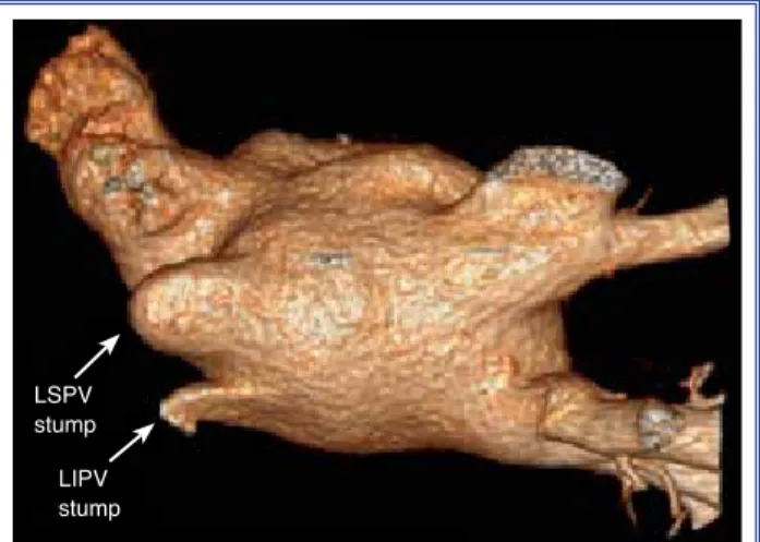
spite of antiarrhythmic treatment. Informed consent was obtained. LA thrombus was excluded with trans-esophageal echocardiography, and 64-slice comput-ed tomography was performcomput-ed to evaluate the PVs. Three-dimensional (3D) image of the LA and PVs showed PV stumps in the superior PV and left-inferior PV (Figure 1).
Ablation was performed under mild anesthesia without intubation. Steerable decapolar catheter was inserted into the coronary sinus for positional ref-erence. Due to unusual postoperative cardiac rota-tion, only 1 transseptal puncture could be performed. EnSite NavX system (St. Jude Medical, Inc., Little Canada, MN, USA) was used for 3D mapping and catheter navigation, and 3D left atrial anatomy was reconstructed (Figure 2). Lasso catheter was insert-ed into the PVs, and PV signals including those of the stumps were recorded. Circumferential radio-Received:August 23, 2015 Accepted:December 23, 2015
Correspondence: Dr. Enes Elvin Gül. 334 Queen Mary Rd, K7M 7E7 K7M7E7 Kingston - Canada. Tel: +1 613 328 2052 e-mail: [email protected]
© 2016 Turkish Society of Cardiology
Abbreviations: 3D Three-dimensional AF Atrial fibrillation LA Left atrium PV Pulmonary vein CASE REPORT
frequency ablation of the ipsilateral PVs was per-formed (30–35 W, 17 mL/min) with irrigated cath-eter (Sprinklr; Medtronic Inc., Minneapolis, MN, USA). Entrance and exit blocks were achieved fol-lowing ablation. Interestingly, both left PV stumps were electrically active prior to ablation, and com-plete isolation was obtained following radiofre-quency ablation. During ablation, weight-adjusted unfractionated heparin was administered to maintain activated clotting time of 300–350 seconds. The pro-cedure was completed without complication. Fluo-roscopy and procedure times were 21 minutes and 125 minutes, respectively. No recurrence of AF was observed during 6-month follow-up period, and no
PV stenosis was observed on computed tomography scan at the sixth month.
DISCUSSION
Catheter ablation is currently the most effective ther-apy for drug-refractory AF.[1] Because the PVs play
an important role as arrhythmia triggers of AF,[2] PV
electrical isolation has become a primary treatment strategy.[3]
Pneumonectomy is defined as removal of the lung and distal section of associated PV. Thus, the segment of the PV attached only to the LA is referred to as the “stump.” The role, if any, of the PV stump in trigger-ing AF is unclear. PV ligation after pneumonectomy should, logically, decrease or minimize the risk of AF, and it has been reported that PV ligation may eliminate and decrease causes of AF.[4] However, results of both a
report by Konstantinidou et al.[5] and a very recent
mul-ticenter study that included 15 patients have demon-strated that PV stumps (remaining following pneumo-nectomy) were electrically active, and that the majority were sites of active firing or triggering.[6] In addition, it
was found that no PV stenosis followed ablation. It was likewise observed in the present patient that all PVs were electrically active, in spite of PV interruptions.
In general, AF ablation is a challenging procedure, with important related complications including PV stenosis, cardiac tamponade, stroke, and atrioesopha-geal fistula.[7] As a complication of AF, PV stenosis is
serious but rare, with a reported incidence of 2–4%, depending on the ablation technique.[8] In patients
with pneumonectomy, PV stenosis may result in det-rimental consequences, even death. The procedure may also be technically difficult due to cardiac dis-placement and rotation.[9] These changes may lead to
difficult transseptal puncture and catheter movement in the LA and around the PVs. In the present patient, only 1 transseptal puncture could be performed due to the aforementioned causes, and intracardiac echocar-diography was not available.
In spite of the technical challenges, AF ablation in patients with pneumonectomy is feasible and safe. It has been shown that PV stumps are electrically active and should be isolated in order to maintain long-term sinus rhythm.
Conflict-of-interest issues regarding the authorship or article: None declared.
Turk Kardiyol Dern Ars 438
Figure 1. Three-dimensional reconstruction of the left atri-um and pulmonary veins in posterior-superior view. Left su-perior and left inferior pulmonary vein stumps are shown (arrows).
LSPV stump LIPV stump
Figure 2. Three-dimensional reconstruction of the left atri-um and pulmonary veins with electroanatomic mapping and circumferential ablation lines around the pulmonary veins. Left superior and left inferior pulmonary vein stumps are shown (arrows).
LSPV stump LIPV stump
Successful radiofrequency pulmonary vein isolation in a patient with pneumonectomy 439
REFERENCES
1. Hakalahti A, Biancari F, Nielsen JC, Raatikainen MJ. Radio-frequency ablation vs. antiarrhythmic drug therapy as first line treatment of symptomatic atrial fibrillation: systematic review and meta-analysis. Europace 2015;17:370–8. Crossref
2. Haïssaguerre M, Jaïs P, Shah DC, Takahashi A, Hocini M, Quiniou G, et al. Spontaneous initiation of atrial fibrillation by ectopic beats originating in the pulmonary veins. N Engl J Med 1998;339:659–66. Crossref
3. Chen SA, Hsieh MH, Tai CT, Tsai CF, Prakash VS, Yu WC, et al. Initiation of atrial fibrillation by ectopic beats originating from the pulmonary veins: electrophysiological characteris-tics, pharmacological responses, and effects of radiofrequen-cy ablation. Circulation 1999;100:1879–86. Crossref
4. Dixit S, Sauer WH, Callans DJ, Marchlinski FE. Arrhythmo-genic potential of pulmonary venous tissue: triggers for atrial fibrillation identified within the remnant of a vein. J Cardio-vasc Electrophysiol 2009;20:441–4. Crossref
5. Konstantinidou M, Schmidt B, Ouyang F, Koektuerk B, Kuck KH, Chun KR. Pulmonary vein isolation after left-sided pneu-monectomy: technically challenging but feasible and
instruc-tive. Europace 2009;11:389–91. Crossref
6. Kanmanthareddy A, Vallakati A, Reddy Yeruva M, Dixit S, Di Biase L, Mansour M, et al. Pulmonary vein isolation for atrial fibrillation in the postpneumonectomy population: a feasibil-ity, safety, and outcomes study. J Cardiovasc Electrophysiol 2015;26:385–9. Crossref
7. Takahashi A, Kuwahara T, Takahashi Y. Complications in the catheter ablation of atrial fibrillation: incidence and manage-ment. Circ J 2009;73:221–6. Crossref
8. Holmes DR Jr, Monahan KH, Packer D. Pulmonary vein stenosis complicating ablation for atrial fibrillation: clinical spectrum and interventional considerations. JACC Cardio-vasc Interv 2009;2:267–76. Crossref
9. Smulders SA, Holverda S, Vonk-Noordegraaf A, van den Bosch HC, Post JC, Marcus JT, et al. Cardiac function and position more than 5 years after pneumonectomy. Ann Thorac Surg 2007;83:1986–92. Crossref
Keywords: Ablation; atrial fibrillation; pneumonectomy; pulmonary vein isolation.
Anahtar sözcükler: Ablasyon; atriyum fibrilasyonu; pnömonektomi; pulmoner ven izolasyonu.