to promote osteoblast alignment and confinement
H. Kenar,1A. Kocabas,2A. Aydinli,2V. Hasirci11METU, BIOMAT, Department of Biological Sciences, Biotechnology Research Unit, Ankara, Turkey 2Department of Physics, Bilkent University, Ankara 06800, Turkey
Received 10 January 2007; revised 21 June 2007; accepted 4 July 2007
Published online 28 September 2007 in Wiley InterScience (www.interscience.wiley.com). DOI: 10.1002/jbm.a.31638
Abstract: Proper cell attachment and distribution, and thus stronger association in vivo between a bone implant and native tissue will improve the success of the implant. In this study, the aim was to achieve promotion of attach-ment and uniform distribution of rat mesenchymal stem cell-derived osteoblasts by introducing chemical and topo-graphical cues on poly(3-hydroxybutyrate-co-3-hydroxyval-erate) (PHBV) film surfaces. As the chemical cues, either alkaline phosphatase was covalently immobilized on the film surface to induce deposition of calcium phosphate minerals or fibrinogen was adsorbed to improve cell adhe-sion. Microgrooves and micropits were introduced on the film surface by negative replication of micropatterned Si wafers. Both chemical cues improved cell attachment and even distribution on the PHBV films, but Fb was more
effective especially when combined with the micropatterns. Cell alignment (<108 deviation angle) parallel to chemi-cally modified microgrooves (1, 3, or 8 lm groove width) and on 10 lm-thick Fb lines printed on the unpatterned films was achieved. The cells on unpatterned and 5 lm-deep micropitted films were distributed and oriented randomly. Results of this study proved that microtopogra-phies on PHBV can improve osseointegration when com-bined with chemical cues, and that microgrooves and cell adhesive protein lines on PHBV can guide selective osteo-blast adhesion and alignment. Ó 2007 Wiley Periodicals, Inc. J Biomed Mater Res 85A: 1001–1010, 2008
Key words: PHBV; photolithography; micropatterned films; osteoblasts; bone tissue engineering
INTRODUCTION
Bone tissue engineering is becoming more promis-ing for the replacement or repair of damaged bone because advanced techniques such as micro and nanotechnology are now being used more exten-sively to form tissues that closely mimic the micro-structure and function of the natural tissue.
Biodegradable polymers are considered to be the most suitable materials for scaffold preparation because of their variety, versatility, and biodegrad-ability. When applied, these materials circumvent the need for device removal from the body.
Poly(3-hydroxybutyrate-co-3-hydroxyvalerate) (PHBV) is one of the naturally derived biodegradable
poly-mers that has improved processing and mechanical properties with respect to PHB,1,2 which is brittle and has a higher melting temperature.3Properties of PHBV such as glass transition temperature (Tg), sta-bility, degradasta-bility, and crystallinity can be mo-dified by changing its copolymer composition and molecular weight.4–6 Therefore, the rate and extent of accumulation of degradation products, such as b-hydroxybutyric acid, which is a normal constituent of blood,7–9 and hydroxyvaleric acid, at the site of implantation is not problematic for PHBV, unlike the faster degrading a-polyhydroxy acids of the poly-lactide family. PHBV can be processed to take on various shapes, forms, and porosities because its ther-moplastic properties are suitable. Studies on using PHBV for bone tissue implants involved attempts to match the mechanical strength and form to that of bone through incorporation of minerals.10,11 It can therefore be assumed that PHBV has a significant potential for use as a support for long-term bone regeneration in vivo. More recently, in vitro growth of osteoblasts on macroporous, three-dimensional PHBV matrices has been reported.12Suitability of the matrices for bone tissue engineering was shown by an increase in osteocalcin expression and alkaline Correspondence to: V. Hasirci; e-mail: [email protected]
Contract grant sponsor: Scientific and Technical Research Council of Turkey (TUBITAK) TBAG; contract grant number: 2288
Contract grant sponsor: METU Graduate School of Natu-ral and Applied Sciences; contract grant number: BAP-2004-07-02-00-15
phosphatase (ALP) activity over a 60-day period.13 In addition, a comprehensive characterization of MC3T3-E1 S14 osteoblast growth and differentiation on nonporous PHBV films was carried out, in which the ability of PHBV to support osteoblast cell func-tion was shown.14 It is well known that initial cell attachment and proliferation are dependent on the functional groups of an implant surface rather than its bulk. Like most of the biodegradable polymers, PHBV lacks functional groups for cell attachment and needs to be modified. Uniform cell attachment, distribution, and thus stronger association between the implant and native tissue in vivo, would improve the success of the implant. Introducing cell adhesive patterns on a polymer surface or creating surface microtopography suitable for cell confinement are two methods that are applied for this purpose. Integ-rin transmembrane receptors of cells bind specific ECM components and the cytoskeleton.15 Integrin-mediated cell adhesion influences subsequent cell processes, including spreading, proliferation, and differentiation via signaling pathways.16,17 Surface topography of a material is known to be important in the cell–material interactions, for cell orientation and migration.18 Osteoblastic phenotype and degree of bone contact were shown to be responsive to topog-raphy; polished surfaces produce less material–bone contact, and bone formation is preferentially ob-served in grooves and crevices.19 Cells appear to recognize surface features and respond accordingly, possibly through reshaping of the actin filaments in filopodia. This is called contact guidance. When filo-podia are presented with a favorable cue, they become stabilized through the action of tubulin microtubules and alignment of actin.19–21 There is evidence that the symmetry or regularity of micro-scopic surface topographies play an important role in cellular responses.22 Lithography has enabled researchers to fabricate surfaces with well-defined chemical and/or topographical patterns at the micro or nanoscale. These patterned materials could be used as templates to transfer these designs on poly-meric surfaces through the use of a variety of meth-ods such as molding, embossing, or solution casting. This could be used to study cell–substrate interac-tions with chemically or topographically patterned polymers of different chemistries, and understand how geometrical features influence osteoblast biol-ogy.23,24 There are strong evidences to claim that micropatterning of osteoblasts in microgrooves leads to their alignment, and improves their activity, another desired property if the patterned film is used as a bone implant. Qu et al. reported that both micronscale pits and grooves stimulated matrix deposition and mineralization, possibly through dif-ferent mechanisms.24 Further evidence about the influence of surface topography on osseointegration
has been provided using in vitro models,25–27 and in vivo tests.28 It was also reported for the first time that osteoblasts can be induced to align with the microgrooves on biodegradable PHBV-P(L/D,L)LA
polymer films when fibrinogen (Fb) adsorbed on the surface was used as a chemical cue, and that this alignment could improve expression of the differen-tiated phenotype.29
The organs and tissues of the body are organized in specific three-dimensional architectures. Repair of such tissues, therefore, would be improved upon provision of 2D and 3D cues, because these would induce cells to assemble and organize in a style typi-cal of the target organ or tissue. Thus, active bioma-terials, carrying the cues required for induction of appropriate morphogenesis, may be designed. Vari-ous researchers have used a range of techniques to pattern substrates with specific surface chemistries to interact with the cells.30–32 Microcontact printing was one of the earliest methods proposed. To our knowledge, there are no preceding studies reported in literature on osteoblast alignment to patterns of chemical cues generated on PHBV films.
This study aimed at promoting mesenchymal stem cell-derived osteoblast attachment to PHBV and their even distribution by introducing chemical and topo-graphical cues on film surface. To serve as a chemi-cal cue for osteoblasts, either Fb was adsorbed or ALP was covalently immobilized on the film surface and subsequently Ca-P minerals were deposited by enzymatic activity. Microgrooves and micropits were generated on the film surface as topographical cues. In addition to that, osteoblast alignment to a pattern was evaluated; either films with micro-grooves of different groove widths (1, 3, or 8 lm) were produced or Fb lines were microcontact printed on the unpatterned film surface. This study demonstrates that 3D and 2D patterns in the form of microgrooves and cell adhesive protein lines, respec-tively, on PHBV can guide osteoblast adhesion and alignment and further highlights the role of surface topography as a modulator of cell distribution.
MATERIALS AND METHODS
Production of micropatterned templates, PDMS stamp, and polymer films
Single crystal silicon wafers were used to create a variety of templates for studying the influence of pattern type on cell adhesion (Table I). The wafers were cut into 1.5 3 1.5 cm2 pieces and cleaned with TCA (trichloroethane), ACE (acetone), and ISO (isopropyl alcohol). Thin films of SiNxwere grown on these slices by plasma-enhanced
chem-ical vapor deposition. Subsequently, the slices were coated with positive photoresist for 40 s at a spin rate of 4000 rpm,
and by using a photomask, exposed to UV light of 6 mW for 25 s in the Karl Suss Mask aligner. The photoresist film was developed for 40 s in a solution of 1:4 developer and deionized water. The pattern on the mask was transferred to the SiNx layer by hydrofluoric acid (HF) etching of the
exposed SiNxfor 60 s. The photoresist was removed off the
wafer with acetone. For the production of microgrooved (MG) Si templates, the exposed parts of the wafers were etched to the desired depth in a KOH solution that was heated to 408C and stirred at 600 rpm. Lastly, the remaining SiNxlayer was removed using dilute HF. The micropitted
(MP) Si template and the template that was used for the preparation of the PDMS stamp were produced by reactive ion etching (RIE) instead of the KOH etching.
PDMS stamp was prepared by pouring the PDMS pre-polymer–catalyst mixture on the MG template and its sub-sequent polymerization at 708C. Resulting MG PDMS poly-mer was removed mechanically from the silicon template.
PHBV (with 8 mol % 3-hydroxyvalerate) films were pre-pared by solvent casting of 6% (w/v) solution of PHBV in dichloromethane on the micropatterned silicon (Si) tem-plates and glass petri tem-plates. Air-dried films were peeled off the template and glass surface.
Chemical modification of film surface
Fb was adsorbed on the air-dried micropatterned and unpatterned films following sterilization in EtOH (70%) for 2 h and PBS (pH 7.2, 10 mM) wash. Fb solution (1 mg/mL in PBS) was applied on the dry films. After 10 min, excess Fb solution was removed from the film surface by a pip-ette, and the films were left at room temperature to dry.
Microcontact printing of Fb to unpatterned PHBV sur-face was done to generate a 2D micropattern of adhesive protein lines. Surface of the PDMS stamp was exposed to 100 W O2 RF plasma for 60 s to make its surface
hydro-philic and then left in 70% ethanol for sterilization. After drying in the laminar hood, the stamp was immersed in Fb solution (1 mg/mL in PBS) for 30 min at room temper-ature. Finally it was dried in the laminar hood and placed in contact with the unpatterned, ethanol sterilized and slightly moist PHBV film, and incubated in the CO2
incu-bator at 378C for 15 min under a 50 g weight.
As the alternate chemical modification, ALP was cova-lently immobilized on micropatterned and unpatterned PHBV films to induce Ca-P deposition on the film surface. To activate the polymer for enzyme immobilization, the films were exposed to UV (k 5 313 nm, 75 W) for 2 h and
then epichlorohydrin solution (4.3 mL 2M NaOH, 1 mL distilled H2O, 0.1 mL epichlorohydrin) was applied on the
film surface. Excess of the epichlorohydrin solution was withdrawn immediately with a pipette. The films were incubated for 15 min at 378C and then washed in PBS (0.1M, pH 7) for 1 h in a shaking water bath at 378C. These epichlorohydrin-modified films were sterilized in 70% EtOH for 2 h, washed with 50% EtOH for 30 min, and left in 2 mg/mL ALP solution (ALP in borate buffer, 0.2M, pH 9) overnight (17 h). Finally, the films were washed in PBS for 4 h and left to dry in a laminar flow cabinet. ALP immobilization was confirmed by indirect immunostaining using monoclonal anti-ALP antibody and FITC-labeled sec-ondary antibody. To induce Ca-P deposition on the film surface by the enzyme ALP, the films were incubated in 1 mL Tris buffer (pH 7.4, 25 mM) solution containing 24 mM Caþ2, 0.5 mM Mgþ2, 142 mM NaCl, and 12.5 mM b-glycerophosphate, at 378C for 90 h. The Tris buffer was refreshed on the second day of incubation. Ca-P deposition was examined using SEM.
In vitro studies
Isolation of bone marrow mesenchymal stem cells
Young adult Sprague–Dawley rats were euthanized by diethyl ether inhalation, their femurs and tibia were excised, and the bone marrow was flushed out with pri-mary medium (DMEM containing 20% FBS and 100 U/mL penicillin and 100 lg/mL streptomycin) using a syringe, centrifuged (5 min, 500 g), resuspended in primary me-dium, and plated in T-75 flasks (cells from one femur per flask).12These primary cultures were incubated in a carbon
dioxide incubator (Sanyo MCO-17 AIC, Japan) at 378C and 5% CO2, washed with PBS after 2 days, and their medium
was changed every other day. First passage cells were cry-opreserved in FBS containing 15% DMSO, thawed, and grown upon use.
Osteoblast culture on the films
Second passage mesenchymal stem cells grown in pri-mary medium were seeded on unpatterned and micropat-terned dry PHBV films (1.5 3 104 cells/cm2), and grown for 24 h in a CO2 incubator. At the end of 3 h following
cell seeding, complete medium (primary medium supple-TABLE I
Properties of the Micropatterned Silicon Wafers
Wafer Parameters
Wafer Micropattern Dimensions (lm)
MG1 MG2 MG3 MCP MP
Groove width 1 1 1 10 4
Ridge width 1 3 8 40 10 and 20
Groove depth 30 30 30 10 5
Inclination angle of the side walls (8) 54.7 54.7 54.7 90 90
MG: microgrooved template; MCP: template used to prepare the stamp for microcontact printing; MP: micropitted template.
mented with 10 mM b-glycerophosphate, 50 mg/mL L-ascorbic acid, and 0.9lM dexamethasone) was added on the films to promote osteoblastic differentiation of the mar-row mesenchymal stem cells. Tissue culture polystyrene (TCPS) served as the positive control. MTS assay, which measures cell metabolism, was carried out 24 h after cell seeding to approximate the number of cells attached on the film surface. The films were transferred into clean wells and the cell number on each sample was determined in triplicate. MTS/PMS reagent (0.5 mL low glucose DMEM containing 10% MTS/PMS) was added on the films and then incubated for 2 h at 378C in the CO2
incu-bator.13Absorbance of the medium from each sample was determined at 490 nm using Elisa Plate Reader (Maxline, Molecular Devices, USA). Cell number was determined using an MTS calibration curve generated by the same protocol.
Osteoblast alignment to the micropattern
Bone marrow mesenchymal stem cells were seeded on PHBV films at low density (at a concentration of 2.53 104 cells/mL, 1000 cells/film) where cell-to-cell contact was minimized, and grown for 4 days in complete medium. The cells were fixed with 2.5% glutaraldehyde (in 0.1M, pH 7.4, sodium cacodylate buffer) for 2 h and stained with acridine orange for 10 min. Using the images recorded at 480 nm excitation, a line was drawn along the long axis of a cell and the angle with respect to the axis of the
micro-pattern was measured (08 indicates perfect alignment, 908 indicates orthogonal to the channel axis). On the unpat-terned films and the films with micropits, an arbitrary line was drawn to serve as the reference axis. Angles of devia-tion from the groove axis (on MG films) or the arbitrary line (on unpatterned and MP films) were determined.
RESULTS AND DISCUSSION
PHBV films with a micropatterned surface were prepared to study the influence of topography and the chemical cues on cell attachment, organization, and alignment. Three MG Si templates with different ridge widths and one MP Si template with alternating pat-tern of two pits of different sizes (Table I) were pro-duced by photolithography and subsequent surface etching. MG templates were produced by wet etching, which results in sloping walls. On the MP template, the isotropic wet etching with KOH did not allow achievement of the desired depth, and therefore, RIE, which leads to vertical walls, was used instead.
Solvent casting of PHBV on micropatterned master templates resulted in an inverted pattern generation on the film surface. The whole approach used to obtain chemical and physical cues on the polymer surface is summarized in Figure 1.
Efficiency of Fb adsorption on MG films was eval-uated using FITC-labeled Fb. Resulting fluorescence
microscopy images verified that Fb adsorbed more intensely on the side walls of the grooves (Fig. 1).
Microcontact printing of FITC-Fb on unpatterned PHBV surface resulted in protein lines that were dis-continuous at some points (Fig. 1). This incomplete transfer of Fb may be attributed to the hydrophobic nature of PHBV, which does not promote protein adsorption in a dry state.
In native bone tissue, the extracellular matrix (ECM) consists mainly of collagen type I and hy-droxyapatite crystals embedded among these collagen fibers. It is known that the ALP enzyme present in the ECM of bone is involved in the formation of hydroxy-apatite crystals by providing free phosphate groups to the environment. To imitate this natural process, we immobilized the ALP to PHBV film surface and induced deposition of Ca-P by this enzyme to gener-ate a natural microenvironment for the osteoblasts. ALP was successfully immobilized on the PHBV films (Fig. 2). It was confined within the grooves of MG films [Fig. 2(d)] while its distribution on MP films could not be discerned clearly [Fig. 2(c)], and on unpatterned films it was randomly distributed. It is the epichlorohydrin spacer that links the ALP enzyme to the polymer film surface, and it was hypothesized that by localizing epichlorohydrin solution only within the grooves and pits it would be feasible to
confine the enzyme at these particular locations. This, in turn, would lead to patterned enzymatic deposition of Ca-P, which is expected to promote selective osteo-blast attachment. The method used to localize the epi-chlorohydrin spacer within the grooves and pits was removal of excess fluid from the film surface by a pip-ette and concentrating the epichlorohydrin in the grooves and pits by surface tension. Although it worked for the deeper, MG films, this was not effec-tive for the shallower, MP PHBV. This result suggests that the important parameters to take into considera-tion in this method for a successful localizaconsidera-tion are the depth and type of the patterns. It was not possible to localize the epichlorohydrin within the pits because they were too shallow (six times more shallow com-pared with the microgrooves) or simply it was the effect of the pattern; removal of excess fluid from the film surface by a pipette may not be an effective method for MP films.
Ca-P deposition on ALP-immobilized films was initiated by phosphate removal from an organic material, b-glycerophosphate, by the immobilized enzyme. Substantial Ca-P deposition was observed in some parts of the film surface [Fig. 2(e,f)] while it was difficult to detect its presence in others. Although ALP was localized within the grooves of MG films, mineralization was observed both in
Figure 2. Images taken at particular steps of the sequential method used for enzymatic Ca-P deposition on micropat-terned PHBV films. (a) Untreated MP film (light micrograph, 3150), (b) untreated MG film (light micrograph, 375), (c) alkaline phosphatase covalently immobilized on MP film (indirect immunostaining with FITC-labeled secondary antibody, 3150), (d) alkaline phosphatase covalently immobilized on MG film (indirect immunostaining with FITC labeled second-ary antibody,3150), (e) enzymatically deposited Ca-P minerals on MP film surface (SEM image, 31000), and (f) enzymati-cally deposited Ca-P minerals on MG film surface (SEM image, 31000). [Color figure can be viewed in the online issue, which is available at www.interscience.wiley.com.]
grooves and on the ridges. ALP activity was not the same on all films; a higher enzyme activity led to a turbid fluid formation due to excess Ca-P generation within that well. It has been reported by Ito et al. that hydroxyapatite can be readily deposited from a solution onto PHBV surfaces.33 These outcomes indi-cate that even though ALP was immobilized within the grooves of MG films, the nonselective Ca-P deposition on the film surface may have been inevi-table in the wells with excess Ca-P. MP films also had Ca-P formed both in the pits and on the ridges [Fig. 2(e)]. There is a need to develop a better method for immobilization of the ALP enzyme, such that the enzyme activity will not be affected, and by this way it will be more feasible to have a control over Ca-P generated and deposited.
Osteoblast morphology and distribution on PHBV films was evaluated using scanning electron micros-copy (Fig. 3). In general, independent of the surface pattern, chemically unmodified films did not sup-port cell attachment. Cells on the unpatterned, unmodified films formed clumps. Very few of the cells on the unmodified, MG films aligned parallel to the groove axis while most formed clumps. On the unmodified, MP films some spread cells were observed, but the majority formed clumps, too.
Chemical modification of film surface greatly improved cell attachment and uniformity of cell
distribution. Improvement in the cell attachment is also revealed by MTS assay carried out 24 h after cell seeding (Fig. 4). Although presence of both Fb and Ca-P on the surface had positive effects, Fb with its RGD sequences for integrin attachment seems to be more effective, especially in the presence of micropatterns. Fb adsorption on MG films led to Figure 3. SEM images of osteoblast-seeded PHBV films, taken 4 days postseeding.
Figure 4. Cell attachment on PHBV films normalized to cell number on TCPS after 24 h, following cell seeding. MG3: Microgrooved 3; MG3 Fb: Fb adsorbed micro-grooved 3; MG3 Ca-P: Micromicro-grooved 3 with Ca-P deposi-tion; MP: Micropitted; MP Fb: Fb adsorbed micropitted; MP Ca-P: Micropitted with Ca-P deposition; UP: terned; UP Fb: Fb adsorbed unpatterned; UP Ca-P: Unpat-terned with Ca-P deposition.
osteoblast alignment along the groove axis. To some extent Ca-P deposition did the same, too. Osteoblasts on the MP films were not confined within the pits, either due to their size being too large relative to the pattern dimensions or due to the presence of Fb or Ca-P over the whole surface. However, regardless of the cell location, the presence of the patterns (micro-pits and microgrooves) on the film surface increased the number of cells attached on Fb-adsorbed films compared with their unpatterned counterparts (Fig. 4). Among the whole range of films prepared, Ca-P MG films showed the lowest cell attachment; however, 2-week MTS data (not shown) indicated that these films supported a cell number as high as the rest.
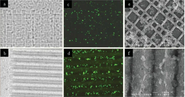
Cell alignment on the MG films and the cell spread-ing on MP and UP films were evaluated by fluores-cence microscopy; fluoresfluores-cence images of cells stained with acridine orange were obtained 4 days postseed-ing [Fig. 5(a,c,d)]. Osteoblasts aligned parallel to groove axis on all MG films; they required 1–3 days to do so. A number of previous studies, including our own, have focused on the contribution of microscale topographical cues to osteoblast alignment,29,34–45and fewer studies explored the same with regard to nano-scale topography.46,47 The results obtained in this study are in accordance with the other reports on osteoblasts on micropatterned surfaces, such as with
the finding by Perizzolo et al. of alignment of rat cal-varial osteoblasts with the grooves after 24 h on Ti and HA coated Si MG templates with dimensions similar to the ones used in this study.39
Microcontact printing led to 2D chemical patterns on the unpatterned films. Only 4 h was enough to Figure 5. Osteoblast morphology on PHBV films with different surface treatment revealed by acridine orange staining. (a) Fb-adsorbed MG film (4 days postseeding, 3150), (b) Fb MCP film (4 h postseeding, 3300), (c) Fb-adsorbed MP film (4 days postseeding,3150), (d) Fb-adsorbed UP film (4 days postseeding, 3150). [Color figure can be viewed in the online issue, which is available at www.interscience.wiley.com.]
Figure 6. Method of (a) alignment determination and (b) the values of deviation angle of cells from groove axis on MG PHBV films and an arbitrary reference line on UP and MP PHBV films. [Color figure can be viewed in the online issue, which is available at www.interscience.wiley.com.]
observe cell alignment to Fb lines on these films. They were effective in aligning the osteoblasts, but the separation between the Fb strips, 40 lm, was not wide enough to prevent cells from crossing the neighboring lines or positioning themselves perpen-dicular to line axis, and this resulted in a decrease in the cell alignment [Fig. 5(b)]. It is possible to deduce from the image that when spread, the rat bone mar-row derived MSC can be as long as about 80–90 lm, and this length should be considered as the mini-mum width of the space between the lines in order to get cells attached and aligned only on the adhe-sive protein strips.
It was observed that nuclei of most of the cells on Fb-adsorbed MP films were localized within the large pits, but still the cell extensions were random and spanning over two or three micropits [Fig. 5(d)]. In a similar study, MG63 osteoblast-like cells cul-tured on titanium surfaces with hemispherical cav-ities with a diameter of 30 lm (ratio of cavity area vs. outside area was 6) were observed to anchor in adjacent cavities, one cell spanning a border between two adjacent cavities.48 In another study, MG63 and SAOS-2 osteoblast-like cells grown on bioactive com-posite surfaces with a microwell width of 50lm and a ridge width of 10 lm aligned along ridges and bridged across wells.37 Thus, osteoblast cells tend to span over the borders of micropits of less than 50 lm width. In contrast, use of gap-cornered micro-wells seems to lead to osteoblast patterning. Rat calvarial osteoblasts seeded on epoxy resins with gap-cornered boxes (wells) of 34 lm width and 10 lm depth were shown to align along the walls of the boxes at 1 and 2 h, but by 4 h additionally began aligning diagonally within the boxes, and at 1 week the cell sheets were aligned diagonally with respect to the boxes.38
Cell alignment was determined quantitatively by measuring the angle of deviation from the groove axis (orientation angle) on MG films and the angle of deviation from an arbitrary reference line drawn on MP and UP film images (Fig. 6).
Although the difference was not significant, the deviation angle was observed to increase as the groove width, and thus the pitch of MG films, increased [Fig. 6(b)]. It was determined from the images of acridine orange stained cells on MG films that the cells attach to the inclined sidewalls of the grooves and only few of them could span the space between the two walls of a groove. Therefore, it was concluded that rather than the groove width (defined as the groove base width here) alone, the whole region between the two ridges is influential on the deviation angles observed with different MG films. Khakbaznejad et al. determined the orientation angle of rat calvarial osteoblasts on titanium-coated MG Si substrates with 30 lm groove depth, 47 lm
pitch width, and inclined walls.43 A nonconfluent monolayer of osteoblast-like cells up to and includ-ing the fourth day of incubation were observed and the mean orientation angle of these cells was 10.78 6 0.88 on the fourth day. The difference between the results of orientation angle on the MG films and the ones in that study can be explained as being due to the difference in cell density on the substrates (1 3 103 cells/cm2in the current study vs. 2 3 105 cells/ cm2), since the higher the density the higher is the cell number not in direct contact with the surface pattern. The difference in the cell carrier materials should be considered, too.
There were too few cells (1–6 in number) on the chemically unmodified MG films (seeding density 1 3 103cells/film) and they were all spread parallel to groove axis. However, it was not possible to determine the deviation angle of the cells on the chemically unmodified MP films, because they all had a circular morphology. In general, the cells on unpatterned surfaces, including TCPS and MP films, spread randomly, which is indicated by the average 458 deviation angle obtained [Fig. 6(b)]. This random osteoblast spreading on unpatterned (smooth) surfa-ces was shown quantitatively in one of our previous publications,29and also by another group.36
CONCLUSION
Polymeric surfaces like PHBV need to be modified with functional groups that are recognized by the cells, in order to support cell attachment. Results of this study proved that microtopographies on PHBV can improve osseointegration when combined with chemical cues. Microgrooves and cell adhesive pro-tein lines on PHBV were able to guide selective osteoblast adhesion and alignment. However, micro-grooves were more effective in restricting cell attach-ment due to the presence of a physical constraint, opposed to Fb lines on unpatterned film surfaces. Use of such microgrooves in combination with bone implants may restore the anisotropic mechanical properties of a damaged bone through generation of an oriented ECM.
References
1. Chun YS, Kim WN. Thermal properties of blends of poly(hy-droxybutyrate-co-hydroxyvalerate) and poly(styrene-co-acry-lonitrile). J Appl Polym Sci 2000;77:673–679.
2. Liu WJ, Yang HL, Wang Z, Dong LS, Liu JJ. Effect of nucleat-ing agents on the crystallization of poly (3-hydroxybutyrate-co-3-hydroxyvalerate). J Appl Polym Sci 2002;86:2145–2152. 3. Anderson AJ, Dawes EA. Occurrence, metabolism, metabolic
role, and industrial uses of bacterial polyhydroxyalkanoates. Microbiol Rev 1990;54:450–472.
4. Gogolewski S. Resorbable polymers for internal fixation. Clin Mater 1992;10:13–20.
5. Gogolewski S, Jovanovic M, Perren SM, Dillon JG, Hughes MK. Tissue response and in vivo degradation of selected polyhydroxyacids: Polylactides (PLA), poly(3 hydroxybuty-rate) (PHB), and poly(3-hydroxybutyrate-co-3-hydroxyvaler-ate) (PHB/VA). J Biomed Mater Res 1993;27:1135–1148. 6. Taylor MS, Daniels AU, Andriano KP, Heller J. Six
bioabsorb-able polymers: In vitro acute toxicity of accumulated degrada-tion products. J Appl Biomater 1994;5:151–157.
7. An YH, Woolf SK, Friedman RJ. Pre-clinical in vivo evalua-tion of orthopaedic bioabsorbable devices. Biomaterials 2000; 21:2635–2652.
8. Hankermeyer CR, Tjeerdema RS. Polyhydroxybutyrate: Plas-tic made and degraded by microorganisms. Rev Environ Contam Toxicol 1999;159:1–24.
9. Holland SJ, Jolly AM, Yasin M, Tighe BJ. Polymers for biode-gradable medical devices. II. Hydroxybutyrate–hydroxyvaler-ate copolymers: Hydrolytic degradation studies. BiomHydroxybutyrate–hydroxyvaler-aterials 1987;8:289–295.
10. Galego N, Rozsa C, Sanchez R, Fung J, Vazquez A, Tomas JS. Characterization and application of poly(b-hydroxyalkanoates) family as composite biomaterials. Polym Test 2000;19:485–492. 11. Coskun S, Korkusuz F, Hasirci V. Hydroxyapatite reinforced
poly(3-hydroxybutyrate) and poly(3-hydroxybutyrate-co-3-hydroxyvalerate) based degradable composite bone plate. J Biomater Sci Polym Ed 2005;16:1485–1502.
12. Kose GT, Kenar H, Hasirci N, Hasirci V. Macroporous poly (3-hydroxybutyrate-co-3-hydroxyvalerate) matrices for bone tissue engineering. Biomaterials 2003;24:1949–1958.
13. Kose GT, Korkusuz F, Korkusuz P, Purali N, Ozkul A, Hasi-rci V. Bone generation on PHBV matrices: An in vitro study. Biomaterials 2003;24:4999–5007.
14. Kumarasuriyar A, Jackson RA, Grondahl L, Trau M, Nur-combe V, Cool SM. Poly( b-hydroxybutyrate-co-b-hydroxyval-erate) supports in vitro osteogenesis. Tissue Eng 2005;11:1281– 1295.
15. Degasne I, Basle MF, Demais V, Hure G, Lesourd M, Grol-leau C, Mercia L, Chappard D. Effects of roughness, fibronec-tin, and vitronectin on attachment, spreading, and prolifera-tion of human-osteoblast like cells (Saos-2) on titanium implants. Calcif Tissue Int 1999;64:499–507.
16. Sinha R, Morris F, Suken A, Shah S, Tuan R. Surface compo-sition of orthopaedic implant metals regulates cell attach-ment, spreading, cytoskeletal organisation of primary human osteoblasts in vitro. Clin Orthop 1994;305:258–272.
17. Juliano RL, Haskill S. Signal transduction from the extracellu-lar matrix. J Cell Biol 1993;120:577–585.
18. Burridge K, Chrzanowska-Wodnick M. Focal adhesions, con-tractility, and signaling. Annu Rev Cell Dev Biol 1996;12:463– 518.
19. Gray C, Boyde A, Jones SJ. Topographically induced bone formation in vitro: Implications for bone implants and bone grafts. Bone 1996;18:115–123.
20. O’Connor TP, Bentley D. Accumulation of actin in subsets of pioneer growth cone filopodia in response to neural and epi-thelial guidance cues in situ. J Cell Biol 1993;123:935–948. 21. Curtis ASG, Wilkinson CDW. Topographical control of cells.
Biomaterials 1997;18:1573–1583.
22. Curtis ASG, Casey B, Gallagher JO, Pasqui D, Wood MA, Wilkinson CDW. Substratum nanotopography and the adhe-sion of biological cells. Are symmetry or regularity of nanoto-pography important? Biophys Chem 2001;94:275–283. 23. Wilkinson CDW, Curtis ASG, Crossan J. Nanofabrication in
cellular engineering. J Vac Sci Technol B 1998;16:3132–3136. 24. Qu J, Chehroudi B, Brunette DM. The use of micromachined
surfaces to investigate the cell behavioural factors essential to osseointegration. Oral Dis 1996;2:102–115.
25. Wieland M, Textor M, Chehroudi B, Brunette DM. Synergistic interaction of topographic features in the production of bone-like nodules on Ti surfaces by rat osteoblasts. Biomaterials 2005;26:1119–1130.
26. Anselme K, Bigerell M. Topography effects of pure titanium substrates on human osteoblast long-term adhesion. Acta Bio-mater 2005;1:211–222.
27. Huang HH, Ho CT, Lee TH, Lee TL, Liao KK, Chen FL. Effect of surface roughness of ground titanium on initial cell adhesion. Biomol Eng 2004;21:93–97.
28. Hallgren C, Sawase T, Ortengren U, Wennerberg A. Histo-morphometric and mechanical evaluation of the bone-tissue response to implants prepared with different orientation of surface topography. Clin Implant Dent Relat Res 2001;3:194– 203.
29. Kenar H, Kose GT, Hasirci V. Tissue engineering of bone on micropatterned biodegradable polyester films. Biomaterials 2006;27:885–895.
30. Mrksich M, Chent CS, Xia Y, Diket LE, Ingbert DE, White-sides GM. Controlling cell attachment on contoured surfaces with self-assembled monolayers of alkanethiolates on gold. Proc Natl Acad Sci USA 1996;93:10775–10778.
31. Matsuda T, Sugawara T. Development of surface photochem-ical modification method for micropatterning of cultured cells. J Biomed Mater Res 1995;29:749–756.
32. Folch A, Toner M. Microengineering of cellular interactions. Annu Rev Biomed Eng 2000;2:227–256.
33. Ito Y, Hasuda H, Kamitakahara M, Ohtsuki C, Tanihara M, Kang IK, Kwon OH. A composite of hydroxyapatite with electrospun biodegradable nanofibers as a tissue engineering material. J Biosci Bioeng 2005;100:43–49.
34. Matsuzaka K, Walboomers XF, Ruijter JE, Jansen JA. The effect of polyL-lactic acid with parallel surface micro groove on osteoblast-like cells in vitro. Biomaterials 1999;20:1293– 1301.
35. Matsuzaka K, Walboomers XF, Yoshinari M, Inoue T, Jansen JA. The attachment and growth behavior of osteoblast-like cells on microtextured surfaces. Biomaterials 2003;24:2711– 2719.
36. Matsuzaka K, Yoshinari M, Shimono M, Inoue T. Effects of multigrooved surfaces on osteoblast-like cells in vitro: Scan-ning electron microscopic observation and mRNA expression of osteopontin and osteocalcin. J Biomed Mater Res A 2004; 68:227–234.
37. Rea SM, Brooks RA, Schneider A, Best SM, Bonfield W. Osteoblast-like cell response to bioactive composites-surface-topography and composition effects. J Biomed Mater Res B Appl Biomater 2004;70:250–261.
38. Hamilton DW, Wong KS, Brunette DM. Microfabricated dis-continuous-edge surface topographies influence osteoblast adhesion, migration, cytoskeletal organization, and prolifera-tion and enhance matrix and mineral deposiprolifera-tion in vitro. Cal-cif Tissue Int 2006;78:314–325.
39. Perizzolo D, Lacefield WR, Brunette DM. Interaction between topography and coating in the formation of bone nodules in culture for hydroxyapatite- and titanium-coated microma-chined surfaces. J Biomed Mater Res 2001;56:494–503. 40. Chehroudi B, McDonnell D, Brunette DM. The effects of
micromachined surfaces on formation of bonelike tissue on subcutaneous implants as assessed by radiography and computer image processing. J Biomed Mater Res 1997;34: 279–290.
41. Lu X, Leng Y. Quantitative analysis of osteoblast behavior on microgrooved hydroxyapatite and titanium substrata. J Biomed Mater Res A 2003;66:677–687.
42. Ber S, Torun Kose G, HasVrcV V. Bone tissue engineering on patterned collagen films: An in vitro study. Biomaterials 2005;26:1977–1986.
43. Khakbaznejad A, Chehroudi B, Brunette DM. Effects of tita-nium-coated micromachined grooved substrata on orienting layers of osteoblast-like cells and collagen fibers in culture. J Biomed Mater Res A 2004;70:206–218.
44. Tan J, Saltzman WM. Biomaterials with hierarchically defined micro- and nanoscale structure. Biomaterials 2004;25:3593– 3601.
45. Wang JH, Grood ES, Florer J, Wenstrup R. Alignment and proliferation of MC3T3-E1 osteoblasts in microgrooved sili-cone substrata subjected to cyclic stretching. J Biomech 2000; 33:729–735.
46. Zhu B, Lu Q, Yin J, Hu J, Wang Z. Alignment of osteoblast-like cells and cell-produced collagen matrix induced by nano-grooves. Tissue Eng 2005;11:825–834.
47. Lenhert S, Meier M-B, Meyer U, Chi L, Wiesmann HP. Osteo-blast alignment, elongation and migration on grooved poly-styrene surfaces patterned by Langmuir-Blodgett lithography. Biomaterials 2005;26:563–570.
48. Zinger O, Zhao G, Schwartz Z, Simpson J, Wieland M, Land-olt D, Boyan B. Differential regulation of osteoblasts by substrate microstructural features. Biomaterials 2005;26:1837– 1847.