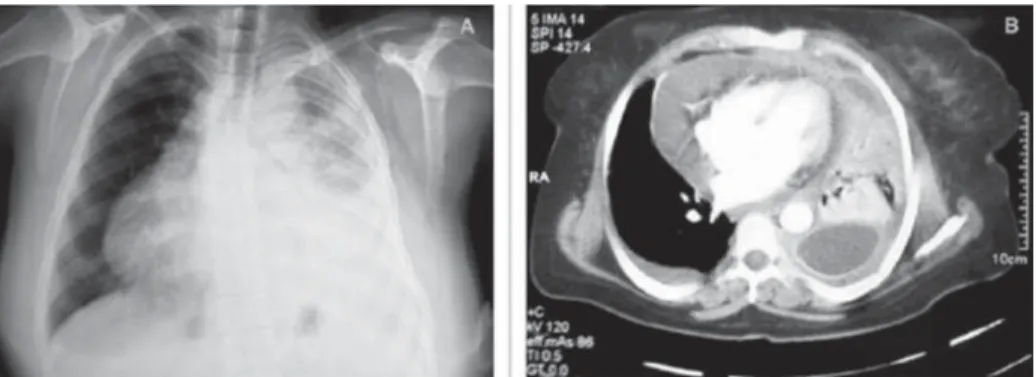
A 46-year-old woman with a previous diagnosis of inoperable stage IV small cell lung cancer presented to the emergency department with shortness of breath. Physical examination showed a pale woman who appeared malnourished. Her vital signs were as follows: pulse rate 115 beats/min, respiration rate 22 breaths/min, body temperature 37.5°C, and blood pressure 95/65 mm Hg. Breath sounds were diminished on the left base and widespread coarse crackles were heard over the left lung. Other system examination fi ndings were within normal limits. A 12-lead electrocardiogram showed sinus tachycardia and lower voltage without marked ST changes. A chest X-ray showed infi ltrates on the left upper zones, and a large left-sided pleural effusion (Figure 1A). Transthoracic echocardiograpy (TTE) showed a large loculated pericardial effusion measuring 3.2 cm in maximal width on the right side of the heart with cardiac tamponade, which was also confi rmed by multislice computed tomography (Figure 1B). Due to the increased risk of general anesthesia, we preferred an echocardiography-guided pericardiocentesis using the parasternal window. As the loculated pericardial effusion was adjacent to the right atrium and ventricle, the right parasternal
approach was chosen. Echocardiography-guided pericardiocentesis was performed under local anesthesia in the operating room owing to the increased risk for pneumothorax. A puncture needle was advanced through the sixth intercostal space at the right parasternal border, and nearly 600 mL of hemorrhagic fl uid was aspirated (Figure 2). After pericardiocentesis, the patient’s vital signs were as follows: pulse rate 85 beats/min, respiration rate 19 breaths/min, body temperature 37.5°C, and blood pressure 115/75 mm Hg. A very small amount of pericardial effusion was seen during TTE. The fl uid cytology was compatible with metastatic disease. She died from respiratory failure three months after the procedure. Pericardiocentesis via the right parasternal approach can be used in a patient with severe comorbidity.
Loküle Perikart Efüzyonunun Sağ Parasternal Perikardiyosentez
ile Başarılı Tedavisi
Mustafa Adem Tatlısu1, Emrah Bozbeyoğlu2, Bülent Aydemir3, Zekeriya Nurkalem4
1 Sivas Numune Hospital, Clinic of Cardiology, Sivas, Turkey 2
Dr. Siyami Ersek Cardiovascular Surgery Center, Clinic of Cardiology, İstanbul, Turkey
3 Dr. Siyami Ersek Cardiovascular Surgery Center, Clinic of Cardiovascular Surgery, İstanbul, Turkey 4
Medipol University Faculty of Medicine, Department of Cardiology, İstanbul, Turkey
Loculated Pericardial Effusion Successfully
Managed with A Right Parasternal
Pericardiocentesis
Mustafa Adem Tatlısu
E-mail: [email protected] Submitted: 06.07.2015 Accepted: 28.09.2015
@ Copyright 2015 by Koşuyolu Heart Journal. Available on-line at
www.kosuyoluheartjournal.com
159
Koşuyolu Heart Journal 2015;18(3):159 • DOI: 10.5578/khj.10234 ●ORIGINAL IMAGE
Correspondence
Figure 1. (A) A chest X-ray shows infi ltrates on the left upper zones and a large left-sided pleural effusion. (B) Multislice
computed tomography shows a large loculated pericardial effusion on the right side of the heart.
Figure 2. Pericardiocentesis via the right parasternal approach.
View publication stats View publication stats