Skewed X Chromosome Inactivation in Blood Cells of
Women With Scleroderma
Zeynep O
¨ zbalkan,
1Sevgi Bag
ˇıs¸lar,
2Sedat Kiraz,
1Cemaliye Boylu Akyerli,
2Hu
¨seyin T. E. O
¨ zer,
3S
¸ule Yavuz,
4A. Merih Birlik,
5Meral C
¸ algu
¨neri,
1and Tayfun O
¨ zc¸elik
6Objective. Scleroderma (SSc) is an autoimmune
disease of unknown etiology. The disease is 3–8 times more frequent in women than in men. The role of X chromosome inactivation (XCI) in the predisposition of women to autoimmunity has been questioned. Until now this has not been illustrated experimentally. This study was undertaken to test the hypothesis that disturbances in XCI mosaicism may be involved in the pathogenesis of the disease in female patients with SSc.
Methods. Seventy female SSc patients and 160
female controls were analyzed for the androgen receptor locus by the Hpa II/polymerase chain reaction assay to assess XCI patterns in DNA extracted from peripheral blood cells. Furthermore, skin biopsy samples were obtained from 5 patients whose blood revealed an extremely skewed pattern of XCI, and the analysis repeated. Since microchimerism in SSc was reported, Y chromosome sequences were investigated in all samples.
Results. Skewed XCI was observed in DNA from
peripheral blood cells in 35 of 55 informative patients (64%), as compared with 10 of 124 informative controls (8%) (P < 0.0001). Extreme skewing was present in 27 of
the patient group (49%), as compared with only 3 of the controls (2.4%) (P < 0.0001). However, XCI was random in all skin biopsy samples. The potential contribution of microchimerism to the random XCI pattern is highly unlikely based on the medical histories of the patients.
Conclusion. Skewed XCI mosaicism may play a
significant role in the pathogenesis of SSc.
Scleroderma (systemic sclerosis; SSc) is an auto-immune connective tissue disease of unknown etiology. Although the pathogenesis is poorly understood, disease progression involves the vasculature, the immune sys-tem, and extracellular matrix deposition (1,2). SSc oc-curs 3–8 times more frequently in women than in men, and the highest incidence of the disease is in individuals ages 45–55 years (3). Based on observations that SSc has clinical features that resemble chronic graft-versus-host disease, microchimerism resulting from transplacental cells (cell passage from child to mother, or in some instances, mother to child) was considered responsible for autoimmune diseases, including SSc (4,5). Subse-quently, fetal DNA and cells were identified in some women with SSc (5–8). Despite these interesting find-ings, microchimerism in SSc could be secondary to the underlying disease, because it offers no explanation for the occurrence of the disease in men or in women who have had no children. In addition, there is no proof that transplant recipients who are chimeric individuals are at increased risk for SSc (9).
Loss of immunologic tolerance to self antigens is an important feature of autoimmune disorders. T cell tolerance, which appears to be broken in many of these diseases, is thought to be established by negative selec-tion against potentially self-reactive T cells in the thymic medulla and cortex–medulla junction. Professional antigen-presenting cells, particularly dendritic cells, me-diate the negative selection process (10,11). It has been demonstrated that risk of autoimmunity could be
in-Supported by grants from the Scientific and Technical Re-search Council of Turkey (TU¨ BI˙TAK-SBAG 2513), International Centre for Genetic Engineering and Biotechnology (ICGEB-CRP/ TUR04-01), and Bilkent University Research Fund (to Dr. O¨ zc¸elik).
1Zeynep O¨ zbalkan, MD, Sedat Kiraz, MD, Meral C¸ algu¨neri,
MD: Hacettepe University Medical School, Ankara, Turkey; 2Sevgi
Bagˇıs¸lar, BSc, Cemaliye Boylu Akyerli, PhD: Bilkent University, Ankara, Turkey; 3Hu¨seyin T. E. O¨ zer, MD: C¸ ukurova University
Medical School, Adana, Turkey;4S¸ule Yavuz, MD: Marmara
Univer-sity Medical School, Istanbul, Turkey;5A. Merih Birlik, MD: Dokuz
Eylu¨l University Medical School, Izmir, Turkey;6Tayfun O¨ zc¸elik, MD:
Bilkent University, Ankara, and Ayhan Sahenk Foundation, Istanbul, Turkey.
Drs. O¨ zbalkan and Bagˇıs¸lar contributed equally to this work. Address correspondence and reprint requests to Tayfun O¨ zc¸elik, MD, Bilkent University, Department of Molecular Biology and Genetics, Faculty of Science, B-242, Bilkent, Ankara 06800, Turkey. E-mail: [email protected].
Submitted for publication September 6, 2004; accepted in revised form February 8, 2005.
creased by a lack of exposure to self antigens in the thymus and the presence of autoreactive T cells (12).
A potential mechanism through which lack of exposure to self antigens could occur in women is a disturbance in the X chromosome inactivation (XCI) process (13–15). XCI is an epigenetic regulation in early development that results in transcription inactivation of 1 of the pair of X chromosomes (16,17). As a result of this physiologic event, the X chromosome inherited from either parent is silenced at random, and normal women are thus a mosaic of 2 cell populations. It is therefore conceivable that skewed XCI could lead to the escape of X-linked self antigens from presentation in the thymus or in other peripheral sites that are involved in tolerance induction, inadequate thymic deletion, and finally loss of T cell tolerance. However, examination of XCI patterns in peripheral blood from female patients with autoimmune diseases (including systemic lupus erythematosus [SLE], juvenile diabetes, multiple sclero-sis, and juvenile rheumatoid arthritis) did not reveal skewed X inactivation patterns (15,18). Conversely, dis-turbances in the XCI process are known to alter the clinical manifestation of X-linked disorders in women (19,20). In addition, high frequency of skewed X inacti-vation has been observed in breast and ovarian cancers (21,22), and in women with recurrent spontaneous abor-tions (23,24).
We hypothesized that extremely skewed XCI, especially in hematopoietic stem cells, may be involved in the pathogenesis of SSc. This hypothesis was based on 2 observations: first, the disease is more prevalent in women than in men. Second, oligoclonal T cell expan-sion has been documented in the skin leexpan-sions of SSc patients (25), which may indicate an inadequate ability to induce tolerance to self antigens, particularly X-linked autoantigens. To test this hypothesis, we ana-lyzed the methylation status of a highly polymorphic CAG repeat in the androgen receptor (AR) gene, and demonstrated that XCI mosaicism is extremely skewed in the blood but not in the skin lesions of female patients with SSc.
PATIENTS AND METHODS
Patients. White women diagnosed as having SSc (n⫽
70), rheumatoid arthritis (n⫽ 12), or SLE (n ⫽ 9), and healthy female controls with no history of autoimmune disease or cancer (n ⫽ 160) were included in the study. All patients fulfilled the American College of Rheumatology (formerly, the American Rheumatism Association) diagnostic criteria for scleroderma (26), SLE (27), or rheumatoid arthritis (28). The
mean (⫾SD) ages were 49 ⫾ 14 years for SSc patients (40 ⫾ 14 years in the nulligravid patients, and 53⫾ 12 years in the gravid patients), 52⫾ 15 years in rheumatoid arthritis patients, 47 ⫾ 15 years in SLE patients, and 46⫾ 10 years in controls. The duration of symptoms, first appearing as Raynaud’s phenom-enon, puffy hands, esophagitis, or dyspnea, was 10⫾ 7 years among the SSc patients. The mean age at diagnosis was 39⫾ 12 years. Five patients had the limited cutaneous form of disease, and the remaining patients had diffuse disease. The duration of the symptoms in the rheumatoid arthritis and SLE patients was 12⫾ 7 and 9 ⫾ 8 years, respectively. All of the patients had attended the rheumatology clinics of the partici-pating institutions for at least 1 year since onset of disease. The SSc patients were chosen randomly for the study. The rheu-matoid arthritis and SLE patients who were subsequently recruited were selected from the group of female patients who were receiving immunosuppressive therapy and were age matched with the SSc patients. The ethics review boards at the participating institutions approved the study protocol. In-formed consent was obtained from all subjects.
X inactivation study. XCI patterns were quantitated by
use of radioactive␣-33P-dCTP (NEN, Boston, MA)
polymer-ase chain reaction (PCR) as described elsewhere (29). Briefly, DNA was extracted from 10 ml venous blood (peripheral blood mononuclear cells), skin biopsy, buccal mucosa, and hair follicle specimens. The methylation status of a highly polymor-phic CAG repeat in the androgen receptor (AR) gene was determined by use of methylation-sensitive restriction enzyme
Hpa II (MBI Fermentas, St. Leon-Rot, Germany). Male DNA
of cytogenetically verified 46XY karyotype was used as control for complete digestion. The PCR products, both before and after digestion, were separated on 8% sequencing gels, dried, and were autoradiographed on Medicalfilm CP-BU (Agfa, Mortsel, Belgium). Densitometric analysis of the alleles was performed using Multi-Analyst software version 1.1 (Bio-Rad, Hercules, CA). The assay was performed at least twice for each sample. In addition, cold PCR followed by electrophoretic separation of the alleles in 4% MetaPhor agarose (FMC BioProducts, Rockland, ME) or 10% polyacrylamide, ethidium bromide staining, and densitometric analysis (as described above) were performed for all samples. The results of the densitometric analyses included normalization of the ratios, based upon the undigested samples. This was deter-mined by dividing the allele ratio of the digested sample by the ratio of the undigested sample from the same specimen. The use of this ratio corrects for preferential amplification of 1 allele, which often occurs for the shorter microsatellite allele.
Y chromosome study. Y chromosome–specific
se-quences were analyzed by PCR and 2 independent sets of primers. First, nested PCR with primers Y1-1 ⫹ Y1-2 and Y1-3⫹ Y1-4 was performed as described (30). Electrophoresis of amplicons in 1.5% agarose gel revealed a 148-bp Y chromosome–specific fragment in positive samples. To con-firm the results, all samples were analyzed with a second set of PCR primers, as previously described (31). Positive (male DNA) and negative (no DNA) controls were included in all reactions. The results from control and test groups in XCI and Y chromosome studies were compared by chi-square test with Yates’ correction.
RESULTS
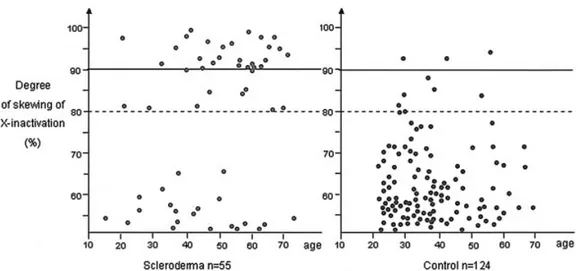
PCR-based X inactivation study of peripheral blood. XCI status was informative in 55 of the 70 SSc patients and in 124 of the 160 controls. Some heterozy-gous individuals were considered uninformative since only those whose alleles resolve adequately for densito-metric analysis were included in the study. Skewed XCI (⬎80% skewing) was observed in 35 of the 55 patients (64%), and 10 of the 124 controls (8%) (P ⬍ 0.0001). More importantly, extremely skewed XCI, defined as ⬎90% inactivation of 1 allele, was present in 27 patients (49%), and in only 3 controls (2.4%) (P ⬍ 0.0001) (Table 1 and Figure 1A). Extremely skewed XCI is a rare event and has been reported in 1–2% of 20–40-year old women, and in 2–4% of 55–72-year-old women (22–24). Since the data for SSc patients is strikingly bimodal, we plotted the distribution of the X inactiva-tion profiles according to age. However, we did not observe a shift toward the skewed range in older patients and controls (Figure 2).
Immunosuppressive therapy and X inactivation. At the time of sample collection, 62 patients were being treated with immunosuppressive therapies (cyclo-phosphamide, 50 mg twice a day, n ⫽ 31; D-penicillamine, 300 mg/day, n ⫽ 16; azathioprine, 50 mg twice daily, n⫽ 9; chloroquine, 200 mg/day, n ⫽ 4; methotrexate, 10 mg once a week, n ⫽ 1; mycopheno-late, 1,000 mg/day, n ⫽ 1). The remaining 8 patients were newly diagnosed and not taking any medications. Although 6 of the 55 informative patients never received immunosuppressive agents, and an additional 16 pa-tients had been receiving immunosuppressive treatment for ⱕ1 week, a major concern with the observed XCI patterns among SSc patients was that concomitant im-munosuppressive therapy could influence the results, as has been observed in feline hematopoietic cells (32). Analysis of the data on XCI patterns versus duration of immunosuppressive therapy did not reveal a statistically
significant correlation. Among the patients with skewed X inactivation, 8 (23%) had received immunosuppres-sive agents for 6–10 years, 14 (40%) for 1–5 years, and 13 (37%) had received no immunosuppressive agents or received them forⱕ1 week. Of the patients with random X inactivation, 4 (20%) had received immunosuppres-sive agents for 6–10 years, 7 (35%) for 1–5 years, and 9 (45%) received no immunosuppressive agents or re-ceived them for ⱕ1 week. Complete ablation of bone marrow or suspended leukopenia was not observed in any of the patients at any stage of the treatment.
In order to demonstrate that immunosuppressive medications do not skew the XCI patterns, we collected peripheral blood samples from patients who were being treated with similar immunosuppressive medications, but who had rheumatoid arthritis (n⫽ 12) or SLE (n ⫽ 9). Skewed XCI was observed in 1 rheumatoid arthritis patient, whereas a random pattern was clearly visible in 17 patients (Figures 1B and C). Three patients were not informative for theAR polymorphism.
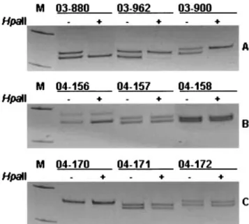
Figure 1. X chromosome inactivation status in scleroderma (A),
rheu-matoid arthritis (B), and systemic lupus erythematosus (C) patients. Polymerase chain reaction products from the androgen receptor methylation assay demonstrate random X chromosome inactivation patterns in samples 04-156 (allele ratio 64.6%:35.4%), 04-157 (52.8%: 47.2%), 04-158 (51.0%:48.4%), 04-171 (53.1%:46.9%), and 04-172 (50.6%:49.4%), and skewed patterns in samples 03-880 (95.4%:4.6%), 03-962 (92.6%:7.4%), and 03-900 (94.1%:5.9%). Sample 04-170 is not informative for the androgen receptor polymorphism. For each sam-ple, DNA was either undigested (⫺) or digested (⫹) with the methylation-sensitive restriction enzymeHpa II. Marker (M; pUC mix 8 [331-bp and 242-bp]) fragments are visible.
Table 1. Proportion of patients and controls with skewed X chromo-some inactivation* Scleroderma patients (n⫽ 55) Female controls (n⫽ 124) Skewed (80–90%) 8 (14.54)† 7 (5.64) Extremely skewed (⬎90%) 27 (49.09)† 3 (2.41) Total 35 (63.63)† 10 (8.05)
* Values are the number (%).
Pregnancy history and Y chromosome analysis. Characteristics of the SSc patients with skewed XCI are shown in Table 2. Y chromosome analysis was per-formed in a blinded manner (without prior knowledge of pregnancy history) in all patients, and Y chromosome– specific DNA was detected in 16 (41%) of the 39 patients who were known to have at least 1 son. There was no history of pregnancy in 23 of the patients. In the control group, 31 women had at least 1 son and Y chromosome–specific DNA was detected in 2 of them (P ⫽ 0.0026) (Table 3). These results are consistent with the previous reports (7,8) of a significant difference in persistence of male DNA in peripheral blood between controls and SSc.
PCR-based X inactivation study of skin biopsy, buccal mucosa, and hair follicle specimens. Skin biopsy samples were obtained from the site of clinically detect-able cutaneous changes in 5 patients (patients 03-894, 03-899, 03-956, 03-963, and 03-1101). Buccal mucosa and hair follicle specimens were also collected from the same patients, excluding patient 03-963. These patients were selected from a group that displayed almost exclu-sive representation for only 1 allele of theAR polymor-phism in their methylation-sensitive PCR assay, which indicated⬎90% skewed XCI. A random pattern of XCI was observed in the skin biopsy samples of all patients, with the allele ratios of 50.5%:49.5% in patient 03-894, 53%:47% in patient 03-899, 51.3%:48.7% in patient 03-956, 65%:35% in patient 03-963, and 54.7%:45.3% for patient 03-1101 (Figure 3). The buccal mucosa and hair follicle samples also revealed a random pattern in all patients analyzed.
It has been reported that microchimeric cells can build tissues as well as attack them (33). Therefore, it is possible that DNA from prior pregnancies contributes to the random XCI profile observed in the skin lesions of the above-mentioned SSc patients. However, several observations provide evidence against this possibility. First, 2 of these patients (03-894 and 03-963) had never been pregnant. Second, none of the patients with skewed XCI in their blood had a twin sibling. Third, careful analysis of the predigested specimens did not reveal 3
AR alleles in any of the informative patients (results not
shown).
DISCUSSION
A reduction in the sex ratio (male:female) is characteristic for most autoimmune diseases, including SSc, and sex differences may be of great relevance. One such sex difference is the number of X chromosomes, which leads to XCI mosaicism in women. In this study we demonstrate skewed XCI patterns in peripheral blood mononuclear cells of a significant proportion (64%) of women with SSc. Approximately 8% of female control subjects and 8% of rheumatoid arthritis patients exhibit skewed X inactivation patterns (ⱖ80:20), which is consistent with previous estimates (15,22–24). The effect is more pronounced for patterns of X inactivation (ⱖ90:10), since nearly half of SSc patients show such skewing, compared with only a small percentage of women in the control group. Furthermore, the skin lesions of 5 patients with extreme skewing in blood displayed random XCI. Our results show that factors
Figure 2. Distribution of X inactivation patterns according to age in scleroderma patients and control
associated with extremely skewed XCI are present in a significant proportion of female patients with SSc, and raise important questions as to the cause and conse-quence of this observation.
Figure 3. Skewed X chromosome inactivation in samples. Polymerase
chain reaction products of undigested (⫺) (lane 1) and Hpa II– digested (⫹) DNA from blood (lane 2) and skin biopsy samples (lane 3) of patients 03-894, 03-899, and 03-1101 are shown. Two alleles are seen in lanes 1 and 3, whereas a single allele resulting from extremely skewed X chromosome inactivation is clearly visible in lane 2 for all 3 samples (see text for allele ratios).
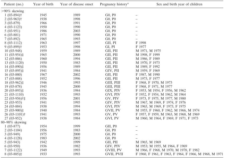
Table 2. Characteristics of the scleroderma patients with skewed X chromosome inactivation
Patient (no.) Year of birth Year of disease onset Pregnancy history* Sex and birth year of children ⬎90% skewing 1 (03-894)† 1945 1989 G0, P0 – 2 (03-963)† 1938 1998 G0, P0 – 3 (03-879) 1966 1991 G0, P0 – 4 (03-1123) 1950 1990 G0, P0 – 5 (03-951) 1986 2003 G0, P0 – 6 (03-881) 1971 1990 G0, P0 – 7 (03-892) 1965 1993 G0, P0 – 8 (03-1112) 1963 1997 GII, PI F 1998 9 (03-899)† 1953 1998 GI, PI F 1977 10 (03-948) 1959 1989 GII, PII M 1971, M 1975 11 (03-954)‡ 1965 2000 GII, PII M 1998, F 1999 12 (03-886) 1960 1994 GII, PII M 1986, F 1989 13 (03-1120) 1950 1983 GII, PII M 1970, F 1973 14 (03-890)‡ 1956 1989 GII, PII M 1989, F 1992 15 (03-895)‡ 1941 1984 GIV, PII M 1959, F 1969 16 (03-880) 1967 2002 GII, PII F 1987, M 1990 17 (03-888) 1952 1996 GII, PII M 1973, F 1977 18 (03-962)‡ 1946 1993 GIII, PIII F 1968, F 1970, M 1973 19 (03-878) 1945 2000 GIII, PIII F 1968, F 1971, M 1977 20 (03-893)‡ 1936 1984 GIX, PIV F 1953, M 1954, F 1956, M 1962 21 (03-1110) 1932 1993 GVI, PIV F 1952, F 1954, M 1962, M 1964 22 (03-956)†‡ 1956 2001 GV, PIV F 1973, F 1975, M 1977, M 1980 23 (03-953) 1941 1995 GIV, PIV M 1967, M 1969, F 1974, F 1976 24 (03-884) 1938 1994 GVI, PIV M 1965, M 1969, F 1973, F 1975 25 (03-900)‡ 1940 1984 GVII, PV M 1955, F 1960, F 1962, M 1966, M 1974 26 (03-1101)† 1941 1993 GV, PV F 1957, F 1959, M 1963, M 1968, M 1969 27 (03-952) 1938 1984 GVI, PV M 1960, M 1964, F 1969, F 1971, F 1973 80–90% skewing 1 (03-877) 1954 1999 GII, P0 – 2 (03-1104) 1956 1983 G0, P0 – 3 (03-949) 1975 2000 G0, P0 – 4 (03-1128) 1983 2002 G0, P0 – 5 (03-943) 1942 1999 GII, PII M 1965, M 1969 6 (03-950) 1936 1982 GIV, PIV M 1953; M 1955, M 1964, F 1969 7 (03-1122) 1949 1993 GVIII, PV M 1966, F 1968, M 1970, M 1978, F 1982 8 (03-885)‡ 1933 1993 GVII, PVII F 1960, F 1961, F 1963, F 1964, F 1966, M 1968, M 1971 * G⫽ gravida; P ⫽ para.
† Skin biopsy sample obtained.
‡ Y chromosome sequence–positive patient.
Table 3. Distribution of Y chromosome sequences in patients and controls who gave birth to male children*
Group
Positive, no. (%)
Negative,
no. (%) Total, no. Scleroderma patients 16 (41.02)† 23 (58.98) 39 Skewed XCI 9 (40.90)† 13 (59.10) 22 Random XCI 4 (36.36)† 7 (63.64) 11 Control females 2 (6.45) 29 (93.55) 31 Skewed XCI 1 (25.00) 3 (75.00) 4 Random XCI 1 (4.16) 23 (95.84) 24 * XCI⫽ X chromosome inactivation.
The causes of skewed XCI are classified into 2 groups: primary and secondary. Bias in the initial choice of which X chromosome to be inactivated, due to germline X-inactive specific transcript mutations, is an example of a primary cause (19). Secondary causes include deleterious X-linked mutations or X chromo-some rearrangements, aging, twinning, or monoclonal expansion of cells (for review, see ref. 34). Existence of deleterious X-linked mutations or X chromosome re-arrangements and their differential expression patterns could provide a disadvantage to affected blood cells, but not to skin cells in SSc patients, and lead to skewed XCI. With respect to the consequences of skewed X inactivation, it is well documented that clinical manifes-tation of X-linked disorders in women could be influ-enced by disturbances in the XCI process (35). In addition, it has been hypothesized that skewed XCI could be a factor influencing the predisposition of women to autoimmunity (13,14). Because we have dem-onstrated skewed XCI patterns in a significant propor-tion of female SSc patients, disturbed X inactivapropor-tion mosaicism could be considered a contributing factor in disease pathogenesis.
Although extremely skewed XCI is rare, it does not lead to the development of SSc in all women. We agree with the hypothesis that a subsequent event, such as environmental exposure to viral, chemical, or other agents, may trigger a cascade that results in SSc (8). In addition, the coinheritance of genetic susceptibility fac-tors, such as functional variants in vital negative regula-tory molecules of the immune system (36), may exacer-bate the effects of skewed XCI and contribute to the development of autoimmune diseases, including SSc.
ACKNOWLEDGMENTS
We gratefully acknowledge Prof. Margaret K. Sands, and Drs. Kamil Can Akcali and Ozlen Konu for critical reading of the manuscript.
REFERENCES
1. Derk CH, Jimenez SA. Systemic sclerosis: current views of its pathogenesis. Autoimmun Rev 2003;2:181–91.
2. Seibold JR. Mixed connective tissue disease, scleroderma, and inflammatory disease. In: Ruddy S, Harris ED, Sledge CB, editors. Kelley’s textbook of rheumatology. Philadelphia: Saunders; 2005. p. 1279–309.
3. Silman AJ, Hochberg MC. Scleroderma. In: Silman AJ, Hochberg MC, editors. Epidemiology of the rheumatic diseases. Oxford: Oxford University Press; 1993. p. 192–219.
4. Mullinax F. Chimerism and autoimmunity. In: Feng PH, Boey ML,
Chng HH, Fong KY, Howe HS, Leong KH, editors. Proceedings of the 4th ASEAN Congress of Rheumatology. Singapore: Com-munication Consultants; 1993. p. 39–40.
5. Nelson JL. Maternal–fetal immunology and autoimmune diseases: is some autoimmune disease auto-alloimmune or allo-auto-immune? Arthritis Rheum 1996;39:191–4.
6. Mullinax F, Mullinax GL. Pregnancy-induced scleroderma: iden-tification of offspring-derived cells in patients with scleroderma [abstract]. Arthritis Rheum 1996;39 Suppl 9:S231.
7. Nelson JL, Furst DE, Maloney S, Gooley T, Evans PC, Smith A, et al. Microchimerism and HLA-compatible relationships of preg-nancy in scleroderma. Lancet 1998;351:559–62.
8. Artlett CM, Smith JB, Jimenez SA. Identification of fetal DNA and cells in lesions from women with systemic sclerosis. N Engl J Med 1998;321:1186–91.
9. Welsh K. Scleroderma: chimerism, the blind man, and the scien-tist. Lancet 1998;351:540–1.
10. Speiser DE, Lees RK, Hengartner H, Zinkernagel RM, Mac-Donald HR. Positive and negative selection of T cell receptor V domains controlled by distinct cell populations in the thymus. J Exp Med 1989;170:2165–70.
11. Laufer TM, DeKoning J, Markowitz JS, Lo D, Glimcher LH. Unopposed positive selection and autoreactivity in mice express-ing class II MHC only on thymic cortex. Nature 1996;383:81–5. 12. Klein L, Klugmann M, Nave KA, Tuohy VK, Kyewski B. Shaping
of the autoreactive T-cell repertoire by a splice variant of self protein expressed in thymic epithelial cells. Nat Med 2000;6: 56–61.
13. Kast RE. Predominance of autoimmune and rheumatic diseases in females. J Rheumatol 1977;4:288–92.
14. Stewart JJ. The female X-inactivation mosaic in systemic lupus erythematosus. Immunol Today 1998;19:352–7.
15. Chitnis S, Monteiro J, Glass D, Apatoff B, Salmon J, Concannon P, et al. The role of X-chromosome inactivation in female predis-position to autoimmunity. Arthritis Res 2000;2:399–406. 16. Lyon MF. Gene action in the X-chromosome of the mouse (Mus
musculus L). Nature 1961;190:372–3.
17. Lyon MF. Sex chromatin and gene action in the mammalian X-chromosome. Am J Hum Genet 1962;14:135–45.
18. Gregersen PK, Chitnis S, Monteiro J, Salmon J. Increased X-in-activation skewing in SLE? Immunol Today 1999;20:152. 19. Puck J, Willard H. X-inactivation in females with X-linked disease.
N Engl J Med 1998;338:325–8.
20. Plenge RM, Stevenson RA, Lubs HA, Schwartz CE, Willard HF. Skewed X-chromosome inactivation is a common feature of X-linked mental retardation disorders. Am J Hum Genet 2002;71: 168–73.
21. Kristiansen M, Langerod A, Knudsen GP, Weber BL, Borresen-Dale AL, Ostravik KH. High frequency of skewed X inactivation in young breast cancer patients. J Med Genet 2002;39:30–3. 22. Buller RE, Sood AK, Lallas T, Buekers T, Skilling JS. Association
between nonrandom X-chromosome inactivation and BRCA1 mutation in germline DNA of patients with ovarian cancer. J Natl Cancer Inst 1999;91:339–46.
23. Lanasa MC, Hogge WA, Kubik C, Blancato J, Hoffman EP. Highly skewed X-chromosome inactivation is associated with idiopathic recurrent spontaneous abortion. Am J Hum Genet 1999;65:252–4.
24. Sangha KK, Stephenson MD, Brown CJ, Robinson WP. Extremely skewed X-chromosome inactivation is increased in women with recurrent spontaneous abortion. Am J Hum Genet 1999;65:913–7. 25. Sakkas LI, Xu B, Artlett CM, Lu S, Jimenez SA, Platsoucas CD. Oligoclonal T cell expansion in the skin of patients with systemic sclerosis. J Immunol 2002;168:3649–59.
Rheu-matism Association Diagnostic and Therapeutic Criteria Commit-tee. Preliminary criteria for the classification of systemic sclerosis (scleroderma). Arthritis Rheum 1980;23:581–90.
27. Hochberg MC. Updating the American College of Rheumatology revised criteria for the classification of systemic lupus erythema-tosus [letter]. Arthritis Rheum 1997;40:1725.
28. Arnett FC, Edworthy SM, Block DA, McShane DJ, Fries JF, Cooper NS, et al. The American Rheumatism Association 1987 revised criteria for the classification of rheumatoid arthritis. Arthritis Rheum 1988;31:315–24.
29. Allen RC, Zoghbi HY, Moseley AB, Rosenblatt HM, Belmont JW. Methylation of HpaII and HhaI sites near the polymorphic CAG repeat in the human androgen-receptor gene correlates with X chromosome inactivation. Am J Hum Genet 1992;51:1229–39. 30. Patri S, Daheron L, Kitzis A, Chomel JC. Evaluation of bone marrow transplantation efficiency by competitive PCR on Y sequences. PCR Methods Appl 1994;3:361–4.
31. Lucotte G, David F, Mariottl M. Nucleotide sequence of p49a, a genomic Y-specific probe with potential utilization in sex deter-mination. Mol Cell Probes 1991;5:359–63.
32. Abkowitz JL, Linenberger ML, Persik M, Newton MA, Guttorp P. Behavior of feline hematopoietic stem cells years after busulfan exposure. Blood 1993;82:2096–103.
33. Johnson KL, Samura O, Nelson JL, McDonnell M d WM, Bianchi DW. Significant fetal cell microchimerism in a nontransfused woman with hepatitis C: evidence of long-term survival and expansion. Hepatology 2002;36:1295–7.
34. Brown CJ. Skewed X-chromosome inactivation: cause or conse-quence? J Natl Cancer Inst 1999;91:304–5.
35. Lyon MF. X-chromosome inactivation and human genetic disease. Acta Paediatr Suppl 2002;91:107–12.
36. Ueda H, Howson JM, Esposito L, Heward J, Snook H, Chamber-lain G, et al. Association of the T-cell regulatory gene CTLA4 with susceptibility to autoimmune disease. Nature 2003;423:506–11.