Correspondence (İletişim adresi): Dr. Hakkı Oktay Seymen. İstanbul Üniversitesi Cerrahpaşa Tıp Fakültesi Fizyoloji Anabilim Dalı, 34098 İstanbul. Tel: 0212 - 414 30 71 Fax (Faks): 0212 - 414 30 72 e-mail (e-posta): [email protected]
© Trakya Üniversitesi Tıp Fakültesi Dergisi. Ekin Tıbbi Yayıncılık tarafından basılmıştır. Her hakkı saklıdır. © Medical Journal of Trakya University. Published by Ekin Medical Publishing. All rights reserved.
Trakya Univ Tip Fak Derg 2009;26(2):95-99
Effects Of Darbepoetin Alpha on Brain Tissue Oxidative Stress in
Experimental Ethanol Administration
Deneysel Etanol Uygulamasında Darbepoetin Alfanın Beyin Dokusu Oksidatif Stresi Üzerine Etkileri
Pınar SEYMEN,1 Erman AYTAÇ,2 Feray BÖLÜKBAŞI,3 Fatih DEMİR,4 Habibe GENÇ,5 Hafize UZUN,5 Tuncay ALTUĞ,6 Hakkı Oktay SEYMEN4
1Department of Nephrology, Haydarpaşa Numune Training and Research Hospital; Departments of 2General Surgery,
3Neurology, 4Physiology, and 5Biochemistry, Cerrahpaşa Medical Faculty of İstanbul University; 6Department of Medical Biology and Genetics, Medical Faculty of İstanbul Bilim University, all in İstanbul
Submitted / Başvuru tarihi: 10.03.2009 Accepted / Kabul tarihi: 02.04.2009
Objectives: The hyperglycosylated erythropoietin
analogue darbepoetin alpha (α) has longer half-life and higher in vivo activity. There is no data about the effects of darbepoetin-α on ethanol-induced oxida-tive stress. In this study, we investigated the effects of darbepoetin-α on brain tissue oxidant/antioxidant status and nitric oxide levels in experimental ethanol administration.
Patients and Methods: Forty-four adult male Wistar
albino rats were randomly divided into groups: saline-treated group (S) (n=10), saline and darbepoetin-treated group (D) (10 μg/kg) (n=10), experimental ethanol-administered [2.5 g/kg (2.6 ml/kg) twice at 2-hr intervals] group (E) (n=12), ethanol-administered and darbepoetin-treated group (ED) (n=12).
Results: Malondialdehyde (MDA) levels of ED group
were significantly lower than E group (p<0.05). Glutathione (GSH) levels of ED group were significantly higher than E group (p<0.001). NO levels of ED group were significantly lower than E group (p<0.001).
Conclusion: We have observed that darbepoetin-α
decreases oxidants and increases antioxidants against ethanol-induced oxidative stress. Darbepoetin-α is protective in ethanol-induced organism via its antioxi-dant activity.
Key words: Darbepoetin alpha; oxidative stress; NO; lipid
peroxidation.
Amaç: Eritropoetin analoğu darbepoetin alfa, yarı
ömrü uzun ve in vivo yüksek aktiviteye sahip bir moleküldür. Alkolün etkili olduğu organizmada dar-bepoetinin etkisi henüz ortaya konulmamıştır. Bu çalışmada, deneysel etanol uygulamasında darbe-poetin alfanın beyin dokusu oksidan/antioksidan denge ve nitrik oksit seviyesi üzerine etkilerini araştırdık.
Hastalar ve Yöntemler: Kırk dört erişkin erkek Wistar
Albino sıçan raslantısal olarak gruplara ayrıldı: %0.9 NaCl uygulanan grup (S) (n=10), %0.9 NaCl ve dar-bepoetin verilen grup (D) (10 μg /kg) (n=10), deneysel etanol uygulaması yapılan [2.5 g/kg (2.6 ml/kg) 2-saat-lik aralıklar ile iki defa] grup (E) (n=12), etanol uygula-nan ve darbepoetin verilen grup (ED) (n=12).
Bulgular: ED grubu malondialdehit (MDA) değerleri
E grubundan anlamlı olarak düşüktü (p<0.05). ED grubu glutatyon (GSH) değeri E grubundan anlamlı olarak yüksekti (p<0.001). ED grubu NO değerleri E grubundan anlamlı olarak düşüktü (p<0.001).
Sonuç: Deneysel etanol uygulaması modelinde
dar-bepoetin alfanın oksidanları düşürdüğünü, antioksi-danları ise artırdığını gözlemledik. Darbepoetin alfa antioksidan aktivitesi ile alkol uygulanan organizmada koruyucu etkilere sahiptir.
Anahtar sözcükler: Darbepoetin alfa; oksidatif stres; NO;
Alcohol consumption is a common habit in many countries. However, ethanol and its oxi-dation products cause oxidative damage and toxicity in various organs such as liver and brain.[1-3] The generation of lipid peroxidation by free radicals has been proposed as a mecha-nism for ethanol induced toxicity. Acetaldehyde is a cytotoxic product of ethanol metabolism. Acetaldehyde is further oxidized to acetate by acetaldehyde dehydrogenase enzyme which is present in the brain and is capable of producing reactive oxygen species (ROS).[1] Acetaldehyde is able to react with protein structures to form adducts which can interact with proteins and fatty acids to cause many adverse metabolic effects.[4] Ethanol increases the NADH/NAD ratio, which causes reduction of ferric iron to ferrous iron, a potent generator of the hydroxyl radical, which is suggested as the cause of lipid peroxidation leading to loss of membrane integ-rity.[1,2]
Recently the neuroprotective and ameliorat-ing effect of recombinant human erythropoietin (rHu-EPO) in ethanol-induced neuronal cell death have been shown.[1] Additionally, benefi-cial effects of erythropoetin have been reported in injured rat brain against oxidative stress.[5] The hyperglycosylated rHuEPO analogue dar-bepoetin-alpha (α) has higher sialic acid content compared to rHuEPO, resulting in longer (3-4 times) half-life and higher invivo activity.[5-7] Although darbepoetin-α is commonly used for correction of anemia in patients with chronic renal disease, it is postulated that darbepoetin-α has neuroprotective effects like that of rHuE-PO because it binds the same receptor with rHuEPO.[8] Likewise, the neuroprotective effect of darbepoetin is shown in rats with cerebral ischemia.[6] However, there is no data about the effects of darbepoetin-α on ethanol-induced oxidative stress. In this study, we investigated the effects of darbepoetin-α on brain tissue oxi-dant/antioxidant status and nitric oxide levels in experimental ethanol administration.
PATIENTS AND METHODS
This study was performed after approval from the Ethics Committee of the Animal Care Review
Board of İstanbul University Cerrahpaşa Medical Faculty. Adult male Wistar albino rats, weigh-ing 300-350 g, were obtained from Cerrahpaşa Medical Faculty Laboratory Animals’ Production Center. The rats were cared for in accordance with the Guide for the Care and Use of Laboratory Animals (NIH Publication no. 86-23, revised 1985), maintained in colony cages of 5 or 6 per cage, and was kept under normal conditions of temperature (23±2 °C), light (10 h light, 14 h dark), humidity (55±15% relative humidity). The rats were permitted ad libitum access to stan-dard laboratory chow and tap water before and after experimental procedures.
Animal design
Forty-four male adult Wistar albino rats were divided randomly into four weight-matched groups: treated group (S) (n=10), saline-darbepoetin-treated group (D) (n=10), experi-mental ethanol-administered group (E) (n=12), ethanol-administered and darbepoetin-treated group (ED) (n=12). 20% ethanol solution pre-pared with sterile saline was administered to rats intraperitoneally (ip) at a dosage of 2.5 g/kg (2.6 ml/kg) twice at 2-hr intervals. Darbepoetin (10 μg/kg) was administered ip. immediately after the second dose of ethanol. Application order is summarized in Table 1. Twenty-four hours after the beginning of the experiment, all the experimental animals were decapitated. The brain was quickly removed excluding the cerebellum, they were immediately immersed in liquid nitrogen and stored at -70 °C until being processed for biochemical investigation.
Biochemical procedures
Tissue homogenization. The brain tissues
were weighed, washed and homogenized in ice-cold 0.9% NaCl, except GSH, which was homog-enized in ice-cold 0.15 M KCl. Homogenates of 20% were obtained and sonicated twice at 30 s intervals at 4 °C. Homogenates were centrifuged at >10 000 g for 15 min at 4 °C. All biochemical parameters in homogenates were studied at the same day. Cu-Zn superoxide dysmutase (SOD), malondialdehyde (MDA), glutathione (GSH) and nitric oxide (NO) were assessed in homog-enized brain tissue.
Assay of MDA. Lipid peroxidation was ascertained by the formation of MDA, which was estimated by the modified thiobarbituric acid method.[9] Thiobarbituric acid-reactive sub-stances (TBARS) concentration was calculated using 1.56_10-5M-1 cm-1 as mol/L extinction coefficient.
Assay of Cu-Zn SOD. Cu-Zn SOD activity
was determined by the method of Sun et al.[10] The assay involves inhibition of nitroblue tet-razolium (NBT) (Sigma chemical Co., St. Louis, USA) and reduction with xanthine-xanthine oxi-dase (Sigma chemical Co., St. Louise, USA) that was used as a superoxide generator. One unit of Cu-Zn SOD is defined as the amount of protein that inhibits the rate of NBT reduction by 50%.
Assay of GSH. Tissue GSH concentration was
determined according to the method of Beutler using metaphosphoric acid for protein precipita-tion and 50-50-dithiobis-2-nitrobenzoic acid for color development.[11] The total protein concen-tration of tissues was measured by the method of Lowry et al.[12]
Assay of NO. NO was measured as its stable
metabolites nitrate (NO3-) and nitrite (NO2-).
Nitrate was first reduced by nitrate reductase to nitrite and then nitrite was determined spectro-photometrically by the Griess reaction (Roche, Cat No 1 756 281).[13] Tissue NO concentrations were expressed as μmol/g wet tissue.
Data analysis
All the values were expressed as the mean ± SD. The data were analyzed by ANOVA test followed by a multiple comparison post hoc test of Tukey. Values were considered as significant when p value was less than 0.05.
RESULTS
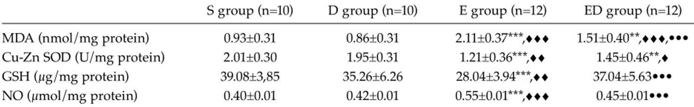
The biochemical parameters of the experimental groups were summarized in Table 2. There were no differences between the biochemical param-eters of the S and D groups. Brain tissue MDA levels of the E group were significantly higher than those of the S and D groups (p<0.05). The brain tissue GSH levels and Cu-Zn SOD activ-ity of the E group were significantly lower than those of the S and D groups (p<0.01). Brain tissue MDA levels of the ED group were sig-nificantly lower than the E group (p<0.05). GSH levels of the ED group were significantly higher
Table 1. Design of the experimental groups
0. hour 2. hour
S (Saline-treated group) (n=10) 2.6 ml/kg saline 2.6 ml/kg saline + 10 μg/kg saline
D (Darbepoetin-treated group) (n=10) 2.6 ml/kg saline 2.6ml/kg saline + 10 μg/kg darbepoetin solution*
E (Ethanol-administered group) (n=12) 2.6 ml/kg 2.6ml/kg 20% ethanol solution + 1 ml/kg saline
20% ethanol solution
ED (Ethanol-administered, 2.6 ml/kg 2.6 ml/kg 20% ethanol solution +
darbepoetin-treated group) (n=12) 20% ethanol solution 10 μg/kg darbepoetin solution*
* 1ml/kg darbepoetin solution includes 10 μg /kg darbepoetin.
Table 2. Results of biochemical paremeters in the experimental groups
S group (n=10) D group (n=10) E group (n=12) ED group (n=12)
MDA (nmol/mg protein) 0.93±0.31 0.86±0.31 2.11±0.37***,ggg 1.51±0.40**,ggg,•••
Cu-Zn SOD (U/mg protein) 2.01±0.30 1.95±0.31 1.21±0.36***,gg 1.45±0.46**,g
GSH (μg/mg protein) 39.08±3,85 35.26±6.26 28.04±3.94***,gg 37.04±5.63•••
NO (μmol/mg protein) 0.40±0.01 0.42±0.01 0.55±0.01***,ggg 0.45±0.01•••
• Significant differences between Sham group and other groups defined with *; (p<0.05)*, (p<0.01)**, (p<0.001)*** • Significant differences between D group and the other groups defined with g; (p<0.05)g, (p<0.01)gg, (p<0.001)ggg • Significant differences between E group and other groups defined with •; (p<0.05)•, (p<0.01)••, (p<0.001)•••
than those of the E group (p<0.001). NO levels of the ED group were significantly lower than those of the E group (p<0.001).
DISCUSSION
Oxidant effects of ethanol have been reported in many studies.[1,2] It has been shown that microsomal oxidation of ethanol generates reac-tive oxygen species, both in vivo and in vitro, via the inducible cytochrome P450 2E1 (CYP2E1) in the the brain.[14,15] Malondialdehyde is one of the products of lipid peroxidation. Lipid per-oxidation occurs when a free radical attacks the fatty-acid side chain of a phospholipid in the cellular membrane.[16] Oxidant and antioxidant status is vital for regulation of homeostasis. Reactive oxygen species (ROS), namely super-oxide and hydroxyl free radicals, together with hydrogen peroxide, are believed to be directly toxic, and ROS can initiate a free-radical-medi-ated chain reaction that causes additional organ damage.[17] Additionally, GSH, a tripeptide and SOD are involved in the antioxidant system and are important for the protection of the tis-sue from oxidative damage. Oxidized form of GSH is a dimer— GSSG, which is involved in the transport of certain amino acids, and is a coenzyme for various enzymes and protects against oxygen radicals and toxic compounds. GSH removes the toxic substances from the environment and protects the tissue from harm-ful substances after biotransformation. High GSH activity protects the cells from oxidative damage by inhibiting lipid peroxidation.[18] SOD that catalyzes the dismutation of superoxide to hydrogen peroxide catalyses the conversion of two O2 molecules into H2O2 and O2. SOD exists in mitochondrial (Mn-SOD) and cytoplasmic forms (Cu-Zn SOD).[19] In our study, MDA and NO levels increased significantly (p<0.001) and the antioxidant parameters (Cu-Zn SOD, GSH) decreased significantly (p<0.001) after ethanol administration.
NO is a free radical gas molecule, which is produced from L-arginine by the catalytic action of enzyme, nitric oxide synthesis (eNOS, iNOS, and nNOS).[20,21] NO is a key factor in a variety of physiological processes such as
neurotransmis-sion and regulation of blood vessel wall in phys-iological levels.[20,22] The role of NO seems to be controversial because in some models of inflam-mation it has been shown that tissue dysfunction or injury could occur after inhibition of NO.[23,24] It has been suggested that increased NO is asso-ciated with oxidative stress. It has been shown that glutamate and NO promote oxidative dam-age by reacting with superoxide anion to form the oxidant compound peroxynitrite.[23,25-27] High production of NO has been suggested as a cause of tissue injury under certain circumstances, may be through the generation of potent free radicals. Studies are not in consensus as to whether NO is cytotoxic or cytoprotective. It may act both as a cytotoxic and/or a cytoprotective agent and the main determinants are its concentration and the environment.[28] We observed that NO levels were parallel with lipid peroxidation levels. Darbepoetin-α treatment after ethanol admin-istration increased antioxidant response and decreased lipid peroxidation in our study.
Many different techniques and drugs are evaluated for the treatment of side effects of ethanol.[29] The data about the effects of darbepoetin-α on oxidative stress is limited. However, some protective effects of EPO against oxidative damage have been reported.[23,30] EPO treatment diminished lipid peroxidation levels and increased glutathione peroxidase activities significantly in brain tissue.[2,31,32] Antioxidant properties of darbepoetin-α in ethanol induced oxidative stress have been shown in our study. Darbepoetin-α protects brain tissue via its anti-oxidant activity.
Acknowledgement
This project was supported by İstanbul University Scientific Research Unit Funds No: 694.
REFERENCES
1. Kumral A, Tugyan K, Gonenc S, Genc K, Genc S, Sonmez U, et al. Protective effects of erythropoietin against ethanol-induced apoptotic neurodegenara-tion and oxidative stress in the developing C57BL/6 mouse brain. Brain Res Dev Brain Res 2005;160:146-56.
2. Masalkar PD, Abhang SA. Oxidative stress and antioxidant status in patients with alcoholic liver disease. Clin Chim Acta 2005;355:61-5.
Taurine restores ethanol-induced depletion of antiox-idants and attenuates oxidative stress in rat tissues. Amino Acids 2004;27:91-6.
4. Upadhya SC, Tirumalai PS, Boyd MR, Mori T, Ravindranath V. Cytochrome P4502E (CYP2E) in brain: constitutive expression, induction by ethanol and localization by fluorescence in situ hybridiza-tion. Arch Biochem Biophys 2000;373:23-34.
5. Oztürk E, Demirbilek S, Köroğlu A, But A, Begeç ZO, Gülec M, et al. Propofol and erythropoietin anti-oxidant properties in rat brain injured tissue. Prog Neuropsychopharmacol Biol Psychiatry 2008;32:81-6. 6. Belayev L, Khoutorova L, Zhao W, Vigdorchik A,
Belayev A, Busto R, et al. Neuroprotective effect of darbepoetin alfa, a novel recombinant erythropoi-etic protein, in focal cerebral ischemia in rats. Stroke 2005;36:1071-6.
7. Egrie JC, Browne JK. Development and character-ization of novel erythropoiesis stimulating protein (NESP). Br J Cancer 2001;84 Suppl 1:3-10.
8. Jelkmann W. Molecular biology of erythropoietin. Intern Med 2004;43:649-59.
9. Buege JA, Aust SD. Microsomal lipid peroxidation. Methods Enzymol 1978;52:302-10.
10. Sun Y, Oberley LW, Li Y. A simple method for clinical assay of superoxide dismutase. Clin Chem 1988;34:497-500.
11. Beutler E, Duron O, Kelly Bm. Improved method for the determination of blood glutathione. J Lab Clin Med 1963;61:882-8.
12. Lowry Oh, Rosebrough Nj, Farr Al, Randall Rj. Protein measurement with the Folin phenol reagent. J Biol Chem 1951;193:265-75.
13. Green LC, Wagner DA, Glogowski J, Skipper PL, Wishnok JS, Tannenbaum SR. Analysis of nitrate, nitrite, and [15N]nitrate in biological fluids. Anal Biochem 1982;126:131-8.
14. Lieber CS, Leo MA, Aleynik SI, Aleynik MK, DeCarli LM. Polyenylphosphatidylcholine decreases alcohol-induced oxidative stress in the baboon. Alcohol Clin Exp Res 1997;21:375-9.
15. Sun AY, Sun GY. Ethanol and oxidative mechanisms in the brain. J Biomed Sci 2001;8:37-43.
16. Koudelová J, Mourek J. The lipid peroxidation in various parts of the rat brain: effect of age, hypoxia and hyperoxia. Physiol Res 1994;43:169-73.
17. Ghoneim AI, Abdel-Naim AB, Khalifa AE, El-Denshary ES. Protective effects of curcumin against ischaemia/reperfusion insult in rat forebrain. Pharmacol Res 2002;46:273-9.
18. Kaplowitz N. Mechanisms of liver cell injury. J Hepatol 2000;32:39-47.
19. Nita DA, Nita V, Spulber S, Moldovan M, Popa DP,
Zagrean AM, et al. Oxidative damage following cere-bral ischemia depends on reperfusion - a biochemical study in rat. J Cell Mol Med 2001;5:163-70.
20. Ates O, Cayli S, Altinoz E, Gurses I, Yucel N, Sener M, et al. Neuroprotection by resveratrol against traumatic brain injury in rats. Mol Cell Biochem 2007;294:137-44.
21. Moncada S, Higgs A. The L-arginine-nitric oxide pathway. N Engl J Med 1993;329:2002-12.
22. Peunova N, Enikolopov G. Amplification of calcium-induced gene transcription by nitric oxide in neu-ronal cells. Nature 1993;364:450-3.
23. Yamasaki M, Mishima HK, Yamashita H, Kashiwagi K, Murata K, Minamoto A, et al. Neuroprotective effects of erythropoietin on glutamate and nitric oxide toxicity in primary cultured retinal ganglion cells. Brain Res 2005;1050:15-26.
24. Kubes P, McCafferty DM. Nitric oxide and intestinal inflammation. Am J Med 2000;109:150-8.
25. Beckman JS, Beckman TW, Chen J, Marshall PA, Freeman BA. Apparent hydroxyl radical production by peroxynitrite: implications for endothelial injury from nitric oxide and superoxide. Proc Natl Acad Sci U S A 1990;87:1620-4.
26. El-Remessy AB, Khalil IE, Matragoon S, Abou-Mohamed G, Tsai NJ, Roon P, et al. Neuroprotective effect of (-)Delta9-tetrahydrocannabinol and can-nabidiol in N-methyl-D-aspartate-induced retinal neurotoxicity: involvement of peroxynitrite. Am J Pathol 2003;163:1997-2008.
27. Gunasekar PG, Kanthasamy AG, Borowitz JL, Isom GE. NMDA receptor activation produces concur-rent generation of nitric oxide and reactive oxygen species: implication for cell death. J Neurochem 1995;65:2016-21.
28. Hunt NC, Goldin RD. Nitric oxide production by monocytes in alcoholic liver disease. J Hepatol 1992;14:146-50.
29. Baydas G, Tuzcu M. Protective effects of mela-tonin against ethanol-induced reactive gliosis in hip-pocampus and cortex of young and aged rats. Exp Neurol 2005;194:175-81.
30. Sakanaka M, Wen TC, Matsuda S, Masuda S, Morishita E, Nagao M, et al. In vivo evidence that erythropoietin protects neurons from ischemic dam-age. Proc Natl Acad Sci U S A 1998;95:4635-40. 31. Calapai G, Marciano MC, Corica F, Allegra A, Parisi
A, Frisina N, et al. Erythropoietin protects against brain ischemic injury by inhibition of nitric oxide formation. Eur J Pharmacol 2000;401:349-56.
32. Tabira T, Konishi Y, Gallyas F Jr. Neurotrophic effect of hematopoietic cytokines on cholinergic and other neurons in vitro. Int J Dev Neurosci 1995;13:241-52.