http://journals.tubitak.gov.tr/biology/ © TÜBİTAK
doi:10.3906/biy-1209-36
Stimulation of dendritic cells with vaccine and vaccine–antibody complex and effect on
immune response
İbrahim HATİPOĞLU1,2,*, İbrahim SÖĞÜT3, Duygu ERCAN1, Soner AKSU1, Hülya SİVAS2, Aynur BAŞALP1
1TÜBİTAK Marmara Research Center, Genetic Engineering and Biotechnology Institute, Gebze, Kocaeli, Turkey 2Department of Biology, Faculty of Science, Anadolu University, Eskişehir, Turkey
3Vocational School of Health Services, İstanbul Bilim University, İstanbul, Turkey
1. Introduction
Dendritic cells (DCs) have a critical role for stimulating innate and adaptive immunity. When DCs are present in peripheral tissue, they encounter and engulf the infectious agents, and they start antigen processing, mature, and migrate to lymph nodes for stimulating naive T cells. T cell activation, especially that of the cytotoxic T cell (Tc), is crucial to overcome the immune tolerance in chronic diseases and cancer. Therefore, DC vaccines are an alternative approach for cell-based immunotherapy (1–5). DCs generally have been derived from peripheral mononuclear cells of human blood or spleen or bone marrow in animal models (6–8), and different methods can be used for loading them. Despite a large number of DC vaccine studies, only one study was approved by the Food and Drug Administration (FDA), which consists of not only DCs but also other antigen presenting cells (9).
Studies for treatment of chronic hepatitis B constitute a crucial part of DC vaccine research. Despite the use of the commercial prophylactic vaccine since 1981, hepatitis B virus (HBV) infection is still a serious health problem
all over the world. The World Health Organization (WHO) indicates that 370 million people are chronically infected with HBV worldwide and each year 1 million people die because of HBV-related diseases (10). Type I interferons and nucleotide analogues have been used for the treatment of chronic HBV infection, but the methods of interest are expensive and their success rate is rather low. Furthermore, nucleotide analogues may lead to the development of resistant types of HBV (11–14). Therefore, the DC vaccines are thought to be an alternative method for treatment and have been used in clinical and animal models. In DC vaccine studies, immature DCs have been loaded with various concentrations of hepatitis B surface antigen (HBsAg), hepatitis B virus c antigen (HBcAg), or complexed other proteins. The experiments have shown that DC vaccines evoke an effective immune response, but the stimulation of immune response in hepatitis B virus e antigen (HBeAg) positive patients is difficult. Thus, more effective loading methods are necessary (15–19).
In the present study, our aim is to overcome HBV tolerance in chronic hepatitis B transgenic (HBV-tg)
Abstract: Dendritic cell (DC) vaccines are a promising and potent therapeutic tool for chronic diseases, autoimmune diseases, and
cancer because of the unique ability of DCs to stimulate T cells. The challenge of DC vaccines is to find an effective form for antigen presentation. Although pure antigens, antigen complexes, plasmids, and mRNA have been used in different studies, no proper application to overcome this problem has been found yet. In this study, we investigated the eligibility of a commercial hepatitis B virus (HBV) vaccine or a vaccine–monoclonal antibody complex for antigen loading of DCs for a therapeutic purpose. DCs were derived from the bone marrow of transgenic hepatitis B (HBV-tg) mice using a granulocyte macrophage-colony stimulating factor and interleukin-4, and then loaded with a commercial HBV vaccine (containing hepatitis B virus surface antigens and aluminum hydroxide adjuvant) or a vaccine–antibody complex. HBV-tg mice were immunized with the vaccine and vaccine–antibody loaded DCs. Optimum HBV vaccine concentration and loading time were determined by 2-(4-iodophenyl)-3-(4-nitrophenyl)-5-(2,4-disulfophenyl)-2H-tetrazolium (WST-1) methods. Therapeutic effects of vaccine–antibody loaded DCs were determined by the evaluation of antibody response and hepatitis B surface expression levels in HBV-tg mice. Our results showed that commercial HBV vaccine loaded DCs induced humoral response in HBV-tg mice but had no effect on cellular immunity.
Key words: Dendritic cell vaccine, hepatitis B virus, immunotherapy
Received: 01.10.2012 Accepted: 10.01.2013 Published Online: 30.07.2013 Printed: 26.08.2013
mice by using a vaccine or vaccine–antibody complex loaded DCs. Akbar et al. showed that commercial HBV vaccines can be used as a source of HBsAg (20); however, the vaccine–antibody complex has not been tested before. The positive effect of antigen–antibody complexes on immunity has been proven by many laboratories. The Fc region of the antibody from the vaccine–antibody complex binds Fc receptors on DCs and enhances antigen uptake, which also improves antigen presentation and Tc response (21–24).
In this study, optimum vaccine dose, incubation time, and DC number were determined by 2-(4-iodophenyl)-3-(4-nitrophenyl)-5-(2,4-disulfophenyl)-2H-tetrazolium (WST-1) cell viability test. DCs were loaded with optimum vaccine or vaccine–antibody complex and HBV-tg mice were immunized twice, intraperitoneally. Therapeutic effects of DC vaccines were determined by investigating humoral and cellular immune responses. Results have shown that vaccine loaded DCs significantly induce anti-HBsAg response but have no effect on cellular immunity. 2. Materials and methods
2.1. Mice
HBV-tg mice were prepared at the Genetic Engineering and Biotechnology Institute of the TÜBİTAK Marmara Research Center as reported before (25), and mice of 6 to 10 weeks old were used in this study. HBsAg is constitutively expressed in transgenic mice and liver HBV DNA, and HBsAg was detected in the sera and livers of HBV-tg mice. All mice were housed under specific pathogen free conditions. Animal experiments were approved by the Ethics Committee of the Genetic Engineering and Biotechnology Institute of the TÜBİTAK Marmara Research Center.
2.2. Generation of dendritic cells and immunization Bone marrow derived DCs were generated as described previously with minor modifications (7). Briefly, bone marrow cells were removed from the femurs of HBV-tg mice and erythrocytes were depleted by using ammonium chloride solution. After counting, the cells (1 × 10⁶/
mL) were seeded onto 10-cm petri dishes in RPMI 1640 medium (Gibco Life Sciences, USA) containing 10 mM N-2-hydroxyethylpiperazine-N’-2-ethanesulfonic acid (HEPES), 10 µg/mL gentamicin, 100 U/mL penicillin, 100 µg/mL streptomycin, 10% fatal bovine serum (FBS), 1% MEM nonessential amino acids, 50 µM β-mercaptoethanol, 20 ng/mL recombinant murine granulocyte/macrophage-colony stimulating factor (mGM-CSF, Gibco Life Sciences), and mouse interleukin-4 (mIL-4, 200 IU/ml, Gibco) and incubated. Every 3 days, two-thirds of the medium was replaced with fresh medium. On day 6, cells were harvested by treating with versene solution (Invitrogen, USA) and DCs were seeded into 6-well culture plates as 1.5 × 10⁶ cells per well, and 100 U/mL rTNF-α was added
in addition to mGM-CSF and IL-4. Twenty-four hours later, the DCs were activated by adding 125 µL vaccine, 125 µL vaccine–antibody complex, and monoclonal antibody (MAb) (5 µg/3.5 mL). Lipopolysaccharide (100 ng/mL) and CpG 1826 (1 µg/well) were also added to each well. Cells were harvested 5 h later and 5 × 10⁵ DCs were
prepared for immunization in 100 µL PBS. Four mice were used in each group and immunizations were performed intraperitoneally twice at weekly intervals with a DC vaccine.
2.3. Flow cytometric analysis
Cells were harvested from petri dishes onday 6 and washed with PBS. The phenotype of the cells was evaluated using fluorochrome-conjugated antibodies, anti-CD11c-PE and anti-CD11b IFTC (BD, USA), and relevant isotype controls were always used. Cells were stained with labeled antibodies for 20 min at 4 °C and washed with cold PBS, and were then suspended in PBS. Data was acquired using the FACScan instrument (BD Biosciences, USA) and analyzed using the Cellquest program.
2.4. Generation of vaccine–antibody complex
Commercial HBV vaccine (Gen Hevac), which contains 40 µg/mL HBsAg, was incubated with anti-HBsAg monoclonal antibody (2G3) at a 2:1 (m/m) ratio for 1 h at 37 °C, then incubated at room temperature overnight. Anti-HBsAg MAb was produced in our laboratory with hybridoma technology as reported previously (26). 2.5. WST-1 assay
This assay is based on the cleavage of the WST salt to soluble formazan by mitochondrial dehydrogenases in viable cells. On day 6, immature dendritic cells were seeded as 5 × 10⁴
or 2.5 × 10⁴ cells per well of 96-well plates in 100 µL. After
24 h of incubation, DCs were loaded with 3.6 µL (144 ng HBsAg) or 0.72 µL (28 ng HBsAg) vaccine, vaccine– antibody complex, and hydrogen peroxide (10 µL/well) for 5, 24, and 48 h at 37 °C. At the end of the incubation, 10 µL/well WST-1 (Roche, Germany) was added to each well, and then the plates were incubated for an additional 2 h at 37 °C. The absorbance was measured at 450 nm.
2.6. Serological assays
Antibody response to HBsAg was measured by indirect ELISA. Microtiter ELISA plates were coated with HBsAg (Fitzgerald, USA; 400 ng/mL) in PBS at 4 °C by overnight incubation. After washing and blocking, the diluted mouse serum was added to each well and incubated for 1 h at 37 °C. The wells were washed and bound antibody was detected with alkaline phosphate conjugated goat antimouse polyvalent antibody (Sigma, at 1/1000 dilution) by incubation at 37 °C for 1 h. Plates were then washed and para-nitrophenylphosphate (PNPP) at 1 mg/mL in substrate buffer (0.1 M glycine, pH 10.4; 1 mM ZnCl2; and 1 mM MgCl2) was added, and the absorbance at 405 nm was measured.
2.7. RNA isolation and quantitative RT-PCR analysis Liver tissues (50–100 mg) were collected from animals and used for RNA isolation. After decapitation, tissues were immediately collected, shock-frozen in liquid nitrogen, and stored at –80 °C until preparation of RNA samples. Total RNA from liver tissues of each mouse was extracted using TRIzol reagent (Invitrogen, USA), chloroform, and isopropanol. cDNA synthesis was carried out using 1 µg whole RNA sample with a Roche Transcriptor High Fidelity cDNA Synthesis Kit according to the manufacturer’s protocol. The primer sequence for the HBs gene of the HBV virus was designed using the Primer 3 program as listed in Table 1. Real-time RT-PCR was performed using the QIAGEN SYBR Green Supermix Kit (QIAGEN, USA) for the selected gene in a Bio-Rad iQ5 thermal cycler (Bio-Rad Laboratories, USA) according to the manufacturer’s instructions. All data were analyzed using the Relative Expression Software Tool (REST 2009 Software, 12/2009, http://rest.gene-quantification.info). The standard REST 2009 algorithm calculates efficiency using the slope from the best-fit standard curve as follows: E = 10–1/slope – 1. The slope value derived from Ct values
of serially diluted samples was automatically calculated by Bio-Rad iQ5 thermal cycler. The estimations of each sample’s expression ratio, an intermediate absolute concentration value, was calculated using the following formula: concentration = Etarget∆Cp target (MEAN control – MEAN sample).
The relative expression of each target gene was calculated by the ratio of the concentration of the target gene to the geometric mean of all reference gene concentrations. 2.8. Statistical analysis
Differences between the groups were analyzed by one-way ANOVA (Tukey’s test) using SPSS 15.0 (IBM, USA). Values were considered to be statistically significant when P < 0.05.
3. Results
3.1. Dendritic cell generation
DCs were derived from mouse bone marrow using GM-CSF and IL-4. On day 6, flow cytometric analyses showed that the frequency of DCs expressing CD 11c+ CD11b+ was
about 80%–85%.
3.2. Determination of the viability of DCs loaded with vaccine and vaccine–antibody complex
To determine the optimum concentration of vaccine for loading of DCs, the WST-1 test was performed. For this,
5 × 10⁴ or 2.5 × 10⁴ DCs were treated with 3.6 µL or 0.72
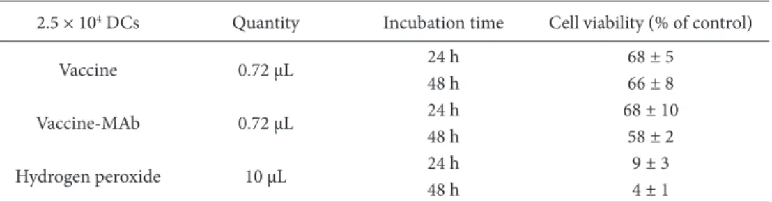
µL vaccine, vaccine–antibody complex, and antibody for 5 h, 24 h, and 48 h. The control group was not treated with a vaccine or antibody. After exposure of all wells, 10 µL of the WST-1 solution was added and incubated for 2 h at 37 °C. The optical density of the solubilized formazan product was determined using a spectrophotometer with a 450-nm and 695-nm filter as a reference. The control group was accepted as 100% live, and cell viability of other groups was determined using the proportion of their absorbance value to the control group. Tables 2 and 3 show the viability of DCs after 24 h and 48 h, treated with 3.6 µL containing vaccine or vaccine–antibody complex, respectively. It was found that the viability of cells decreased by about 50% with respect to the cell number in these groups. Tables 4 and 5 show the viability of DCs treated with 0.72 µL HBsAg containing vaccine and vaccine antibody for 24 h and 48 h. We found that this amount of HBsAg is less cytotoxic than 3.6 µL HBsAg, but it contains nearly less than 5 times the HBsAg. For increasing the percentage of loaded DC, 5 × 10⁴ DCs were loaded with 3.6 µL HBsAg containing vaccine
or vaccine–antibody complex for 5 h, and cell viability was found at 77 ± 7% and 74 ± 13% respectively (Table 6). This experimental protocol was chosen and adapted for vaccine or vaccine-antibody complex immunizations in HB-tg mice.
3.3. Detection of immune response in DC immunized mice To determine the effect of the HBV vaccine, HBV-tg mice (n = 4) were immunized with vaccine, vaccine– antibody, and antibody loaded and unloaded DCs 2 times, intraperitoneally. The concentration of loaded vaccine and vaccine–antibody complex was determined according to WST-1 test results. The WST-1 test was performed in a 96-well plate, but DCs were loaded in 6-well plates for immunization and DC number and the vaccine and vaccine–antibody complex concentration were increased in accordance with the volume of medium in the well.
HBV-tg mice sera were collected 3 weeks after the second immunization. An ELISA test was performed to detect anti-HBsAg levels in mice. We found that all DC immunized mice had an induced anti-HBsAg response, but vaccine loaded DCs induced better antibody response compared to others. The anti-HBsAg level in this group was nearly 3 times higher than in control groups (Figure).
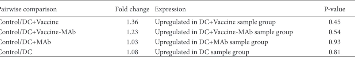
As is generally accepted, the liver is the primary site of HBV infection (27). A decrease in HBs gene expression
Table 1. Primer sequences for RT-qPCR.
Primers Nucleotide sequence Expected size (bp)
was expected after stimulation of T cell response in the liver. HBs gene expression level in HBV-tg mice liver was determined by RT-qPCR, but there was no difference between the groups (Table 7).
4. Discussion
Discovery of the DC immune effect in the early 1970s was the milestone in DC studies (2). Following studies have shown that DCs have an important role in innate and
Table 2. Incubation of 5 × 10⁴ DCs for 24 and 48 h with 3.6 µL vaccine or vaccine–antibody complex;
10 µL hydrogen peroxide was used as a positive control. The experiments were performed in triplicate. 5 × 10⁴ DCs Quantity Incubation time Cell viability (% of control)
Vaccine 3.6 µL 24 h48 h 49 ± 348 ± 6 Vaccine-MAb 3.6 µL 24 h48 h 49 ± 643 ± 6
Hydrogen peroxide 10 µL 24 h 9 ± 5
48 h 5 ± 1
Table 3. Incubation of 5 × 10⁴ DCs for 24 and 48 h with 0.72 µL vaccine or vaccine–antibody complex;
10 µL hydrogen peroxide was used as a positive control. The experiments were performed in triplicate. 5 × 10⁴ DCs Quantity Incubation time Cell viability (% of control)
Vaccine 0.72 µL 24 h48 h 93 ± 365 ± 6
Vaccine-MAb 0.72 µL 24 h 94 ± 6
48 h 74 ± 6
Hydrogen peroxide 10 µL 24 h 9 ± 5
48 h 5 ± 1
Table 4. Incubation of 2.5 × 10⁴ DCs for 24 and 48 h with 3.6 µL vaccine or vaccine–antibody complex;
10 µL hydrogen peroxide was used as a positive control. The experiments were performed in triplicate. 2.5 × 10⁴ DCs Quantity Incubation time Cell viability (% of control)
Vaccine 3.6 µL 24 h 53 ± 11
48 h 58 ± 13
Vaccine-MAb 3.6 µL 24 h 54 ± 17
48 h 55 ± 8
Hydrogen peroxide 10 µL 24 h48 h 9 ± 34 ± 1
Table 5. Incubation of 2.5 × 10⁴ DCs for 24 and 48 h with 0.72 µL HBsAg containing vaccine or vaccine–
antibody complex; 10 µL hydrogen peroxide was used as a positive control. The experiments were performed in triplicate.
2.5 × 10⁴ DCs Quantity Incubation time Cell viability (% of control)
Vaccine 0.72 µL 24 h48 h 68 ± 566 ± 8
Vaccine-MAb 0.72 µL 24 h 68 ± 10
48 h 58 ± 2
Hydrogen peroxide 10 µL 24 h 9 ± 3
adaptive immune response, especially in T cell response. DC vaccines are promising alternative therapeutic methods to overcome immune tolerance. A large number of DC vaccine studies have been performed for more than 30 years, but there are no exact protocols and more studies are necessary. The method for loading DCs is a special issue in DC vaccine studies. Purified antigens, tumor lysate, viral vectors, mRNA, or DNA have been used for loading the DCs (17,28–31).
In the current study, the HBV vaccine, which consists of HBsAg and aluminum hydroxide adjuvant, was used as an alternative source for HBsAg. The vaccine was complexed with anti-HBsAg for enhancing loading success. We determined the optimum loading concentrations of vaccine for DCs by WST-1 cell viability assay due to the potential cytotoxic effect of the vaccine (32–34). We found that 125 µL vaccine was the optimal dose for 5 h of incubation. After deciding the optimal vaccine concentration, DCs were loaded with vaccine or vaccine–antibody complex and HBV-tg mice were immunized. Before our study, Akbar et al. showed that HBV vaccine loaded human peripheral blood DCs induce anti-HBsAg response and T cell proliferation in HBV vaccine nonresponders (20), and Flach et al. immunized alum pretreated DCs to C57BL/6 mice along with OVA and induced anti-OVA immune response (35). However, in our study, vaccine loaded DCs were used with a therapeutic purpose in HBV-tg mice for the first time.
After the second immunization, anti-HBsAg response was followed for 3 weeks in the groups. Only vaccine loaded DCs induced significant humoral response. This result is correlated with those of Akbar et al. and Flach et al. (20,35). However, we did not find any significant antibody response in mice immunized with the vaccine– antibody complex loaded DCs. Alum binds lipids of DC
(35). This mechanism may inhibit Fc receptor mediated phagocytosis and effects of the vaccine–antibody complex on humoral immune response.
Three weeks after the second immunization, decrease of HBs gene expression was not observed. These results can be related to T cell stimulation capacity of the vaccine or vaccine-antibody loaded DCs. Although aluminum adjuvant generally induces a protective immune response, the mechanism of action of aluminum adjuvant on the immune system and DCs is still being investigated (35– 39). While some studies indicated that alum adjuvant did not induce expression of CD40, CD80, CD86, or major histocompatibility complex class II, others showed that expression of CD86 receptor was induced (37,38). In our study, we found that there were no differences in the
Figure 1. Humoral immune response. Antibody response in sera
of HBV-tg mice immunized with vaccine, vaccine–antibody, and antibody loaded and nonloaded DCs. DC+Vaccine: vaccine loaded DCs, DC+Vaccine-MAb: vaccine–antibody complex loaded DCs, DC+MAb: antibody loaded DCs, DC: nonloaded DCs. The experiments were performed in quadruplicate and results are shown as mean ± standard deviation. *: P < 0.05
Table 6. Incubation of 5 × 10⁴ DCs for 24 and 48 h with 3.6 µL vaccine or vaccine–antibody complex.
The experiments were performed in triplicate.
5 × 10⁴ DCs Quantity Incubation time Cell viability (% of control)
Vaccine 3.6 µL 5 h 77 ± 7 Vaccine-MAb 3.6 µL 5 h 74 ± 13 0.0 0.2 0.4 0.6 0.8 1.0 1.2 Control DC+
Vaccine DC+Vaccine-MAb DC+MAb DC
OD 405 nm
Week 1 Week 2 Week 3 *
*
Table 7. HBs gene expression profile 3 weeks after the last immunization in DC immunized groups. The experiments were
performed in quadruplicate and results are shown as mean ± standard deviation.
Pairwise comparison Fold change Expression P-value
Control/DC+Vaccine 1.36 Upregulated in DC+Vaccine sample group 0.45 Control/DC+Vaccine-MAb 1.23 Upregulated in DC+Vaccine-MAb sample group 0.54 Control/DC+MAb 1.03 Upregulated in DC+MAb sample group 0.93
expression levels of CD40, CD80, and CD86 (data not shown).
In our study, we showed that HBV vaccine loaded DCs induced humoral response. We also showed that depending on the concentration of vaccine and incubation time, the vaccine might have a cytotoxic effect on DCs. We believe that DC vaccine studies will be more important in the future and that the major proof of the idea will lie in phase studies.
Acknowledgments
This work was supported by a grant from a scientific research project of Anadolu University via contract 1002F59. We would like to thank Şakir Sekmen and Gazi Turgut for their excellent technical assistance and Melis Savaşan Söğüt for editing the manuscript.
References
1. Steinman RM, Cohn ZA. Identification of a novel cell type in peripheral lymphoid organs of mice. I. Morphology, quantitation, tissue distribution. J Exp Med 137: 1142–62, 1973.
2. Banchereau J, Steinman RM. Dendritic cells and the control of immunity. Nature 392: 245–52, 1998.
3. Ardavin C, Martinez del Hoyo G, Martin P et al. Origin and differentiation of dendritic cells. Trends Immunol 22: 691–700, 2001.
4. Wykes M, Pombo A, Jenkins C et al. Dendritic cells interact directly with naive B lymphocytes to transfer antigen and initiate class switching in a primary T-dependent response. J Immunol 161: 1313–19, 1998.
5. Cella M, Scheidegger D, Palmer-Lehmann K et al. Ligation of CD40 on dendritic cells triggers production of high levels of interleukin-12 and enhances T cell stimulatory capacity: T-T help via APC activation. J Exp Med 184: 747–52, 1996. 6. Brasel K, De Smedt T, Smith JL et al. Generation of murine
dendritic cells from flt3-ligand-supplemented bone marrow cultures. Blood 96: 3029–39, 2000.
7. Shimizu Y, Guidotti LG, Fowler P et al. Dendritic cell immunization breaks cytotoxic T lymphocyte tolerance in hepatitis B virus transgenic mice. J Immunol 161: 4520–29, 1998. 8. Spranger S, Javorovic M, Burdek M et al. Generation of Th1-polarizing dendritic cells using the TLR7/8 agonist CL075. J Immunol 185: 738–47, 2010.
9. FDA. Approval Letter – Provenge, 2010. Food and Drug Administration. Washington DC; 2010. Available at http://www. fda.gov/BiologicsBloodVaccines/CellularGeneTherapyProducts/ ApprovedProducts/ucm210215.htm
10. WHO. Hepatitis B vaccines WHO position paper, 2006. World Health Organization. Geneva; 2006. Available at http://www. who.int/immunization/topics/WHO_position_paper_HepB. pdf.
11. WHO. Hepatitis B, 2000. World Health Organization. Geneva; 2000. Available at http://www.who.int/mediacentre/factsheets/ fs204/en/.
12. Michel ML, Deng Q, Mancini-Bourgine M. Therapeutic vaccines and immune-based therapies for the treatment of chronic hepatitis B: perspectives and challenges. J Hepatol 54: 1286–96, 2011.
13. Dienstag JL. Hepatitis B virus infection. N Engl J Med 359: 1486–1500, 2008.
14. Zoulim F, Locarnini S. Hepatitis B virus resistance to nucleos(t) ide analogues. Gastroenterology 137: 1593–1608, 2009. 15. Furukawa S, Akbar SMF, Hasabe A et al. Production of
hepatitis B surface antigen-pulsed dendritic cells from immunosuppressed murine hepatitis B virus carrier: evaluation of immunogenenicity of antigen-pulsed dendritic cells in vivo. Immunobiology 209: 551–57, 2004.
16. Jiang WZ, Fan Y, Liu Y et al. Therapeutic potential of dendritic cell-based immunization against HBV in transgenic mice. Antivir Res 77: 50–5, 2008.
17. Luo J, Li J, Chen RL et al. Autologus dendritic cell vaccine for chronic hepatitis B carriers: a pilot, open label, clinical trial in human volunteers. Vaccine 28: 2497–504, 2010.
18. Lou Y, Liu C, Kim GJ et al. Plasmacytoid dendritic cells synergize with myeloid dendritic cells in the induction of antigen-specific antitumor immune responses. J Immunol 178: 1534–41, 2007. 19. Chen W, Zhang Z, Shi M et al. Activated plasmacytoid dendritic
cells act synergistically with hepatitis B core antigen-pulsed monocte-derived dendritic cells in the induction of hepatitis B virus-specific CD8 cell response. Clin Immunol 129: 295–303, 2008.
20. Akbar SMF, Furukawa S, Yoshida O et al. Induction of anti-HBs in HB vaccine nonresponders in vivo by hepatitis B surface antigen-pulsed blood dendritic cells. J Hepatol 47: 60–6, 2007. 21. Randall RE, Young DF, Southern JA. Immunization with solid
matrix-antibody-antigen complexes containing surface or internal virus structural proteins protects mice from infection with the paramyxovirus, simian virus 5. J Gen Virol 69: 2517– 26, 1988.
22. Yao X, Zheng B, Zhou J et al. Therapeutic effect of hepatitis B surface antigen-antibody complex is associated with cytolytic and non-cytolytic immune responses in hepatitis B patients. Vaccine 25: 1771–79, 2007.
23. Wen YM, Wu XH, Hu DC et al. Hepatitis B vaccine and anti-HBs complex as approach for vaccine therapy. Lancet 345: 1575–76, 1995.
24. Wen Y. Antigen–antibody immunogenic complex: promising novel vaccines for microbial persistent infections. Expert Opin Biol Th 9: 285–91, 2009.
25. Bagis H, Arat S, Mercan HO et al. Stable transmission and expression of the hepatitis B virus total genome in hybrid transgenic mice until F10 generation. J Exp Zool Part A 305: 420–27, 2006.
26. Başalp A, Yücel F. Development of mouse hybridomas by fusion of myeloma cells with lymphocytes derived from spleen, lymph node and bone marrow. Hybridoma Hybridom 22: 329– 31, 2003.
27. Guidotti LG, Chisari FV. Immunobiology and pathogenesis of viral hepatitis. Annu Rev Pathol-Mech 1: 23–61, 2006. 28. Asavaroengchai W, Kotera Y, Mulé JJ. Tumor lysate-pulsed
dendritic cells can elicit an effective antitumor immune response during early lymphoid recovery. P Natl Acad Sci USA 99: 931–36, 2002.
29. You H, Liu Y, Cong M et al. HBV genes induce cytotoxic T-lymphocyte response upon adeno-associated virus (AAV) vector delivery into dendritic cells. J Viral Hepatitis 13: 605–12, 2006.
30. Lesterhuis WJ, De Vries IJ, Schreibelt G et al. Immunogenicity of dendritic cells pulsed with CEA peptide or transfected with CEA mRNA for vaccination of colorectal cancer patients. Anticancer Res 30: 5091–97, 2010.
31. Steele JC, Rao A, Marsden JR et al. Phase I/II trial of a dendritic cell vaccine transfected with DNA encoding melan A and gp100 for patients with metastatic melanoma. Gene Ther 18: 584–93, 2011.
32. Bishop NJ, Morley R, Day JP et al. Aluminum neuro toxicity in preterm infants receiving intravenous-feeding solutions. N Engl J Med 336: 1557–1561, 1997.
33. Wang M, Chen JT, Ruan DY et al. The influence of develop-mental period of aluminum exposure on synaptic plasticity in the adult rat dentate gyrus in vivo. Neuroscience 113: 411–419, 2002.
34. Tomljenovic L, Shaw CA. Aluminum vaccine adjuvants: are they safe? Curr Med Chem 18: 2630–37, 2011.
35. Flach TL, Ng G, Hari A et al. Alum interaction with dendritic cell membrane lipids is essential for its adjuvanticity. Nat Med 17: 479–87, 2002.
36. Glenny AT, Pope CG, Waddington H et al. Immunological notes: XVII–XXIV. J Pathol Bacteriol 29: 31–40, 1926. 37. Marrack P, McKee AS, Munks MW. Towards an understanding
of the adjuvant action of aluminium. Nat Rev Immunol 9: 287– 93, 2009.
38. Sun H, Pollock KG, Brewer JM. Analysis of the role of vaccine adjuvants in modulating dendritic cell activation and antigen presentation in vitro. Vaccine 21: 849–55, 2003.
39. Morefield GL, Sokolovska A, Jiang D et al. Role of aluminum-containing adjuvants in antigen internalization by dendritic cells in vitro. Vaccine 23: 1588–95, 2005.