ORIGINAL ARTICLE
Argon laser-assisted hypotony model in the rabbit
Gokhan Gurelik1&Sabahattin Sul2&Pınar Uyar Gocun3&Safak Korkmaz4&Cemal Ozsaygili5
Received: 24 April 2018 / Accepted: 21 June 2018 / Published online: 29 June 2018 # Springer-Verlag London Ltd., part of Springer Nature 2018
Abstract
To investigate whether ocular hypotony formation with 360 degrees endocyclophotocoagulation is possible. Twelve male New Zealand White rabbits were used. Entire ciliary body epithelium was destructed with green laser photocoagulation after pars plana lensectomy and anterior vitrectomy in six rabbits. Endocyclophotocoagulation was not performed to the remaining six rabbits (control group). Intraocular pressure (IOP) was measured preoperatively and followed up everyday in the first week and weekly until the end of month one. All of the rabbits were sacrificed and ciliary bodies were left for gross and light microscopic examination. Mean baseline IOPs were similar in laser and non-laser group (14.8 ± 1.4 (range 12.2–17.3) vs 14.4 ± 1.4 (range 12.2–15.9), p = 0.650). Mean IOP was 6.6 ± 0.45 mmHg (range 5.9–7.1) in the laser group and 11.5 ± 1.2 mmHg (range 10.2– 13.4) in the non-laser group in postoperative day 1. IOP was below 4 mmHg in all eyes on the second day and after in laser group. In the macroscopic evaluation, the entire ciliary body had a white (loss of pigmentation) and atrophic appearance in all of the eyes in the laser-treated group compared to non-laser group. In the laser group, light microscopic examination demonstrated a severe 360 degrees disruption of ciliary processes. Ciliary processes were covered with fibrin exudation consisting of fibroblasts. There was a mild inflammation with disruption or atrophy of ciliary body epithelium with cystic vacuolar degeneration. Three hundred sixty degrees endocyclophotocoagulation yielded severe ciliary epithelium damage. IOP reduction started very early and con-tinued in hypotonic levels during follow up period.
Keywords Ciliary body . Hypotony . Argon laser . Endocyclophotocoagulation . Intraocular pressure
Introduction
Chronic hypotony is defined as intarocular pressure (IOP) less than 6 mmHg. Increased uveoscleral outflow and reduced aqueous production are the two mechanisms of chronic hy-potony development [1]. Several animal studies have been designed to better delineate the mechanism and identify the treatment modalities of hypotony [2–5]. The most challenging
prob lem is treating red uc ed aqueous product ion. Inflammation, traction of non-pigmented ciliary epithelium due to epiciliary membrane, ciliary epithelium atrophy, and vascular compromise are the reasons of decreased aqueous production [6]. Hypotony may reverse after removal of epiciliary membrane traction with surgery [7]. However, sur-gery would be inadequate if the ciliary body is atrophic. Therefore, there is still need for detailed studies to increase the identification and management strategies of ciliary body atrophy.
Cyclophotocoagulation have been used to treat recalcitrant glaucoma to the surgical interventions. Cyclophotocoagula-tion cause the disrupCyclophotocoagula-tion and atrophy of ciliary body epitheli-um [8]. Transscleral or endocyclophotocoagulation are the techniques to ablate ciliary body processes [9–11]. Endocyclophotocoagulation has been defined as a more re-fined procedure that gives a chance to directly visualize the ciliary body.
The aim of this study is whether formation of chronic hy-potony is possible with the application of 360 degrees endocyclophotocoagulation of ciliary body epithelium.
* Sabahattin Sul [email protected]
1
Department of Ophthalmology, Gazi University Medical School, 48000 Ankara, Turkey
2
Department of Ophthalmology, Muğla Sıtkı Koçman University, Muğla, Turkey
3 Department of Pathology, Gazi University Medical School,
Ankara, Turkey
4
Department of Ophthalmology, Düzce State Hospital, Düzce, Turkey
5
Department of Ophthalmology, Kayseri Research and Training Hospital, Kayseri, Turkey
Lasers in Medical Science(2019) 34:11–14
https://doi.org/10.1007/s10103-018-2570-1
CrossMark
Materials and methods
Twelve male New Zealand White rabbits were used (age 3 months, average weight 12 kg). The study was approved by the animal research ethics board of the Medical School of Gazi University. The Principles of Laboratory Animal Care (NIH publication No. 8623, revised 1985) and the ARVO Statement for the Use of Animals in Research were followed. Rabbits were deeply anesthetized with intramuscular injection of 35 mg/kg ketamine and 5 mg/kg xylazine. Conjunctiva was opened with Westcott scissors at upper temporal, upper nasal and lower temporal sites and three sclerotomies (upper tem-poral, upper nasal and lower temporal) 2 mm from limbus were opened with 20 gauge microvitreoretinal blade (Fig. 1a). Pars plana lensectomy and anterior vitrectomy (DORC Associate, Dutch Ophthalmic, USA) were performed only to right eyes of rabbits. A 20 gauge standart angled green laser probe (Iridex, Mountain View, CA) was inserted through the both upper sclerotomies to facilitate the laser application to the ciliary body. Continuous wave green color laser (532 nm) light was used. Treatment power and duration of the laser beam were 1000 mW and 0.2 s with an estimated 500μm spot
size. Three hundred sixty degrees endocyclophotocoagulation was performed to the ciliary bodies of the right eyes of six rabbits with scleral indentation (Fig.1b, c). The power settings were adjusted and yielded to obtain a white concave burn, on the targeted part of the tissue. Microbubbles and pigment dis-persion were observed during treatment. Entire ciliary body epithelium was destructed with laser photocoagulation.
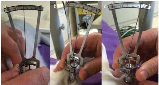
Endocyclophotocoagulation was not performed to the re-maining six rabbits which were defined as control group. Sclerotomy sites were sutured with 7/0 prolen suture in a watertight manner. IOP was measured preoperatively and followed up everyday in the first week and weekly until the end of month one. IOP was measured with Schiotz tonometer with a 5.5-g weight (Fig.2a). Scale readings were converted to mmHg. We could not measure aqueous production before and after laser procedure due to inadequate infrastructure. All of the rabbits were sacrificed with an overdose of xylazine and eyes were enucluated at the end of 1 month. The eyes were fixed for 1 week in 4% formaldehyde solution for macroscop-ic and histopathologmacroscop-ic examinations. Posterior portion of the globe was cut away after fixation procedure. The remaining eye cup including ciliary body was left for gross examination.
Fig. 1 Pars plana lensectomy, anterior vitrectomy and endolaser application to the right eye of a rabbit.a Three-port pars plana lensectomy. b Ciliary body visualization with scleral depression.c Endolaser application through the sclerotomy
Fig. 2 Intraocular pressure (IOP) measurement with schiotz tonometer.a Preoperative IOP measurement.b Postoperative IOP measurement of laser treated eye (day 1).c. Postoperative IOP measurement 4 weeks after laser treatment
12 Lasers Med Sci (2019) 34:11–14
Three hundred sixty degrees small slices including only ciliary body were done and stained with hematoxylin and eosin for light microscopy examination. The sections were blindly ex-amined with the light microscope Olympus BX51 (Japan) by a pathologist.
Results
In the immediate postoperative period, there was no fluid leakage from the wound sites in all of the eyes. Mean baseline IOPs were similar in laser and non-laser group (14.8 ± 1.4 (range 12.2–17.3) vs 14.4 ± 1.4 (range 12.2–15.9), p = 0.650) (Fig. 2a). Mean IOP was 6.6 ± 0.45 mmHg (range 5.9–7.1) in the laser group and 11.5 ± 1.2 mmHg (range 10.2–13.4) in the non-laser group in postoperative day 1 (Fig.2b). IOP was within normal range (11.2–14.6 mmHg)) in the non-laser group, however, below 4 mmHg in all eyes on the second day and after in laser group (Fig.2c).
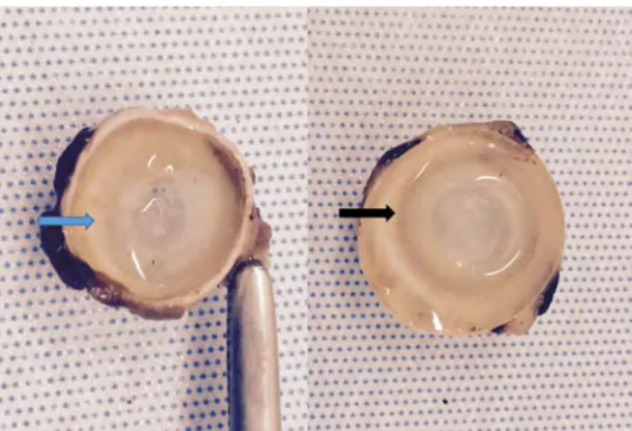
At the end of month one, all eyes were enucluated. In the macroscopic evaluation, the entire ciliary body had a white
(loss of pigmentation) and atrophic appearance in all of the eyes in the laser-treated group compared to non-laser group (Fig.3).
Histopathologic examination
In the laser group, light microscopic examination demonstrat-ed a severe 360 degrees disruption of ciliary processes (Fig.4). Ciliary processes were covered with fibrin exudation consisting of fibroblasts (Fig.4). There was a mild inflamma-tion with disrupinflamma-tion or atrophy of ciliary body epithelium with cystic vacuolar degeneration (Fig.4). No ciliary epithelium regeneration was detected in histopathologic sections.
Discussion
I n t h i s a n i m a l s t u d y, w e p e r f o r m e d 3 6 0 d e g r e e s endocyclophotocoagulation to the ciliary body epithelium of normal rabbit eyes. The IOP was significantly lower than baseline values of the treated eyes and untreated eyes during follow up period and remained lower at the end of month 1. Histological findings demonstrated the disruption of both pigmented and non-pigmented ciliary body epithelium, and severe loss of ciliary processes along with the significant re-duction of IOP. Furthermore, we detected fibrinous exudation that covered the entire surface, suggesting possible later for-mation of cyclitic epiciliary membranes. We did not detect ciliary body epithelium regeneration in microscopic sections. Ciliary body epithelium regeneration has been proposed the main reason for failure of laser treatment. Lee et al. per-formed transpupillary argon laser cyclophotocoagulation with the aid of gonioscopy to their refractory galucoma patients [12]. IOP increased gradually and they have ascribed increase in IOP to the regeneration of secretory mechanisms of ciliary processes, which was then hypothesized as the reason of long
Fig. 3 Entire ciliary body had a white and atrophic appearance in the laser-treated eye (left) compared to untreated eye (right)
Fig. 4 Light microscopic view of the laser treated and untreated eyes.a Disruption of the ciliary processes, fibrin exudation (blue arrow), and disruption of the ciliary epithelium (black arrows).b Normal view of the ciliary body processes
Lasers Med Sci (2019) 34:11–14 13
.
.
...
.
.
.
.
.
.
.
.
.
.
... :..
... .
.
.
. .
.
.
.. ..
.
.
.
.
.
.
.
.
.
.
.
. .
.
.
.
.
.
.
. .
.
. . .
.
.
.
.
. . .
.
.
. .
.
.
.
. . .
.
.
. .
.
.
.
.
.
.
.
.
. .
-
..
.
.
.
.·
::
:::
: :
::
:
::
:::
::
:::: :::::
::
::::
:
.
.
·::.·:::: ::::::
.
.
.
.
.
.
.
.
.
...
.
.
:
.
:
.
::::::.·::::::::::
.
.
.
. .
.
.
. .
.
.
. .
.
.
.
.
.
• ..,a.:.;~L .,.I".!."!..:' •~•~•~• :·~ ··.:•:,:.• • • .• • •• •; :;;;;;;;:;:::;:: ~ Springerterm failure of transscleral cyclophotocoagulation in cannine eyes [13]. Therefore, we performed laser application after lensectomy and anterior vitrectomy. The probability of inade-quate visualization due to the close relation of lens and ciliary body in rabbits was the main reason of lensectomy. Inadequate visualization of the ciliary processes was speculated as the reason of the regeneration of ciliary processes previously [14]. Additionally, laser power is important to achieve ade-quate burns on the ciliary body processes. We produced a concave burn with gas bubble formation and pigment disper-sion which was suggested by previous reports for success [14,
15]. Besides, hypotony may develop due to the leakage from the sclerotomy sites as well as ciliary body damage. However, IOP was similar with baseline values in the untreated eyes during follow-up period. We did not encounter serious adverse effects due to cilioblation such as severe inflammation, hypopion, vitreous hemorrhage, or retinal detachment.
Today, hypotony is a challenging problem after vitreoretinal surgery and in eyes with chronic uveitis. Development of ciliary traction due to epiciliary membranes in anterior proliferative vitreoretinopathy (PVR) and chronic uveitis is proposed for reduced aqueous production. Zarbin et al. removed epiciliary membranes and noted IOP elevation following removal of membranes [7]. However, in such cases, IOP may not increase despite removal of these membranes due to the development of atrophy of the ciliary processes owing to permanent damage which has no definitive treatment in humans. Intraocular viscoelastic injections are being used; however, these are temporary managements and require re-peated interventions [16]. Silicone oil may be a permanent intraocular tamponade; nevertheless, permanent structural changes can develop due to the absence of aqueous humor which is necessary for the survival of ocular tissues. This animal study achieved to form hypotony with ciliary process damage similar to the hypotony which is encountered in an-terior PVR or chronic uveitis patients during 1 month follow-up period. There are some limitations of this study. First, lon-ger period of follow-up may be required to evaluate the dura-tion of the effect. Second, we could not measure aqueous production before and after laser procedure and third, out-comes of endocyclophotocoagulation may be compared with the outcomes of transscleral cyclophotocoagulation. However, this new chronic hypotony model creates an analo-gous environment for this challenging situation, which would be helpful in developing new strategies in the treatment of chronic ocular hypotony.
In conclusion, 360 degrees endocyclophotocoagulation yielded permanent ciliary body epithelium damage. IOP re-duction started very early and remained in hypotonic levels during follow-up period.
Compliance with ethical standards
Conflict of interest The authors declare that they have no conflict of interest.
References
1. Costa VP, Arcieri ES (2007) Hypotony maculopathy. Acta Ophthalmol Scand 85:586–597
2. Streeten BW, Belkowitz M (1967) Experimental hypotony with silastic. Arch Ophthalmol 78(4):503–511
3. Pederson JE, MacLellan HM (1928) Medical therapy for experi-mental hypotony. Arch Ophthalmol 100:815–817
4. Kim HC, Hayashi A, Shalash A, de Juan E (1998) A model of chronic hypotony in the rabbit. Graefes Arch Clin Exp Ophthalmol 236:69–74
5. Dellaporta A, Obear MF (1964) Hyposecretion hypotony: experi-mental hypotony through detachment of the uvea. Am J Ophthalmol 58:785–789
6. Coleman DJ (1995) Evaluation of ciliary body detachment in hy-potony. Retina 15:312–318
7. Zarbin MA, Michels RG, Green WR (1991) Dissection of epiciliary tissue to treat chronic hypotony after surgery for retinal detachment with proliferative vitreoretinopathy. Retina 11:208–213
8. Assia EI, Hennis HL, Stewart WC, Legler UF, Carlson AN, Apple DJ (1991) A comparison of neodymium: yttrium aluminum garnet and diode laser transscleral cyclophotocoagulation and cyclocryotherapy. Invest Ophthalmol Vis Sci 32:2774–2778 9. Iliev ME, Gerber S (2007) Long-term outcome of trans-scleral
di-ode laser cyclophotocoagulation in refractory glaucoma. Br J Ophthalmol 91:1631–1635
10. Haller JA (1996) Transvitreal endocyclophotocoagulation. Trans Am Ophthalmol Soc 94:589–676
11. Kaplowitz K, Kuei A, Klenofsky B, Abazari A, Honkanen R (2015) The use of endoscopic cyclophotocoagulation for moderate to ad-vanced glaucoma. Acta Ophthalmol 93:395–401
12. Lee PF (1979) Argon laser photocoagulation of the ciliary process-es in casprocess-es of aphakic glaucoma. Arch Ophthalmol 97:2135–2138 13. Nadelstein B, Wilcock B, Cook C et al (1997) Clinical and histo-pathologic effect of diode laser transcleral cyclophotocoagulation in the normal canine eye. Veterinary Opthalmology 7:155–162 14. Shields MB (1986) Intraocular cyclophotocoagulation. Trans
Ophthalmol Soc UK 105:237–241
15. Shields HB, Chandler DB, Hickingbotham D et al (1985) Intraocular cyclophotocoagulation: histopathologic evaluation in primates. Arch Ophthalmol 103:1731–1735
16. Küçükerdönmez C, Beutel J, Bartz-Schmidt KU, Gelisken F (2009) Treatment of chronic ocular hypotony with intraocular application of sodium hyaluronate. Br J Ophthalmol 93(2):235–239
14 Lasers Med Sci (2019) 34:11–14