© by PSP Volume 21 – No 12. 2012 Fresenius Environmental Bulletin
EFFECTS OF MAGNESIUM AND CALCIUM
CATIONS ON BIOFILM FORMATION BY Sphingomonas
paucimobilis FROM AN INDUSTRIAL ENVIRONMENT
Nur Ceyhan Guvensen1,*, Serdar Demir2 and Guven Ozdemir31
Mugla University, Faculty of Sciences, Department of Biology, Mugla 48170, Turkey 2Mugla University, Faculty of Sciences, Department of Statistics, Mugla 48170, Turkey 3Ege University, Faculty of Sciences, Department of Biology, Izmir 38100, Turkey
ABSTRACT
Bacterial biofilms may form on all surface-associated natural and many industrial environments. Biofilm forma-tion requires particular notice due to its associated risks for human health and its impact on environmental con-tamination and pollution. In this work, we investigated the
effects of Mg2+ and Ca2+ ions on biofilm formation by
Sphingomonas paucimobilis from an industrial
environ-ment. The biofilm formation on coupons within nutrient broth medium was significantly enhanced after addition of 0, 100, 250 and 500 µM Mg2+. Similarly, the addition of Ca2+ caused a significant increase in S. paucimobilis bio-film formation when the above concentration levels for
Ca2+ were tested. In contrast, the same concentrations of
these ions had no effect on growth of free-living (planktonic)
S. paucimobilis cells in the medium. Hence, Mg2+ and Ca2+
increased the biofilm formation as adherent-cells on the
coupons. Both ion types were significantly effective on S.
paucimobilis biofilm formation, particularly at 100 and
250 µM (P≤0.05). These firstly reported data for S. pau-cimobilis biofilms are important in the elucidation of the roles of divalent cations, such as Mg2+ and Ca2+, in
bacte-rial adhesion to the environmental surfaces for biofilm
formation, and prevention of environmental contamina-tion by this bacterium.
KEYWORDS: Biofilm, industrial environment, bacterial adhesion,
Sphingomonas paucimobilis,magnesium ions,calcium ions
1 INTRODUCTION
In environment, bacteria often grow as populations attached to surfaces in complex structures called biofilms. A biofilm is an aggregate of bacteia in which cells adhere to each other and/or to a surface. Bacterial adhesion involves the attachment (or deposition) of bacteria on the surface (solid, gel layer, etc.). In nature, microorganisms usually
* Corresponding author
attach to solid surfaces, especially on the liquid–solid inter-face. After attachment, they form micro-colonies, and a bio-film which is highly resistant to antimicrobials and some physical treatments [1, 2], and it is also a serious problem for infectious diseases [3].
Biofilm contamination and biofouling occurs on nearly every environmental water-contacted surface [4]. Environ-mental systems by their nature are comprised of interfaces that provide chemical and mechanical stimuli for micro-bial attachment and biofilm accumulation. Often, attach-ment to a surface is the way in which microbes respond to environmental stimuli, and this attachment effects numerous environmental and health problems [5, 6]. Virtually any sur-face - animal, mineral, or vegetable (i.e., biotic or abiotic) - is fair game for bacterial colonization and biofilm for-mation, including contact lenses, ship hulls, dairy and petroleum pipelines, rocks in streams, and all varieties of biomedical implants and transcutaneous devices [7-9].
Understanding the environmental (e.g., temperature, pH, ionic strength, electrolyte type), interfacial (e.g., sur-face charge and hydrophobicity), and physiological (e.g., bacterial growth stage and metabolic activity) factors that govern adhesion mechanisms defines one of the most important challenges in the microbial and interfacial sci-ences [10, 11]. Environmental factors, such as electrolyte con-centrations [12] and medium composition [13], have important impacts on biofilm formation. Bacterial exopoly-saccharides are the main component of the biofilm matrix. Their production and surface adhesion, and, hence, surface colonization are influenced by electrostatic and hydrophobic interactions, steric hindrance, van der Waals forces, tem-perature, and hydrodynamic forces, to name a few [14, 15]. Divalent cations can potentially initiate bacterial ad-hesion and biofilm formation directly through effects on electrostatic interactions, and indirectly by affecting physi-ology-dependent attachment processes [16, 17]. From these divalent cations, magnesium (Mg2+) is the molecular key that activates many important enzymes responsible for diverse biochemical reactions in living cells [18, 19]. Mg is regarded as one of the intracellular bulk elements [20]
© by PSP Volume 21 – No 12. 2012 Fresenius Environmental Bulletin
and is involved in enzyme catalysis, which performs a variety of roles, such as structure stabilization, charge neu-tralization, and control of osmotic pressure [21].
In spite of this potentially important role, the effect of Mg2+ or Ca2+ concentrations on bacterial growth and ad-hesion have not been studied in Sphingomonas
paucimo-bilis biofilms. The aim of this study was to investigate the
effects of Mg2+ and Ca2+concentrations on the planktonic and sessile growth in biofilm formation by S.
paucimobi-lis. This bacterium was selected because it commonly
ex-ists in biofilms in natural, clinical, and industrial environ-ments e.g. [22-24]. S. paucimobilis is a yellow-pigmented, aerobic, motile with polar flagellum, non-fermentative, gram-negative bacterium. This organism is widely distri-buted in various environments, especially in water and soil [25], and has also been recovered from hospital envi-ronments [26]. S. paucimobilis is an opportunistic patho-gen and has been implicated in a variety of community-acquired and nosocomial infections, including bacteremia, catheter-related sepsis, meningitis, peritonitis, cutaneous infections, visceral abscesses, urinary tract infections, ade-nitis, and diarrheal disease [27]. S. paucimobilis has been reported to cause outbreaks of bacteremia; these outbreaks are possibly related to bacterial colonization of water sys-tems [23, 26]. Gellan, an exopolysaccharide (EPS), is produced by S. paucimobilis and is effective on its bacte-rial colonization and biofilm formation [28], but the effects of divalent cations, such as Mg2+ and Ca2+, on S.
paucimo-bilis biofilms are largely unknown. Attachment and biofilm
formation by opportunistic pathogens, such as S.
paucimo-bilis, are of public health and cross-contamination concern.
2 MATERIALS AND METHODS
2.1 Bacterium and medium
S. paucimobilis, isolated from the biofilms in the
cool-ing tower water of a petrochemical industrial plant, was used as the test strain in this study. This strain was reported to be a biofilm-forming bacterium [24]. Because of continu-ous circulation of water, the warm temperatures and con-tinuous scrubbing of nutrients, cooling towers are a prime environment for the build-up of microorganisms [29, 30]. Identification of the isolate was carried out using Standard biochemical tests as outlined in Bergey’s Man-ual [31]. API 20 E and API 20 NE commercial kits (Bio-Me´rieux, France), and 16S rRNA sequencing analysis were used to further identify this isolate [32]. The bacterium was cultured in Modified Tryptone Soya Broth (TSB) (Oxoid CM0989, pancreatic digest of casein 17.0 g/L, papaic digest of soybean meal 3.0 g/L, sodium chloride 5.0 g/L, di-potassium hydrogen phosphate 2.5 g/L, glucose 2.5 g/L, bile salts 1.5 g/L, distilled water 1 L; pH 7.4±0.2.
2.2 Preparation of test surfaces
Bacterial adhesion and biofilm accumulation were per-formed on stainless steel coupons (30x10x15 mm). Stainless
steel coupons were prepared according to Nandakumar et
al. [21] with modifications. In the study, six stainless steel
coupons were used for treatment group (3 different con-centrations of MgCl2 and CaCl2), and two coupons were used for control group (without MgCl2 and CaCl2). Prior to the biofilm experiments, the coupons were smoothed with emery papers and washed with a commercial surfactant solution, and then deionized water. They were dried in a Pasteur oven at 50 °C.
2.3 Formation of biofilms and cultural conditions
Bacterial cells were grown in TSB supplemented with 0, 100, 250, 500 µM of MgCl2, or the same concentrations of CaCl2. Bacterial growth of the cells was carried out in shaken flasks at 100 rpm for 24 h at 30 °C. The flasks with 200 ml of the experimental medium, a flask with 200 ml of deionized water, and a flask with 200 ml of TSB were taken and sterilized. One ml of late exponential phase S.
paucimobilis culture (at an optical density of 1.0,
corre-sponding to ca. 1.0x10⁷ cells/ml) was inoculated into each of these flasks. The coupons were sterilized by autoclav-ing for 20 min at 121 °C and transferred into the flasks under aseptic conditions. These flasks were incubated at 30 °C for 7 days. The coupons were removed after 7-days incubation, and the adherent cells on them were finally in-vestigated for biofilm formation. The experiment was not continued further because the number of bacteria in the medium with coupons was reduced considerably by this time so that the experiment could not be continued. Three replicates were tested for each experiment.
2.4 Determination of cell counts in the medium and biofilm
In this study, free-living biomass (planktonic) was considered as cell count (colony forming units (CFU)) in TSB (cell count medium), and adherent-living biomass (sessile) on the coupons was regarded as cell count of biofilms (cell count biofilm). Free-living and sessile bac-teria were collected simultaneously with retrieval of cou-pons. Determinations of the biofilm counts were achieved on the coupons in TSB supplemented separately with Mg+2 and Ca+2. The flasks with 200 ml of TSB each were in-oculated with S. paucimobilis as mentioned above. After 7 days, each coupon was rinsed with 100 ml of sterile 0.1% peptone water to remove unattached cells. It was then placed in a sterile glass jar containing 250 ml TSB. After that, the side of the coupon in contact with the prod-uct was repeatedly scraped about 120 times by using a sterile spatula in order to recover attached cells. The cells were placed in 250 ml of TSB and the resulting attached cell suspension, which was later also used for pre-en-richment of cells, was thoroughly shaken and decimal dilu-tions were immediately prepared with sterile 0.1% peptone water. Dilutions were plated using TSB + 1.5% Tryptone Soya Agar (TSA) (pH 7.4±0.2) for the biofilm forming total viable counts and incubated at 35 °C for 48 h. After the incubation period, colonies were enumerated and the number of biofilm-forming cells per cm2 was calculated (modified, Gunduz and Tuncel [33]). Sterile TSB was
© by PSP Volume 21 – No 12. 2012 Fresenius Environmental Bulletin
used as negative control. A minimum of three assays was performed in each experiment.
To control the scraping technique, replicate coupons were examined before and after scraping by fluorescent microscopy to get a second estimate of cell numbers, and the percentage removed. The detection limit for removal of cells was determined to be 99.4± 0.5 %.
2.5 Standard plate count
Cell counts medium and cell counts biofilm were per-formed by a modified standard plate count method [34]. One ml of the sample was placed on the centre of a sterile Petri dish (100 mm diameter) by using a sterile pipette. Sterile, molten (44-46 °C) TSA was added and mixed with the sample by swirling the plate. The samples were allowed to cool at room temperature until being solidified, and then were inverted and incubated at 35 °C for 48 h. Colo-nies formed in or on the TSA within 48 h were counted as described in this standard method, and the results were re-ported as CFU/ml. Where applicable, this value was multi-plied by the dilution factor to obtain the corrected CFU/ml.
2.6 Statistical analysis
Statistical analysis was performed using SPSS soft-ware (version 16.0). One-way variance analysis (ANOVA) was used to test the differences among all effects of Mg+2 and Ca+2 concentrations on biofilm formation. Levene`s test was used to examine homogeneity of variances. Fisher’s least significant difference (LSD) test was performed for multiple comparisons. All tests were considered to be sig-nificant at the level P<0.05. Error bar plots in figures are shown as one standard deviation. The values for each ex-periment are the mean values of results from three experi-ments unless otherwise stated.
3 RESULTS AND DISCUSSION
A number of researches have demonstrated that envi-ronmental conditions play an important role for biofilm formation [35]. Biofilm development is a complex proc-ess and can be regulated by different factors, such as cell surface structure, growth medium, oxygen limitation, or substratum [36]. However, it has not been obtained any data with S. paucimobilis among previous works on the effecting factors of adhesion to surfaces and the end of this
biofilm formation, except from the study of Venugopalan
et al. [37]. These researches declared that changing
hydro-dynamic conditions had discernible influence on the char-acteristics of Sphingomonas biofilms during development. In our study, evaluation of the effect of Mg+2 and Ca+2 sepa-rately was done using an assay based on cell counts in me-dium (TSB) and biofilm by S. paucimobilis. In the second part of the study, the effects of these cations on bacterial growth and biofilm development were evaluated statistically.
A biofilm can be defined as a sessile bacterial commu-nity of cells that live attached to each other and to surfaces [38]. Hence, we detected the numbers of sessile cells for biofilm formation. Significant differences between plank-tonic counts in medium and sessile counts on the coupons at a given point (P≤0.05) are shown in Table 1. Asterisks indicate a significant (P≤0.05) reduction or enhancement in mean CFU counts of planktonic S. paucimobilis in liquid culture when Mg+2 and Ca+2 was supplemented on the coupons.
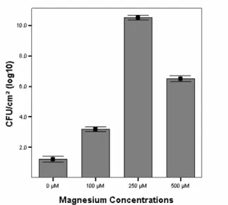
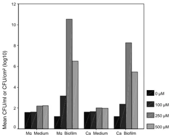
After 7 days, biofilm development with a significant enhancing in S. paucimobilis log CFU/cm2 was noted at 100 µM (3.1x10⁷, P = 0.000), at 250 µM (10.4x10⁷, P = 0.000), at 500 µM (6.5x10⁷, P = 0.000) of Mg2+ compared with cell counts at 0 µM (1.2x10⁷). Conversely, non-significant enhancing planktonic S. paucimobilis counts/ ml of the medium containing Mg2+ was noted at 100 µM (1.7x10⁷, P = 0.866), at 250 µM (2.3x10⁷, P = 0.092), at 500 µM (2.2x10⁷, P = 0.120) compared with planktonic cell counts at 0 µM (1.6x10⁷) (Figs. 1 and 2, Table 2). Similarly, biofilm development with Ca+2 was significantly enhanced in log CFU/cm2 at 100 µM (2.3x10⁷, P = 0.000), at 250 µM (8.3x10⁷, P = 0.000), at 500 µM (5.2x10⁷, P = 0.000) compared with cell counts at 0 µM (1.2x10⁷). On the contrary, non-significant enhancing planktonic S.
pau-cimobilis counts/ml of the medium containing Ca+2 was noted at 100 µM (1.6x10⁷, P = 0.866), at 250 µM (2.0x10⁷, P = 0.092), at 500 µM (2.0x10⁷, P = 0.120) with regard to planktonic cell counts at 0 µM (1.6x10⁷) (Table 2). In other two works, Tamura et al. [39] showed that magnesium had no significant effect on adherence of Streptococci at physio-logical concentrations (2mM Mg2+) while higher concentra-tions enhanced adherence to a small degree. Dunne and Burd [40] found that magnesium (as low as 16 mM) sig-nificantly enhanced the in vitro adhesion of
Staphylococ-cus epidermidis to plastics.
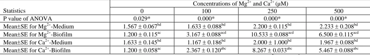
TABLE 1 - The results of ANOVA and LSD multiple comparison test over cell counts in medium supplemented with Mg2+ or Ca2+ ,and mean counts of S. paucimobilis cells in biofilms formed in the presence of Mg2+ or Ca2+.
Concentrations of Mg2+ and Ca2+ (µM)
Statistics 0 100 250 500
P value of ANOVA 0.029* 0.000* 0.000* 0.000*
Mean±SE for Mg2+-Medium 1.567 ± 0.067bd 1.633 ± 0.088bd 2.200 ± 0.115bd 2.233 ± 0.208bd
Mean±SE for Mg2+-Biofilm 1.200 ± 0.115ac 3.167 ± 0.088acd 10.533 ± 0.088acd 6.500 ± 0.115acd
Mean±SE for Ca2+-Medium 1.633 ± 0.145bd 1.167 ± 0.186bd 2.000 ± 1.000bd 1.967 ± 0.088bd
Mean±SE for Ca2+-Biofilm 1.200 ± 0.058ac 2.367 ± 0.120abc 8.267 ± 0.033abc 5.467 ± 0.088abc
*Significantly different (P≤0.05). Each assay was carried out in triplicate (n=3). a There is a significant difference from Mg2+-Medium. b There is a significant difference from Mg2+-Biofilm. c There is a significant difference from Ca2+-Medium. d There is a significant difference from Ca2+-Biofilm
© by PSP Volume 21 – No 12. 2012 Fresenius Environmental Bulletin
FIGURE 1 - Effects of Mg2+ concentrations on the cell counts in
medium (Error bars indicate one standard deviation). FIGURE 2 - Effects of different Mg
2+ concentrations on the cell counts of biofilms formed on the coupon (Error bars indicate one standard deviation).
TABLE 2 - The results of ANOVA and LSD multiple comparison tests over mean counts of S. paucimobilis cells in the different concentra-tions of Mg2+ or Ca2+.
Statistics MgM MgB CaM CaB
P value of ANOVA 0.002* 0.000* 0.187 0.000*
Mean±SE for 0 µM 1.567 ± 0.067cd 1.200± 0.115bcd 1.633± 0.145 1.200± 0.058bcd
Mean±SE for 100 µM 1.633± 0.088cd 3.167 ± 0.088acd 1.667± 0.186 2.367± 0.120acd
Mean±SE for 250 µM 2.200± 0.115ab 10.533± 0.088abd 2.000 ±0.100 8.267 ± 0.033abd
Mean±SE for 500 µM 2.233± 0.120ab 6.500± 0.115abc 1.967 ± 0.088 5.467 ± 0.088abc
*Significantly different (P≤0.05). Each assay was carried out in triplicate (n=3). a There is a significant difference from 0 Μm. b There is a significant difference from 100 µM. c There is a significant difference from 250 µM. d There is a significant difference from 500 µM.
FIGURE 3 - Effects of Ca2+ concentrations on the cell counts in medium (Error bars indicate one standard deviation).
FIGURE 4 - Effects of different Ca2+ concentrations on the cell counts of biofilms formed on the coupon (Error bars indicate one standard deviation).
Previous researches with the gram-negative bacteria
P. aeruginosa and Escherichia coli and the gram-positive
bacteria S. epidermidis and S. aureus revealed that diva-lent cations are stimulating attachment to surfaces, espe-cially at low or moderate concentrations of them [41-44].
Similarly, in this study, the mean CFU values of S.
pau-cimobilis in biofilm cultures were 3.1x10⁷ and 2.3x10⁷, respectively, with Mg+2 and Ca+2. The mean CFU of S.
paucimobilis increased dramatically within the biofilm
with 250 µM of Mg+2 (10.4x10⁷, P = 0.000; Table 2, Fig. 2)
s
....
m ,g_ E 3 IL u 2.0 1.5 I.D D.5 D.O 2.00000 ~ 1.50000 m ,g_ E :5 1.00000 IL u 0.50000 0.00000s
....
m ,g_ " E u 3 IL u O µM 100 µM 250 µM 500 µM Magnesium Concentrations 0 µM 100 µM 250 µM 500 µM Calcium Concentrations 10.0 8.0 6.D 4.0 2.0 OµM 8.00000 - 6.00000 ~ m ,g_ N ~ 4.00000 3 IL u 2.00000fol
100 µM 250 µM 500 µM Magnesium Concentrations 0 µM 100 µM 250 µM 500 µM Calcium Concentrations© by PSP Volume 21 – No 12. 2012 Fresenius Environmental Bulletin
but only moderately with 500 µM (6.5x10⁷, P = 0.000) (Table 2, Fig. 2). Moreover, the mean of bacterial counts increased also dramatically in the biofilm with 250 µM of Ca+2 (8.3x10⁷, P = 0.000) (Table 2, Figs. 4 and 5). Con-versely, this effect on planktonic bacterial growth in liq-uid culture was not seen at these concentrations of Mg+2 or Ca+2 (Table 2, Figs. 1, 3 and 5). High levels of these cations might screen cross-linking electrostatic interac-tions [45], and have resulted in a reduction of attached cells in 500 µM. In addition, these cations might enhance the production of EPS by the bacterium [41]. EPS are often associated to bacteria growing in biofilms in which they play a key role in the architecture [46].
Ca_Biofilm Ca_Medium Mg_Biofilm Mg_Medium Mean C F U /ml or C F U /cm² ( log10) 12 10 8 6 4 2 0 0 µM 100 µM 250 µM 500 µM
FIGURE 5 - Comparison of cell counts in Mg2+-Medium, Mg2+ -Biofilm, Ca2+-Medium, and Ca2+-Biofilm.
Physical and chemical environment has a significant impact on the way of biofilm formation and its persistence. Diva-lent cations are regarded to be important constituents of microbial aggregates, since they bind to negatively charged groups present on bacterial surfaces, in EPS, and on inorganic particles entrapped in biofilms [41]. It has
been reported that extraction of calcium (Ca2+) from
bio-films by displacement with monovalent cations or by chela-tors, such as EDTA and EGTA, resulted in the destabiliza-tion of biofilms. These observadestabiliza-tions suggest that divalent cations may be important in maintaining the biofilm struc-ture, acting in the bridging of a 3-D polymeric matrix [15].
Similar with these investigations, in the absence of Mg+2 and Ca+2 in TSB, sessile bacterial counts on the coupons did not increase significantly (P = 1.000) in this study (Ta-ble 1, Figs. 6 and 7). On the other hand, the mean number of S. paucimobilis CFU on the coupons increased signifi-cantly in the presence of Mg+2 or Ca+2 (P<0.05) (Table 1, Fig. 7), whilst Mg+2 or Ca+2, at all concentrations examined, had no significant effects on S. paucimobilis numbers in the medium (P>0.05) (Table 1, Fig. 7). When all corresponding concentrations of these cations were compared, the statis-tical analyses showed highly significant differences between attached cell counts by adding Mg+2 and attached cell counts by adding Ca+2 into TSB (Table 1, Figs. 5-8). According to
500 µM 250 µM 100 µM 0 µM M ean CFU/m l or CFU/c m ² (log10) 12 10 8 6 4 2 0 Mg_Medium Mg_Biofilm Ca_Medium Ca_Biofilm
FIGURE 6 - Comparison of cell counts in medium or biofilm when different concentrations of Mg2+ and Ca2+ are considered.
FIGURE 7 - Comparison of cell counts determined in different concentrations of Mg2+-Medium, Mg2+-Biofilm, Ca2+-Medium, Ca2+ -Biofilm with each other.
FIGURE 8 - Comparison of cell counts in each concentration of Mg2+-Medium, or Mg2+-Biofilm, or Ca2+-Medium, or Ca2+-Biofilm with others.
•
•
•
Dfol
10 6" ll 6 -DµM filHI IIo
+---~---~---1
llilg__Mcdum Mg__BaUm C>_PAl>dlm Ca_Bollm
S' 10
- C Bit>fim
o
o~
+ - - - ~ - - - ~ - - - 1,
~
© by PSP Volume 21 – No 12. 2012 Fresenius Environmental Bulletin
these findings, S. paucimobilis increased attachment and biofilm formation.
Also in other studies, divalent cations have been shown to influence adherence to surfaces (specifically, using
Pseudomonas sp. and sand columns) [47]. Furthermore, for Aeromonas hydrophilia, mutations in Mg2+ transport sys-tems result in reduction of swarming and biofilm formation [48]. For Pseudomonas fluorescens, Mg2+ increased initial attachment and altered subsequent biofilm formation and structure [49]. Dunne and Burd [40] demonstrated that increasing Mg2+ levels enhanced biofilm production by S.
epidermidis, while EDTA caused a dose-dependent
de-crease in the accumulation of cells on a plastic surface. Additionally, although Ca2+ had a much stronger affinity towards alginate produced by P. aeruginosa than Mg2+ [50],
P. fluorescens produces an acidic galactoglucan, and its
surface colonization and biofilm depth increased with in-creasing Mg2+ concentrations [49]. All these studies have found that divalent cations have varying effects on bacte-rial adhesion [38, 40, 49], which might be due to the dif-ference in bacterial species and cation concentrations.
Furthermore, increase in surface hydrophobicity of E.
coli in the presence of Mg2+ excess was found by
Latra-che et al. [51]; on the other hand, antimicrobial effects of
Ca2+against various microorganisms isolated from oral
cavity were described by Pezelj-Ribari et al. [52].
4 CONCLUSION
We detected significant effects of Mg2+ and Ca2+ on biofilm formation activity of S. paucimobilis. These diva-lent cations showed significant stimulation of the adhe-sion of S. paucimobilis cells onto coupons. The numbers of S. paucimobilis cells on the stainless steel coupons were nearly 1.0x10⁷ CFU/cm2, and these numbers in-creased significantly (P≤0.05) after addition of Mg2+ and Ca2+,particularly at 100 and 250 µM levels. This influ-ence might contribute to an increase in the hydrophobicity of planktonic cells, which may have altered their ability to attach to surfaces [53]. Moreover, S. paucimobilis in a variety of environments is frequently mentioned. Its sur-vival in an environment could be associated with its ca-pacity to colonize on abiotic/biotic surfaces because of its production of gellan [37]. In industrial or other environ-ments, biofilm formation may be undesirable for con-tamination and safety reasons concerning the attachment of opportunistic pathogenic microorganisms, such as S.
paucimobilis, to surfaces.
REFERENCES
[1] Mah, T.F. and O’Toole, G.A. (2001). Mechanisms of biofilm resistance to antimicrobial agents. Trends in Microbiology, 9: 34-39.
[2] O’Toole, G.A. and Stewart, P.S. (2005). Biofilms strike back. Nature Biotechnology, 23: 1378-1379.
[3] Furukawa, S., Kuchma, S.L. and O’Toole, G.A. (2006). Keeping their options open: Acute versus persistent infec-tions. Journal of Bacteriology, 188: 1211-1217.
[4] Mittelman, M.W. (1998). Structure and functional character-istics of bacterial biofilms in fluid processing opera-tion.Journal of Dairy Science, 81: 2760-2764.
[5] Ginn, T.R., Wood, B.D., Nelson, K.E., Scheibe, T.D., Mur-phy, E.M. and Clement, T.P.(2002). Processes in microbial transport in the natural subsurface. Advances in Water Re-sources, 25: 1017-1042.
[6] Camesano, T.A., Liu, Y. and Data, M. (2007).Measuring bac-terial adhesion at environmental interfaces with single-cell and single-molecule techniques. Advances in Water Re-sources, 30:1470-1491.
[7] Carpentier, B. and Cerf, O. (1993). Biofilms and their conse-quences, with particular reference to hygiene in the food in-dustry. J Appl Bacteriol,75:499-511.
[8] Costerton, J.W., Lewandowski, Z., Caldwell, D.E., Korber, D.R. and Lappin-Scott, H.M. (1995).Microbial biofilms. An-nual Review of Microbiology,49:711-745.
[9] Elder, M.J., Stapleton, F., Evans, E. and Dart, J.K.G. (1995). Biofilm related infections in ophthalmology. Eye,9:.102-109. [10] Stepanovic´, S.,Vukovic´, D., Ježek, P., Pavlovic´, M. and Šva-bic-Vlahovic, M. (2001).Influence of dynamic conditions on biofilm formation by Staphylococci.European Journal of Clini-cal Microbiolology and Infectious Diseases, 20: 502-504. [11] Kerchove, A.J. and Elimelech, M. (2007). Impact of alginate
conditioning film on deposition kinetics of motile and non-motile Pseudomonas aeruginosa strains.Appl Environ Mi-crobiol, 15: 5227-5234.
[12] Fletcher, M. (1998). Attachment of Pseudomonas fluorescens to glass and influence of electrolytes on bacterium-substratum separation distance. Journal of Bacteriology, 170: 2027-2030.
[13] McEldowney, S. and Fletcher, M. (1986). Variability of the-influence of physicochemical factors affecting bacterial adhe-sion to polystyrene substrata. Applied and Environmental Microbiology, 52: 460-465.
[14] An, Y.H., Dickinson, R.B. and Doyle, R.J. (2000). Mecha-nisms of bacterial adhesion and pathogenesis of implant and tissue infections, pp. 1-27. In: Y.H. An and R.J. Friedman (eds.) Handbook of bacterial adhesion: principles, methods, and applications. Humana Press, Totowa, N.J.
[15] Flemming, H.C., Windenger, J., Mayer, C., Körstgens, V. and Borchard, W. (2000).Cohesiveness in biofilm matrix polymers, pp. 294-307. In: D.G. Allision, P. Gilbert, H. Lap-pin-Scott, M. Wilson (Eds): SGM Symposium 59: Commu-nity Structure and Cooperation in Biofilms.Cambridge Uni-versity Press, Cambridge (UK).
[16] Koerstgens, V.,Flemming, H.C., Wingender, J. and Borchard, W. (2001). Influence of calcium ions on the mechanical properties of a model biofilm of mucoid Pseudomonas
aeru-ginosa. Water Science and Technology, 43:49-57.
[17] Vu, B., Chen, M., Crawford, R.J. and Ivanova, E. P. (2009).
Bacterial Extracellular Polysaccharides Involved in Biofilm Formation.Molecules,14: 2535-2554.
[18] Hughes, M.N. and Poole, R.K. (1989). Metals and microor-ganisms.pp. 412. London/NY:Chapman and Hall.
[19] Todar, K. (1997). Nutrition and growth of bacteria. Bacteriol-ogy, http://www.bact.wisc.edu/Bact303/Nutrition and Growth, pp. 1-9.
© by PSP Volume 21 – No 12. 2012 Fresenius Environmental Bulletin
[20] Beveridge, T.J., Hughes, M.N., Lee, H., Leung, K.T., Poole, R.K., Savvaidis, I., Silver, S. and Trevors, J.T. (1997). Met-al–microbe interactions: contemporary approaches. Advances in Microbial Physiology, 38: 177-243.
[21] Nandakumar, K., Sreekumari, K.R. and Kikuchi, Y. (2002). Antibacterial properties of magnesium alloy AZ31B:In-vitro studies using the biofilm-forming bacterium Pseudomonas
sp. Biofouling, 18: 129-135.
[22] Ashtaputre, A.A. and Shah, A.K. (1995). Studies on a Vis-cous, Gel-Forming Exopolysaccharide from Sphingomonas
paucimobilis GS1.Applied and Environmental Microbiology,
61: 1159-1162.
[23] Perola, O., Nousiainen, T., Suomalainen, S., Aukee, S., Karkkainen, U.M. and Kauppinen, J. (2002). Recurrent
Sphingomonas paucimobilis bacteraemia associated with a
multi-bacterial water-borne epidemic among neutropenic pa-tients. Journal of Hospital Infection, 50: 196-201.
[24] Ceyhan, N. and Ozdemir, G. (2008). Extracellular polysac-charides produced by cooling water tower biofilm bacteria and their possible degradation. Biofouling, 24:129-135. [25] Hsueh, P.R., Teng, L.T., Yang, P.C., Chen, Y.C., Pan, H.J., and
Ho, S.W. (1998). Nosocomial infections caused by
Sphingo-monas paucimobilis: clinical features and microbiological
characteristics. Clinical Infectious Diseases, 26:676-681. [26] Kilic, A., Senses, Z., Kurekci, A.E., Aydogan, H., Sener, K.
and Kismet, E. (2007). Nosocomial outbreak
ofSphingomo-nas paucimobilis bacteremia in a hemato/oncology
unit.Japanese Journal of Infectious Diseases, 60: 394-396. [27] Reina, J., Bassa, A., Liompart, I., Portel, D. and Borrell, N.
(1991). Infections with Pseudomonas paucimobilis: report of four cases and review. Reviews of Infectious Diseases, 13: 1072-1076.
[28] Fialho, A.M., Martins, L.O., Donval, M.L., Leitao, J.H., Ri-dout, M.J., Jay, A.J., Morris, V.J. and Sa-Correia, I. (1999). Structures and properties of gellan polymers produced by
Sphingomonas paucimobilis ATCC 31461 from lactose
com-pared with those produced from glucose and from cheese whey. Applied Environmental Microbiology, 65: 2485-2491. [29] Bott, T.R. (1998). Techniques for reducing the amount of bi-ocide necessary to counteract the effects of biofilm growth in cooling water systems. Applied Thermal Engineering, 18: 1059-1066.
[30] Turetgen, I. (2004). Comparison of the efficacy of free resid-ual chlorine and monochloramine against biofilms in model and full scale cooling towers. Biofouling, 20: 81-85. [31] Krieg, N.R. and Holt, C.G. (1984). Bergey’s Manual of
Sys-tematic Bacteriology, pp 787. Williams&Wilkins Press, Bal-timore.
[32] Ceyhan, N. (2008). Determination of biodegradation and bi-odegradation capability of polysaccharides of extracellular polysaccharides producing microorganisms which cause problems in several systems (PhD Thesis). Ege University, Faclty of Sciences, Izmir, pp. 314. (In Turkish)
[33] Gunduz, G.T. and Tuncel, G. (2006).Biofilm formation in an ice cream plant. Antonie van Leeuwenhoek, 89: 329-336. [34] American Public Health Association, Section 9215A and B.
Standard methods for the examination of water and wastewa-ter, 19th ed. (1995). American Public Health Association, Washington, D.C. pp. 250.
[35] O'Toole, G.A., Kaplan, H.B. and Kotler, R. (2000). Biofilm formation as microbial development. Annual Review of Mi-crobiology,54: 49-79.
[36] Dunne, W.M. (2002). Bacterial adhesion: seen any good bio-films lately. Clinical Microbiology Reviews, 15: 155-166. [37] Venugopalan, V.P., Kuehn, M., Hausner, M., Springael, D.,
Wilderer, P.A. and Wuertz, S. (2005). Architecture of a nas-cent Sphingomonas sp. biofilm under varied hydrodynamic conditions. Applied and Environmental Microbiology, 71: 2677-2686.
[38] Movassagh, M.H. and Karami, A.R. (2010). Biofilm forma-tion of Escherichia coli O111 on food contact glass surfaces. Global Veterinaria, 4: 222-224.
[39] Tamura, G.S., Kuypers, J.M., Smith, S., Raft, H. and Rubens, C.E. (1994). Adherence of group B Streptococci to cultured epithelial cells: roles of environmental factors and bacterial surface components. Infection and Immunity, 62: 2450-2458. [40] Dunne, W.M. and Burd, E.M. (1992). The effects of magne-sium, calcium, EDTA, and pH on the in vitro adhesion of
Staphylococcus epidermidis to plastic. Microbiology and
Immunology,36:1019-1027.
[41] Ozerdem Akpolat, N., Elçi, S., Atmaca, S., Akbayin, H. and Gül, K. (2003). The effects of magnesium, calcium and EDTA on slime production by Staphylococcus epidermidis strains. Folia Microbiol (Praha), 48: 649-653.
[42] Kerchove, A.J. and Elimelech, M. (2008). Calcium and mag-nesium cations enhance the adhesion of motile and nonmotile
Pseudomonas aeruginosa on alginate films. Langmuir, 24:
3392-3399.
[43] Dass, C.L., Walsh, M.F., Seo, S., Shiratsuchi, H., Craig, D.H. and Basson, M.D. (2009). Irrigant divalent cation concentra-tions influence bacterial Adhesion. Journal of Surgical Re-search, 156: 57-63.
[44] Lin, Y., Yu, J., Lu, Q. and Lin, L. (2009). Effects of Magnesium ions on the mucoid Pseudomonas aeruginosa biofilm. Chinese Journal of Microecology, 6: 515-518. [45] Chen, X. and Stewart, P.S. (2002). Role of electrostatic
inter-actions in cohesion of bacterial biofilms. Applied Microbiol-ogy and BiotechnolMicrobiol-ogy, 59: 718-720.
[46] Donlan, R.M. (2002). Biofilms: microbial life on surfaces. Emerging Infectious Diseases, 8: 881-890.
[47] Simoni, S.F., Bosma, T.N.P., Harms, H. and Zehnder, A.J.B. (2000). Bivalent cations increase both the subpopulation of adhering bacteria and their adhesion efficiency in sand col-umns. Environmental Science and Technology, 34: 1011-1017.
[48] Merino, S., Gavin, R., Altarriba, M., Izquierdo, L., Maguire, M.E. and Tomas, J.M. (2001). The MgtE Mg+ transport pro-tein is involved in Aeromonas hydrophilia adherence.FEMS Microbiology Letters, 198: 189-195.
[49] Song, B. and Leff, L.G. (2006). Influence of magnesium ions on biofilm formation by Pseudomonas fluorescens. Microbi-ological Research, 161: 355-361.
[50] Lattner, D., Flemming, H.C. and Mayer, C. (2003). 13C-NMR study on the interaction of bacterial alginate with biva-lent cations. International Journal of Biological Macromole-cules, 33: 81-88.
[51] Latrache, H., Bourlioux, P., Karroua, M., Zahir, H. and Hak-kou, A. (2000). Effects of subinhibitory concentrations of ni-troxoline on the surface properties of Escherichia coli. Folia Microbiology,45: 485-490.
© by PSP Volume 21 – No 12. 2012 Fresenius Environmental Bulletin
[52] Pezelj-Ribari, S., Brekalo, I., Abram, M., Dori, M., Mileti, I. and Karlovi, Z. (2002). Influence of calcium hydroxide root-canal sealer on microbial growth in vitro. Folia Microbiol-ogy, 47: 458-460.
[53] Song, B. (2004). Identification and characterization of bacte-rial isolates from the Mir Space Station and the impact of Mg2+ concentration on biofilm formation of Pseudomonas
fluorescens. MS Thesis, Kent State University, Kent, OH, pp.
107.
Received: February 29, 2012 Accepted: June 13, 2012
CORRESPONDING AUTHOR
Nur Ceyhan Guvensen
Mugla University Faculty of Sciences Department of Biology 48170 Mugla TURKEY Phone: +90 252 211 31 37 Fax: +90 252 211 14 72 E-mail: [email protected]
FEB/ Vol 21/ No 12/ 2012 – pages 3685 - 3692