African Journal of Biotechnology Vol. 8 (14), pp. 3118-3127, 20 July, 2009 Available online at http://www.academicjournals.org/AJB
ISSN 1684–5315 © 2009 Academic Journals
Review
A molecular-based fast method to determine the extent
of DNA damages in higher plants and fungi
Murat Dikilitas
1*, Abdurrahim Kocyigit
2and Fahri Yigit
31Department of Plant Protection, Faculty of Agriculture, Harran University, S. Urfa, Turkey. 2Department of Clinical Biochemistry, Faculty of Medicine, Harran University, S. Urfa, Turkey.
3Vocational High School, Mugla University, Fethiye, Turkey.
Accepted 21 May, 2009
Comet assay also called ‘single cell gel electrophoresis is a technique for the detection of DNA damage and repair at the level of single cells, which is one of the most advanced techniques introduced to the agricultural sciences in recent years. The assay is one of the most popular tests of DNA damage detection (e.g. single and double-strand breaks, oxidative-induced base damage and DNA-DNA/DNA-protein cross linking) by electrophoresis. The assay is very sensitive, rapid, easy to handle, non-invasive, visual and inexpensive compared to most conventional techniques to detect DNA damage, there is also little amount of cell samples required and it is applicable for most eukaryotic cells, thus, it has rapidly gained importance in the fields of genetic toxicology, medicine, environmental studies and agriculture. Isolated DNA from cells are embedded in a thin agarose gel on a microscope slide and unwound in a suitable buffer and exposed to a weak electric field to attract broken, negatively-charged DNA towards the anode. After electrophoresis, migrated DNA fragments stained with a fluorescent dye would resemble a shape of a comet observed by a fluorescence microscopy. The extent of comet-like shapes would indicate the level of DNA damage in cells. The intensity of comet tail relative to the head would also reflect the extent of DNA damage in numerical.
Key words: The alkaline cell gel electrophoresis, comet assay, single cell gel electrophoresis, SCGE, DNA damage, DNA repair.
INTRODUCTION
As a result of rapid urbanization, air pollution and environmental quality deterioration have been affecting our daily lives as well as the nature. Under these circum-stances, organisms might suffer from the damages and
*Corresponding author. E-mail: [email protected]. Tel.: +90 4142470384. Fax: +90 414 2474480.
Abbreviations:TM, Tail Moment; NMP, Normal Melting Point;
LMP, Low Melting Point; PBS, Phosphate Buffered Saline; tBr, Ethidium Bromide; MH, Maleic Hydrazide; EMS, Ethyl ethanesulfonate, SPSS, Statistical Package for the Social Sciences.
show various defense responses. However, for the deter-mination of stress levels in living organisms various mole-cular and biochemical tests are required. Some of them are quite expensive and need various meticulous and tedious works. The case, however, is not different in agricultural sciences. In fact, laboratory facilities and financial supports are poorer than the other areas of science in many places. For example, crop plants, as well as other living organisms are exposed to various types of abiotic and biotic stress factors such as disease, drought and salinity etc., either deliberately as in the case of wrong agricultural practices or accidentally as com-pounds present in polluted air, soil or water (Gichner and Mühfeldova, 2002). Assays to quick determination and measurement of the genotoxicity for these stress factors
are not available at present for many plants. In fact, only specific tester lines could be used and these tester lines are not available for many of the plant species either (Gichner and Mühlfeldova, 2002). For example, only a few mutagenicity assays have been performed for a couple of plants such as Arabidopsis thaliana and Nicotiana tabacum (Gichner et al., 1994; Gichner and Plewa, 1998). This limitation, of course, hinders or in most cases, prevents the detection of levels of DNA damage and stress level in many crop plants and micro-organisms living in soil.
In many biochemical and molecular tests, metabolites synthesized in living organisms such as hormones, enzymes, carbohydrates and stress proteins etc. have given the indication of severity of stress and the extent of organism resistance against such stress agents. How-ever, the actual site, DNA, where these metabolites are encoded and triggered to be synthesized have long been ignored due to lack of technical facilities and skills. But, introduction of a new technique enabling a quick deter-mination of the severity of stress, especially in DNA level, would open a new research area in many crop plants as well as microorganisms that have not been previously studied.
So far, a number of techniques for the detection of DNA damage have been used to identify substances with genotoxic activity. Of these, the most frequently used methods involved either the detection of DNA repair synthesis in individual cells, or the detection of DNA single strand breaks or alkali-labile sites in pooled cell populations using the alkaline elution assay (Tice et al., 2000). The first method provided information at the level of individual cells, however, the method is technically difficult to perform and requires the use of radioactivity and is not very sensitive. On the other hand, the second assay ignored the critical importance of intercellular diffe-rences in DNA damage and required relatively large number of cells (Tice et al., 2000). In recent years, a new molecular-based assay, the Comet or single cell gel electrophoresis (SCGE) has been introduced to plant and mycological sciences for detecting the induced DNA damage (Collins and Harrington, 2002; Gichner et al., 2003; Lin et al., 2007) to overcome this limitation. Although this technique has been primarily applied to human and animal cells (Sing et al., 1988; Mitchelmore and Chipman, 1998) such as sperm and blood cells, the incorporation of this technique with plant tissues has enabled us to fast determination of level of DNA dama-ges in plants. Use of this technique also extends the utility of plants in basic and applied studies in environ-mental mutagenesis. In theory, comet assay can be applied to every type of eukaryotic plant cell. The basic principle of this assay is to determine the DNA breaks by measuring the DNA damage which is quantified by the proportion of DNA, which migrates out of the nuclei towards the anode when individual cells or isolated nuclei
Dikilitas et al. 3119
embedded in a thin agarose layer (Menke et al., 2001). The formation of comet or “comet-like” shape (with a head, the nuclear region and a tail which contains DNA fragments) of nuclei followed by electrophoresis enables quantification of DNA in comet tails after staining with an appropriate fluorochrome such as propidium iodide or ethidium bromide (Bhanoori and Venkateswerlu, 1998; Olive and Banath, 2006). Diameter of nuclei of the studied species and the degree of DNA denaturation under assay conditions would indicate the condition of DNA, which is responsible for many metabolic activities.
Comet assay was first described by Swedish resear-ches Östling and Johansson (1984), then it was modified by Singh et al. (1988) as ‘alkaline comet assay’ and after that numerous modifications have been made to date (Fairbairn et al., 1995; Lin et al., 2007; Gichner et al., 2008). Comet assay has 2 commonly used versions; neutral (neutral unwinding/neutral electrophoresis, N/N) and alkaline (alkaline unwinding/alkaline electrophoresis, A/A). In recent studies, alkaline-neutral (alkaline unwinding/neutral electrophoresis, A/N) assay was also employed (Lin et al., 2007). The N/N assay (pH of lysing and electropheretic solutions are approximately 9) is useful to assess DNA double strand breaks (Östling and Johanson, 1984). This method was then developed by Olive et al. (1990) to detect single strand breaks. Alkaline version of the comet assay, A/A, (pHs of lysing and electrophoretic solutions are 10 and 13, respectively) can quantitatively measure DNA damage, including single strand breaks, double strand breaks, alkali labile sites (primarily aprunic and apyrimidinic sites) incomplete excision repair sites and DNA cross links (Singh et al., 1988; Gichner and Plewa, 1998; Lin et al., 2007). The Singh and Olive methods are identical in principle and similar in practice, but Singh method appears to be one or two orders of magnitude more sensitive. The A/N method (pHs of unwinding and electrophoresis solutions are 10 and 8.5, respectively) also useful to measure both double- and single strand breakages of DNA (Lin et al., 2007).
In many works, various combinations of neutral and alkali pH solutions prior to and during electrophoresis or addition of antioxidant to the lysing/electrophoretic buffer, and precipitation of DNA with ethanol and the use of sensitive dyes (e.g. YOYO-1, DAPI) have enhanced the sensitivity of assay techniques to screen for low level DNA damages in variety of cells (Singh, 1996; Angelis et al., 1999).
Since its first application to Vicia faba (Koppen and Verschaeve, 1996), the comet assay has also been applied to A. thaliana (Menke et al., 2001), to onion (Navarrete et al., 1997), tobacco (Gichner and Plewa, 1998; Gichner et al., 2008), carrot (Jiang et al., 1998), barley (Jovtchev et al., 2001), agronomic plants (Gichner et al., 2003), weeds (Gichner and Mühlfeldova, 2002) and potato (Gichner et al., 2006) etc.
3120 Afr. J. Biotechnol.
THE AREA OF USE OF COMET ASSAY In environmental monitoring
Air pollutants generated from traffic and industrial plants are believed to be one of the major causes of DNA damage in living species. Effects of the combination of these pollutants on living organisms have not been clarified in detail. In many cases, some indicator plants have been chosen and their responses to stress agents have been evaluated. Particularly, their biomass values followed by the harvest were considered at first place. For example, bioindicators exposed to air pollutants and natural environmental stresses such as water stress, nutrient deficiency and temperature stress may undergo some biochemical and physical alterations. However, some of which are able to accommodate large amount of pollutants without undergoing damage. In this case, only a chemical analysis may allow the determination of toxic elements. However, in the growth season no valid tests, especially non-destructive tests, have been carried out in situ conditions. In some cases, biological stress agents such as fungi or bacteria might also suffer from the environmental stresses while they are infecting the host (Dikilitas, 2003). Under these circumstances, comet assay would be helpful to elucidate the each stress agents in detail on organisms while aiming to solve the negative sides of other methods. Therefore, the results obtained from the molecular and biochemical analysis would be more meaningful. For example, in a study carried out by Sriussadaporn et al. (2003), comet assay was performed on mature plants located in the road-side and non-roadside environments in which the road-side samples showed significantly higher degrees of DNA damage than non-roadside samples. The comet assay is also able to determine the low level of DNA damage, which cannot be determined by other assays.
In soil monitoring
Pesticides are by definition toxicants intended to control pest populations and although the benefits associated with their use in agriculture are unquestionable, many of their active substances have potentially adverse effects on living organisms including human and non-target organisms. However, their toxicity, at both the genetic and metabolic level, has not been adequately described. Preliminary results on a broad series of compounds belonging to different biological classes (herbicides, fungicides, insecticides) seem to indicate that pesticides are toxic as shown by negative or weakly positive results on different organisms (Garaj-Vrhovac and Zeljezic, 2000). In recent years, “environmentally-friendly” called substances have been on the market. The impression given from the suppliers is that these chemicals in right
concentrations and right amounts are not harmful to the human beings or crop plants. However, tests with comet assay showed that the DNA damages, even the lowest degree, were able to be detected in living organisms facing the low level concentrations of such chemicals (Piperakis et al., 2000, 2003).
In fungi biomonitoring
Single cell gel electrophoresis assay is also becoming more significant in this area because of its simplicity, sensitivity, speed and reliability.
Fungi are ubiquitous in natural environment and can be regarded as valuable biomonitors of genotoxic effects caused by environmental pollutants like toxic metals, metalloids, organometalloid compounds and salts (Bhanoori and Venkateswerlu, 1998; Dikilitas and Smith, 2004). Therefore, physiological and biochemical meta-bolisms such as growth and cell differentiations of fungi could be affected depending on the species of minerals and organisms (Gadd, 1993; Trevors et al., 1986). For example, the mechanisms by which cadmium induced toxicity are multiple and lead to increased lipid peroxida-tion level. Cadmium can also cause genotoxicity by directly binding to DNA bases or by enhancing reactive oxygen species production (Waalkes and Poirier, 1984). Interactions between toxic chemicals, pathogens and plants are quite complex. Each organism could be affec-ted differentially by toxic elements while the pathogen could continue its infection. However, the effect of toxicants on the virulence of pathogens or on the resistance of plants would vary according to their con-centrations and species. Under these circumstances, it would be more appropriate and beneficial to evaluate the biochemical mechanisms by studying the DNA damage in each organism. For example, studies carried out by Dikilitas and Smith (1997) and Dikilitas (2003) showed that the combined effect of NaCl and the wilt fungus Verticillium albo-atrum was more detrimental on tomato and lucerne plants than those of each individual stress agents. Devastating effects of the combined stress on those plants could be well studied by looking at the DNA damages both on the fungus and the plants under normal or saline conditions. Thus, biochemical pathways for the fungus and NaCl would be clarified both under normal and stressed conditions. It is also important to determine the level of damage occurred in DNA structures since the repairing could only be determined by knowing the sites of damaged areas. The efficacy of any ameliorative chemicals, on the other hand, no DNA would then also be evaluated via comet assay. In many studies, the application of comet assay to fungal protoplasts were used to estimate the bioavailability and genotoxic damage of pollutants to fungal and other microorganisms (Gadd, 1993; Bhanoori and Venkateswerlu, 1998).
Other areas of the use of assay can be arranged in order as apoptosis, genetic toxicology, DNA repair, nutritional toxicology, cell cycle analysis and clinical applications etc.
METHODOLOGY OF THE ASSAY
In general, comet involves the following steps (Gichner et al., 2006; Mancini et al., 2006; Lin et al., 2007; Baysal et al., 2009). As a general rule, all buffers and reagents used in the assay should be fresh if possible and should not exceed more than 2 weeks. All operations should be conducted under dim or yellow light to avoid DNA damage induced by light.
Isolation of nuclei from leaves or other plant parts
It is quite important to isolate the nuclei from plant parts mechanically since the plant cell walls cannot be removed as the animal cell membrane by lysis in high concentrations of detergents and salts. After experi-mental treatments, a small part of leaf or root is placed in a tilted petri dish kept on iced surface and spread with 200 - 300 µl of cold 0.4 M tris buffer, pH 7.5, using a fresh razor blade. In some studies, 200 - 300 µl of cold PBS buffer (130 mmol L-1NaCl, 7 mmol L-1 Na2HPO4, 3 mmol
L-1 NaH2PO4 L, 50 mmol L-1 Na2EDTA, pH 7.5) was also
used for the same purpose (Lin et al., 2007). The plant parts are then gently sliced into fringes so the nuclei should be collected in the buffer solution (Gichner et al., 2004). Here, slicing the leaves with a new razor edge rather than chopping is of utmost importance in obtaining low control values such as low comet tail moment (TM).
Isolation of nuclei from plant callus or cell cultures
After the treatment period, the cell suspensions are poured onto a pad of multiple layers of cheese-cloth which is placed onto absorbent paper. The cells are then rinsed with a suitable buffer and scraped from the cheese-cloth and placed into a micro centrifuge tube using a spatula. The cells are gently agitated with small amounts of sand and buffer. After the settlement, the mix-ture is filtered through the mesh nylon filter to obtain the nuclei which are then collected in a tube on ice for the comet assay (Stavreva and Gichner, 2002).
Isolation of nuclei from fungal cell cultures
Primarily, fungal protoplasts should be isolated from fungal cells to measure the extent of DNA damages. In this step, lysis buffer and cell wall degrading enzymes
Dikilitas et al. 3121
(e.g. Novozyme 234) could be used to release protoplasts. Protoplasts viability should be tested by using trypan blue technique. A detailed protocol for fungal protoplast isolation was reported in detail in the works of Unkles et al. (1989) and Bhanoori and Venkateswerlu (1998). Enzymatic release of protoplasts from fungal or plants could be sometimes tedious and require an ample amount of time. DNA damage in nuclei obtained after the isolation of protoplasts could be too high to achieve good and reliable results. However, in the study of Hahn and Hock (1999) this step was omitted and the extent of DNA damages in fungal cells exposed to toxic chemicals or irradiations were directly measured.
Preparation of slides for plants
The ultimate goal of slide preparation is to obtain uniform and sufficiently stable gels which survive throughout the experimental procedure and to ensure easily visualized comets with minimal background noise. The slides should also be preserved for data collection. A number of different techniques have been used to prepare comet slides. Initially, fully frosted slides were used commonly due to their increased gel bonding and stability. However, either conventional microscope slides (Klaude et al., 1996; Hahn and Hock, 1999) or slides specifically modified to increase gel stability (frosted end slides) have also been used (Tatli et al., 2008). There are also comm-ercially available comet assay slides, which shortens assay time and allowing rapid and reliable analysis for large number of samples (Trevigen Inc., 2000). There is couple of procedures for preparing the microscope slides. In the single-layer procedure, cells are suspended in low melting point (LMP) agarose (generally at 37 - 40°C) and placed directly on a slide (Tice et al., 2000).
In the 3-layer procedure; microscope slides with frosted ends are dipped into a solution of 1% normal melting point (NMP) agarose prepared with water at 50ºC (Gichner et al., 2006). The bottom of the slides are wiped to remove the agarose and placed horizontally on a level surface then and covered with a coverslip and finally, it is dried for 5 min at 4ºC (Kocyigit et al., 2005). At this point, the slides could be stored in slide boxes at 4ºC until use for unwinding and electrophoresis. After this, the cover-slip is gently removed from the slide, subsequently, 50 µl of the nuclear suspension and 50 µl of 1% molten low melting point (LMP) agarose prepared in PBS (2 mmol L-1 KCl, 1 mmol L-1 KH2PO4, 136 mmol L-1 NaCl, 8 mmol L-1
Na2HPO4.12H2O, pH 7.4) is added to the slide at 40ºC.
The nuclei and LMP agarose are gently mixed by repeated pipetting using a cut micropipette tip and immediately covered with a coverslip to flatten out each molten agarose layer. The slide is then placed horizontally on an iced surface for a minimum of 5 min to enhance gelling of the agarose after which the coverslip
3122 Afr. J. Biotechnol.
is removed (Gichner, 2003; Gichner et al., 2004). Finally, a third layer of 100 µl molten 0.5% LMP agarose is placed on the second layer to fill any residual holes and to increase distance between cells and the gel surface and should be kept on for further 5 min. However, in a recent study of Kocyigit et al. (2005) and Gichner et al. (2008), the top layer of agarose in the 3 layer method was found unnecessary, so this step is omitted.
Preparation of slides for fungi
Although there are limited publications on DNA damages of fungi with comet assay (Bhanoori and Venkateswerlu, 1998; Hahn and Hock, 1999; Miloshew et al., 2002), here a detailed protocol was summarized.
It could be difficult to work with cell wall-degrading enzymes especially with such sensitive methods as comet assay. Therefore, microgels, a compact test sys-tem for growing, treating and analyzing fungi for DNA damage (Hahn and Hock, 1999) was introduced here. First, microgels are prepared by dipping microscope slides in 1% NMP agarose and following dryness of the slides 150 µl of 0.8% LMP agarose in a defined growth medium should be applied and covered with a coverslip and incubated for a period of 15 min at 4ºC to allow gelling to occur. The coverslips should then be removed and the gels should be inoculated by transferring a small piece from a well-grown colony onto the gel film. The slides, if needed, are placed in petri dishes and incubated for a required period of time at optimum temperature for fungal growth. After this stage, chemicals or other test agents are applied directly onto the slides (90 µl) and covered with a coverslip and incubated depending on the treatment period, then washed off with distilled sterile H2O. Slides are then submerged in lysis buffer (0.3 mol L -1 NaOH, 30 mmol L-1 Na
2EDTA, 0.1% SDS) for 30 min at
room temperature, all subsequent steps until microscopic analysis should be performed at 4ºC (Hahn and Hock, 1999).
If treatments are made on fungi in liquid cultures with various concentrations of toxic chemicals, the viability test should be performed on fungal cells. After the preparation of slides as described above, fungal cells must be collected by centrifugation in an Eppendorf microcentrifuge tubes and washed with distilled sterile water and resuspended in growth medium or sorbitol buffer (1 mol L-1 Sorbitol, 25 mM KH2PO4 pH 6.5) to avoid
from osmotic shock. Aliquots of cells should be mixed with LMP agarose containing cell wall-degrading enzymes and spread over the slides, covered with coverslips and incubated as a period of time (e.g. 20 min) at room temperature to disintegrate the cell wall and obtain protoplasts.
The concentrations of cells in agarose, as well as the concentration of agarose itself, are important issues for
ensuring a successful analysis. Higher cell densities can result in a significant proportion of overlapping comets, especially at high levels of DNA migration. Higher agarose concentrations can also affect the extent of DNA migration (Hartmann et al., 2003). On the other hand, DNA migration also depends on the pH, temperature and duration of unwinding and electrophoresis, as well as voltage and amperage.
Unwinding, electrophoresis and neutralization of DNA
In this stage, the slides are dipped into the related buffer and the nuclei are incubated for a required period of time to allow the DNA to unwind. The length of time for unwinding here mainly depends on the plant or fungi species used. After unwinding, the slides without cover-slips must be placed on a platform in the electrophoresis tank and covered by electrophoresis buffer followed by the electrophoresis at a required voltage and time. In general, electrophoresis solution should be cooled if it is not specified (Gichner et al., 2003). Due to large variability in the size of commercially available electro-phoresis units, the electric field strength should be expressed as V cm-1 with accompanying amperage. Once
the electrophoretic conditions have been established, the same electrophoresis unit and power supply should be used throughout the study. Slide to slide variation should be kept at minimum by maintaining a constant tempera-ture during electrophoresis. The optimal electrophoresis duration depends on the extent of DNA migration desired in control cells.
Although there are several comet assay protocols for macro- and microorganisms as well as plants and fungi, here, 3 different comet assay protocols for plants and fungi were summarized.
Electrophoresis conditions for plant cells; in A/A comet assay protocol, the slides are put in freshly prepared cold alkaline buffer (300 mmol L-1 NaOH, 1 mmol-1 Na2EDTA,
pH > 13) at 4ºC for 10 - 15 min to allow DNA to denature (Navaratte et al., 1997; Lin et al., 2007). Electrophoresis is generally conducted at 4ºC in the alkaline buffer for 15 min at 300 mA. Following electrophoresis, if the condi-tions are alkaline during unwinding stage, the gels must be neutralized by rinsing the slides at least three times with a suitable buffer such as Tris (pH 7.5) for 5 min each. An increased rinsing may be useful in situations where a high background is seen during scoring (Rojas et al., 1999; Kocyigit et al., 2005; Gichner et al., 2008).
After electrophoresis, the slides must be neutralized with a neutralization buffer (0.4 mol L-1 Tris-HCl, pH 7.5) at room temperature for 15 min.
In A/N Comet assay protocol, the slides are subjected to cold alkaline buffer (contents given above) at 4ºC for 10 min to unwind DNA, then neutralized for 5 min in 0.4
mol Tris-HCl (pH 7.5) followed by equilibration in TBE buffer (90 mmol L-1 Tris-borate, 2 mmol L-1 Na
2EDTA, pH
8.4) at least there times for 5 min each. Electrophoresis is generally conducted in TBE buffer at room temperature for 4 min at 13 mA (Angelis et al., 2000; Lin et al., 2007).
In N/N protocol, the slides are initially subjected to lysis in high salt solution (2.5 mol L-1 NaCl, 10 mmol L-1
Tris-HCl, pH 7.5, 100 mmol L-1 Na2EDTA) for 20 min at room
temperature. Equilibration is made in TBE buffer as above and the electrophoresis is conducted at room temperature in the same buffer for 6 min at 15 - 17 mA (Koppen et al., 1999; Lin et al., 2007).
Electrophoresis conditions for fungal cells; after the preparation of slides, a 10 min preincubation step is required in half strength of TBE electrophoresis buffer (50 mmol L-1 Tris-borate, 10 mmol L-1 Na2EDTA, pH 8), then
the slides were transferred to the electrophoresis tank and subjected to electrophoresis with a current ranging from 10 - 20 mA. Neutralization and washing steps in the half strength of TBE for 5 min is generally enough. The slides should be dried as described below (Hahn and Hock, 1999; Miloshew et al., 2002).
After neutralization, the slides should be stained and the comets should be scored within 6 h or so. If time is not convenient for immediate scoring, the slides should be incubated for 10 - 15 min in cold distilled water followed by dehydration in 70 and 100% ethanol before staining and left overnight to dry (Klaude et al., 1996; Gichner et al., 2008). The slides could then be stored up to several months in slide boxes in dry and dust-free conditions. The comets by this way do no suffer from high background noise (personal communication with Prof. Abdurrahim Kocyigit, 2009).
Staining and scoring
The most frequently used dyes are fluorescent dyes including ethidium bromide (Singh et al., 1988; Lin et al., 2007), propidium iodide (Olive et al., 1990), 4,6-diamid-ino-2-phenylindole (DAPI) and YOYO-1 (benzoxazolium-4-quinolinum oxazole yellow homodimer) (Singh et al., 1994; Gichner et al., 2006). Non-fluorescent techniques for visualizing comets based on staining with silver nitrate are also in use. The most common magnifications used have been between 200X and 400X. Slides with stained nuclei (e.g. 80 - 100 µl ethidium bromide, 20 µg ml-1)
should be washed at least 3 times with ice cold water to remove excess dye then scoring should be made on slides followed by covering with a coverslip within 6 h.
For scoring, the slide preparations should contain sufficient amount of cells. Generally, 50 randomly chosen cells per slide at least with 2 replicates should be analyzed with a fluorescence microscope with an exci-tation filter of BP 546/10 mm and a barrier filter of 590 nm (in case of staining DNA with ethidium bromide). Slides
Dikilitas et al. 3123
should be coded before analysis unless fully automated analysis is used. However, in fully automated image analysis, which is commonly used, some parameters such as the percentage of DNA in the head (H-DNA, %), the percentage of DNA in the tail (% of migrated DNA), tail length (TL, µm) and tail moment (TM expressed in µm, which is the fraction of migrated DNA multiplied by the tail length divided by 100) are easily measured. Of these, tail moment and/or tail length measurements are the most commonly reported, but there is much to recommend the use of percent DNA in tail, as this gives a clear indication of the appearance of the comets and in addition, is linearly related to the DNA break frequency over a wide range of levels of damage (Gichner et al., 2008; Collins and Harrington, 2002). Several companies supply software which, linked to a closed circuit digital camera mounted on the microscope, automatically ana-lyses individual comet images. The programs are designed to differentiate comet head from tail and to measure a variety of parameters including cell area, comet area, % H-DNA, % tail DNA, olive TM, TL, TM etc.
It is also possible to analyze comets quantitatively without image analysis software. The human eye can discriminate comets representing different levels of damage, therefore, visual scoring is performed due to its speed and simplicity. Comets must be selected without bias and must represent the whole gel, so it is important to scan the whole gel either in computer-based analysis or visual scoring. The migration of DNA could be cate-gorized according to its head and tail shape by visually. For this, a generally accepted DNA damage-index has been used in many cited articles (Kobayashi et al., 1995; Gichner et al., 2003; Kocyigit et al., 2005). According to this; different levels of DNA damage is classified from 0 (no tail) to 4 (almost all DNA in tail). The scale used is as follows: 0 = no cometting; 1 = comet < 0.5 times the width of nucleus; 3 = Comet greater than width of nucleus; 4 = Comet > twice the width of the nucleus. Scoring cells in this manner has been shown to be as accurate and precise as using computer image analysis.
If 100 comets are scored and each comet assigned a value of 0 - 4 according to its class (Figure 1), the total score for the sample will be between 0 - 400 ‘arbitrary units’ (Collins et al., 1997).
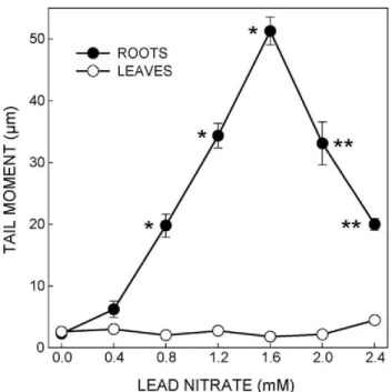
It is also possible to measure TL by using an eyepiece micrometer to come to a conclusion about the condition of DNA. The extent of DNA damage can also be evaluated quantitatively by determining the TM values for each treat-ment group. For example, in a study carried out by Gichner et al. (2006 and 2008), the DNA damage on tobacco plants was expressed in numerical by calculating TM values. They were able to determine the individual effect of genotoxic stress agents such as MH (maleic hydrazide), EMS (ethyl methanesulfonate) or lead nitrate (Figure 2).
The DNA-specific dye and the magnification used for comet visualization depend largely on investigator’s needs
3124 Afr. J. Biotechnol.
Figure 1. Photomicrographs of EtBr-stained DNA from protoplasts of Neurospora crassa processed for alkaline comet assay. (A) Untreated showing no DNA damage (length:width ratio 1:1) and (B) CdSO4 (100 µM) treated showing DNA migration towards anode (length:width ratio 3:2). [Adopted from the work of Bhanoori and Venkateswerlu (1998) with permission of Elsevier].
and presumably have little effect on assay sensitivity or reliability. For some fluorescent dyes, anti-fade could be used to greatly reduce the rate of signal quenching (Tebbs et al., 1999), allowing the same slide to be scored multiple times.
Statistical analysis of comet assays
Generally data are analyzed using the basic versions of any statistical and graphical package programs such as sigmaplot or SPSS. TM or % of tail DNA values are analyzed with a 1-way analysis of variance test. If a significant F-value of P < 0.05 is obtained, then multiple comparison test between the treated and control group is conducted. Differences between 2 groups are statistically evaluated by using the paired t-test (Gichner et al., 2006; Mancini et al., 2006).
Figure 2. The average median tail moments (TMs) in root and leaf nuclei after a 24 h treatment of tobacco (Nicotiana tabacum var. xanthi) seedlings with lead nitrate. The error bars represent the standard error of the mean. *Significantly (P < 0.05) different from the control. **Significantly (P < 0.05) lower than the TM after treatment with 1.6 mmol L-1 lead nitrate. [Adopted from the work of Gichner et al. (2008) with permission of Elsevier].
Preparation and storage conditions of the reagents used in the assay
i) Tris buffer: 0.4 M, pH 7.5-store at 4ºC.
ii) NMP agarose (ROTH Germany or Sigma): dissolve NMP in dH2O at 50ºC store at 4ºC.
iii) LMP agarose (ROTH Germany or Sigma): dissolve LMP in PBS at boiling temperature- store at 4ºC.
iv) PBS: Contents were given in the text. Store at 4ºC. v) Electrophoresis buffer: Store at 4ºC.
vi) Fluorescent DNA stains e.g. EtBr (80-100 µl, 20 µg l
-1). Store at 4ºC.
Equipments
i) Large-bed gel electrophoresis tank and a power supply unit.
ii) Fluorescence microscope equipped with an excitation filter of 515 - 560 nm and a barrier filter of 590 nm. iii) Computerized image analysis system linked to a charge-coupled device (CCD) camera.
iv) And general laboratory equipments such as water bath for melting agarose, heamocytometer for counting cells, frosted-end slides and coverslips for nuclei. Fully frosted-end slides could also be used but drying and storing after staining could damage to the cells.
+ 50
--e-
ROOTS--er-
LEAVES 40E
2;ı-z
30 w ~o
~ ....J 204:
ı-+ 10 O O.O 0.4 0.8 1.2 1.6 2.0 2.4 LEAD NITRATE (mM)Timing
i) Preparation of reagents, 30 min.
ii) Slide preparation, cell-sample preparation and agarose embedding: 1 h.
iii) Unwinding and electrophoresis, neutralization: 2 h. iv) Staining: 30 min.
v) Scoring and analysis: 2 h.
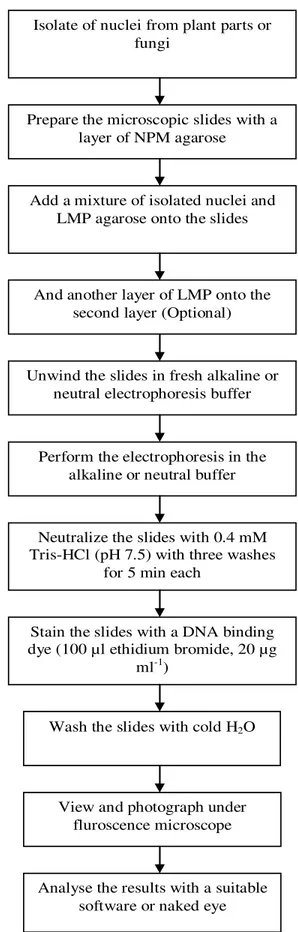
vi) The above steps for the comet assay protocol are also illustrated in Figure 3.
LIMITATIONS OF THE METHOD
The main limitation to the application of this technique to all eukaryotic cells is the fact that solid tissues need a previous treatment to free the individual cells (Singh et al., 1988; Tice, 1995; Demirbag et al., 2005). Preparation of the samples for the assay is one of the most important steps. If this step is performed rapidly and accurately, then any DNA damage caused by the procedure could be minimized significantly. There are, however, practical limitations to the number of cells and samples that can be analyzed. If the automated system to be used maximally 500 - 600 comets per hour can be scored and analyzed (Olive and Banath, 2006). Manually, 50 slides per day could be evaluated. If there is heterogeneity in the sample, more slides should be counted. Viability of cell suspension could be checked if necessary. On the other hand, necrotic and apoptic cells could be mixed in a population, this could prevent the workers from having accurate information on detection of single and double strand breaks as well as base damage. If one is suspicious about this case, these cell types could be distinguished by using DNA diffusion assay method (Singh, 2000).
It is also difficult to come to a conclusion about the degree of severity of the stress in any organism by just looking at the extent of DNA damage under the micro-scope. Comparing comet assay results with other mole-cular or biochemical methods could be very useful to have an idea about the resistance of target organism and the proposed damaging agents. It is important to note that DNA damage is not only caused by a direct effect of genotoxicity (Olive and Banath, 2006) but membrane damage, oxidative stress or unsuitable isolation methods for nuclei could also cause extensive DNA damages.
Conclusion
The main issue in comet assay is to measure increased DNA migration values in treated cells without affecting the migration in controls and make dose-response comparisons between the groups and conclude the extent of damage in cells. Compared with other genotoxicity assays, the
Dikilitas et al. 3125
Isolate of nuclei from plant parts or fungi
Prepare the microscopic slides with a layer of NPM agarose
Add a mixture of isolated nuclei and LMP agarose onto the slides
And another layer of LMP onto the second layer (Optional)
Unwind the slides in fresh alkaline or neutral electrophoresis buffer
Perform the electrophoresis in the alkaline or neutral buffer
Neutralize the slides with 0.4 mM Tris-HCl (pH 7.5) with three washes
for 5 min each
Stain the slides with a DNA binding dye (100 µl ethidium bromide, 20 µg
ml-1)
Analyse the results with a suitable software or naked eye Wash the slides with cold H2O
View and photograph under fluroscence microscope
Figure 3. General illustration of the comet assay protocol.
ı
+
+
ı
+
+
ı
+
ı
ı
3126 Afr. J. Biotechnol.
advantages of the technique include: its demonstrated sensitivity for detecting low levels of DNA damage, the requirement for small numbers of cells per sample, flexibility, low costs, ease of application, the ability to conduct studies using relatively small amounts of a test substance and the relatively short time period (half a day) compared to most conventional cytogenetics which take a few days needed to complete an experiment (Lin et al., 2007; Gichner et al., 2008). For each species, e.g. plant, fungus, etc., the comet assay protocol has to be modified, especially during unwinding stage. Voltage and amperage of electrophoresis should be rearranged if needed. Not only seedlings, but also isolated leaves or roots of various mature plants could also be used in the assay. In many molecular assays, calli or cell cultures are preferred due to their sensitivity and required amount of work, which is less time consuming than working on whole-plant organisms. However, establishment of a laboratory with good facilities such as illuminated incubators and thermo-stable orbital shakers for studying cell and callus cultures is not always easy and quick, especially in the early stages of laboratory establishment. With this assay, research programs and projects would be more valuable and reliable. On the other hand, it is not always possible to work on callus or cell cultures for every plant species. In most cases, in vivo conditions have been preferred. Therefore, this assay would provide a new opportunity for scientists working on biochemical stress mechanisms and pathways.
CONFLICT OF INTEREST STATEMENT
The authors declare that they have no conflict of interest.
COMPETING INTEREST STATEMENT
The authors declare that no endorsement of named products is intended nor any criticism implied of similar products which are not mentioned.
ACKNOWLEDGEMENTS
We are thankful to Prof. Tomas Gichner and his colleagues for introducing, developing and adapting the comet assay protocols for the agricultural sciences. The authors are grateful to the source Elsevier for giving permissions of the figures.
REFERENCES
Angelis KJ, Dusinska M, Collins AR (1999). Single cell gel electrophoresis: detection of DNA damage at different levels of sensitivity. Electrophoresis 20:2133-2138.
Angelis KJ, McGuffie M, Menke M, Schubertz I (2000). Adaptation to alkylation damage in DNA measured by the comet assay. Environ. Mol. Mutagen. 36:146-150.
Baysal Z, Cengiz M, Ozgonul A, Cakir M, Celik H, Kocyigit A (2009). Oxidative status and DNA damage in operating room personnel. Clin. Biochem. 42 (3): 189-193.
Bhanoori M, Venkateswerlu G (1998). The alkaline single cell gel electrophoresis: a new test for assessing DNA single strand breaks in Neurosporacrassa. Mutat. Res. 405:29-34.
Collins AR, Dobson VL, Dusinska M, Kennedy G, Stetina R (1997). The comet assay: what can it really tell us? Mutat. Res. 375:183-193. Collins AR, Harrington V (2002). Repair of oxidative DNA damage:
assessing its contribution to cancer prevention. Mutagenesis 17:489-493.
Demirbag R, Yılmaz Y, Kocyigit A (2005). Relationship between DNA damage, total antioxidant capacity and coronary artery disease. Mutat. Res. 570:197-203.
Dikilitas M (2003). Effect of salinity & its interactions with Verticillium albo-atrum on the disease development in tomato (Lycopersicon esculentum Mill.) and lucerne (Medicago sativa & M. media) plants. Ph.D. Thesis, University of Wales, Swansea.
Dikilitas M, Smith CJ (2004). Bitki, patojen ve tuz arasındaki ili kilerin de erlendirilmesi. I. Bitki Koruma Kongresi 8-10 Eylül 2004, Samsun. Dikilitas M, Smith CJ (1997). The effect of spore concentration of
Verticillium albo-atrum and salt tolerant on infection and disease progression in tomato plants. Presidential Meeting 1997 at the University of York, UK, 16-18 December 1997.
Fairbairn DW, Olive PL, O’Neill KL (1995). The Comet assay: a comprehensive review. Mutat. Res. 339:37-59.
Gadd GM (1993). Interactions of fungi with toxic metals. New. Phytol. 124:25-60.
Garaj-Vrhovac V, Zeljezic D (2000). Evaluation of DNA damage in workers occupationally exposed to pesticides using single-cell gel electrophoresis (SCGE) assay: Pesticide genotoxicity revealed by comet assay. Mutat. Res. 469:279-285.
Gichner T (2003). Differential genotoxicity of ethyl methanesulphonate, N-ethyl-N-nitrosourea and maleic hydrazide in tobacco seedlings based on data of the Comet assay and two recombination assays. Mutat. Res. 538: 171-179.
Gichner T, Mühlfeldova Z (2002). Induced DNA damaged measured by the Comet assay in 10 weed species. Biol. Plant. 45:509-516. Gichner T, Patkova Z, Kim JK (2003). DNA damage measured by the
Comet assay in eight agronomic plants. Biol. Plant. 47:185-188. Gichner T, Patkova Z, Szakova J, Demmerova K (2006). Toxicity and
DNA damage in tobacco plants growing on soil polluted with heavy metals. Ecotox. Environ. Safe. 65:420-426.
Gichner T, Patkova Z, Szakova J, Demnerova K (2004). Cadmium induces DNA damage in tobacco roots, but no DNA damage, somatic mutations or homologous recombinations in tobacco leaves. Mutat. Res. 559:49-57.
Gichner T, Plewa MJ (1998). Induction of somatic DNA damage as measured by single cell gel electrophoresis and point mutation in leaves of tobacco plants. Mutat. Res. 401:143-152.
Gichner T, Znidar I, Szakova J (2008). Evaluation of DNA damage and mutagenicity induced by lead in tobacco plants. Mutat. Res. 652:186-190.
Gichner, T, Badayev SA, Demchenko SI, Relichova J, Sandhu SS, Usmanov PD, Usmanova O, Veleminsky J (1994). Arabidopsis assay for mutagenicity. Mutat. Res. 310:249-256.
Gichner, T, Mukherjee A, Veleminsky J (2006). DNA staining with the fluorochromes EtBr, DAPI and YOYO-1 in the comet assay with tobacco plants after treatment with ethyl methanesulphonate, hyperthermia and DNase-I. Mutat. Res. 605:17-21.
Hahn A, Hock B (1999). Assessment of DNA damage in filamentous fungi by single cell gel electrophoresis, comet assay. Environ. Toxicol. Chem. 18 (7): 1411-1424.
Hartmann A, Agurell E, Beevers C, Brendler-Schwaab S, Burlinson B, Clay P, Collins A, Smith A, Speit G, Thybaud V, Tice RR (2003). Recommendations for conducting the in vivo alkaline Comet assay. Mutagenesis 18:45-51.
Jiang XF, Zhu ZH, Zhou J (1998). Application of comet assay in plant protoplast apoptosis detection. Acta Bot. Sinica 40:928-932.
adaptation to MNU-induced DNA damage in barley. Mutat. Res. 493:95-100.
Klaude M, Ericson S, Nygren J, Ahnstrom G (1996). The comet assay: mechanisms and technical considerations Mutat.Res. 363:89–96. Kobayashi H, Sugiyama C, Morikawa Y, Hayashi M, Sofuni T (1995). A
comparison between manual microscopic analysis and computerized image analysis in the single cell gel electrophoresis assay. MMS Commun. 3:103-115.
Kocyigit A, Keles H, Selek S, Guzel S, Celik H, Erel O (2005). Increased DNA damage and oxidative stress in patients with cutaneous leishmaniasis. Mutat. Res. 585:71-78.
Koppen G, Toncelli LM, Triest L, Verschaeve L (1999). The Comet assay: a tool to study alteration of DNA integrity in developing plant leaves. Mech. Ageing Dev. 110:13-24.
Koppen G, Verschaeve L (1996). The alkaline comet test on plant cells: A new genotoxicity test for DNA strand breaks in Vicia faba root cells. Mutat. Res. 360:193-200.
Lin A, Zhang X, Chen M, Cao Q (2007). Oxidative stress and DNA damages induced by cadmium accumulation. J. Environ. Sci. 19:596-602.
Mancini A, Buschini A, Restivo FM, Rossi C, Poli P (2006). Oxidative stress as DNA damage in different transgenic tobacco plants. Plant Sci. 170:845-852.
Menke M, Chen Peng I, Angelis KJ, Schubert I (2001). DNA damage and repair in Arabidopsisthaliana as measured by the comet assay after treatment with different classes of genotoxins. Mutat. Res. 493:87–93.
Miloshew G, Mihaylov I, Anachkova B (2002). Application of the single cell gel electrophoresis on yeast cells. Mutat. Res. 513: 69-74. Mitchelmore CL, Chipman JK (1998). DNA strand breakage in aquatic
organisms and potential value of the comet assay in environmental monitoring. Mutat. Res. 399:135-147.
Navarrete MH, Carrera P, Demiguel M, Delatorre C (1997). A fast comet assay variant for solid tissue cells: The assessment of DNA damage in higher plants. Mutat. Res. 389:271-277.
Olive PL, Banath JP (2006). The Comet assay: a method to measure DNA damage in individual cells. Nat. Prot. 1:23-29.
Olive PL, Banath JP, Durand RE (1990). Heterogeneity in radiation-induced DNA damage and repair in tumor and normal cells measured using the “comet” assay. Radiat. Res. 122:86-94.
Östling O, Johanson KJ (1984). Microelectrophoretic study of radiation-induced DNA damages in individual mammalian cells. Biochem. Biophys. Res. Commun. 123:291-298.
Piperakis SM, Petrakou E, Tsilimigaki S (2000). Effects of air pollution and smoking on DNA damage of human lymphocytes. Environ. Mol. Mutagen. 36:243–249.
Piperakis SM, Petrakou E, Tsilimigaki S, Sagnou M, Monogiudis E, Haniotakis G, Karkaseli H, Sarikaki E (2003). Biomonitoring with the comet assay of Greek greenhouse workers exposed to pesticides. Environ. Mol. Mutagen. 41:104-110.
Rojas E, Lopez MC, Valverde M (1999). Single cell gel electrophoresis assay: methodology and applications. J. Chromat. B. 722:225-254.
Dikilitas et al. 3127
Singh NP (1996). Microgel electrophoresis of DNA from individual cells: Principles and Methodology. In: GP Pfeifer (Ed.): Technologies for Detection of DNA Damage and MutationsPlenum Press, New York, pp. 3-24.
Singh NP (2000). A simple method for accurate estimation of apoptotic cells. Exp. Cell Res. 256:328-337.
Singh NP, McCoy MT, Tice RR, Schneider EL (1988). A simple technique for quantitation of low levels of DNA damage in individual cells. Exp. Cell Res. 175:184-91.
Singh NP, Stephens RE, Schneider EL (1994). Modification of alkaline microgel electrophoresis for sensitive detection of DNA damage. Int. J. Radiat. Biol.66:23-28.
Sriussadaporn C, Yamamoto K, Fukushi K, Simazaki D (2003). Comparison of DNA damage detected by plant comet assay in roadside and non-roadside environments. Mutat. Res. 541:31-44. Stavreva DA, Gichner T (2002). DNA damage induced by hydrogen
peroxide in cultured tobacco cells is dependent on the cell growth stage. Mutat. Res. 514:147-152.
Tatli MM, Minnet C, Kocyigit A, Karadag A (2008). Phototherapy increases DNA damage in lymphocytes of hyperbbilirubinemic nonates. Mutat. Res. 654 (1): 93-95.
Tebbs RS, Pederson RA, Cleaver JE, Hartmann A (1999). Modification of the comet assay for the detection of DNA strand breaks in extremely small samples. Mutagenesis 14:437-438.
Tice RR (1995). The single cell gel/comet assay: a micro gel electrophoretic technique for the detection of DNA damage and repair in individual cells, in: DH Phillips, S Venitt. Bios (Eds.), Environmental Mutagenesis. Bios Scientific Publishers, Oxford, 1995, pp. 315-319. Tice RR, Agurell E, Anderson D, Burlinson B, Hartmann A, Kobayashi H
(2000). The single cell gel/comet assay: guidelines for in vitro and in vivo genetic toxicology testing. Environ. Mol. Mutagen. 35:206-21. Trevigen Inc. (2000). TREVIGENTM Instructions for Comet AssayTM. Trevors JT, Stration GW, Gadd GM (1986). Cadmium transport,
resistance and toxicity in bacteria, algae and fungi, Can. J. Microbiol. 32:447-464.
Unkles SE, Campbell EI, Carrez D, Grieve C, Contreas R, Fiers W, Van den Hondel CAMJJ, Kinghorn JR (1989). Transformation of Aspergillus niger with the homologous nitrate reductase gene. Gene 78:157-166.
Waalkes MP, Poirier LA (1984). In vitro cadmium-DNA interactions: cooperativity of cadmium binding and competitive antagonism by calcium, magnesium and zinc. Toxicol. Appl. Pharmacol. 75:539-546.