DOI: 10.5455/annalsmedres.2019.11.703 2020;27(3):814-8
Plasma and salivary irisin levels in lepramatous leprosy
patients
Emine Tugba Alatas1, Mehmet Kalayci2, Asude Kara Polat3, Gursoy Dogan1, Asli Akin Belli4 1Mugla Sitki Kocman University, Faculty of Medicine, Department of Dermatology, Mugla, Turkey
2Elazig Training and Research Hospital, Clinic of Biochemistry, Elazig, Turkey 3Istanbul Training and Research Hospital, Clinic of Dermatology, Istanbul, Turkey 4Erdem Hospital, Clinic of Dermatology, Istanbul, Turkey
Copyright © 2020 by authors and Annals of Medical Research Publishing Inc. Abstract
Aim: We aimed to investigate the plasma and salivary irisin levels in leprosy patients, as well as to evaluate the role of the irisin in
the leprosy pathogenesis. Leprosy is a granulomatous disease with the peripheral nerves, mucous membrane, and skin involvement primarily. Irisin, a novel protein, has been associated with several inflammatory diseases and demonstrated in the peripheral nerve cells, deep sebaceous glands, and saliva excessively. To our knowledge, there is no study investigating the irisin levels in patients with leprosy.
Material and Methods: We conducted a case-control study on 20 patients with lepromatous leprosy and 20 healthy controls between
January 2016 and January 2017. Of the participants, the demographic data and disease history including duration of the disease, any current treatments, and presence of any other family members with leprosy were recorded. The plasma and salivary irisin levels of the participants were measured by enzyme-linked immunosorbent assay (ELISA). Additionally, the serum levels of C-reactive protein (CRP), glucose, hemoglobin A1c (HbA1C), triglyceride, cholesterol, high-density lipoprotein (HDL) and low-density lipoprotein (LDL) were evaluated in both groups.
Results: The plasma and salivary irisin levels were significantly lower in the patient group than those in the control group (P = 0.001
and P = 0.03, respectively). In patients with leprosy, the serum and salivary irisin levels had a positive correlation (r: 576, p: 0.008). In addition, there was a negative correlation between the plasma irisin and CRP levels (r: −468, p: 0.037).
Conclusion:The irisin may act an immunomodulatory role in leprosy. The irisin production is probably suppressed by the increased inflammatory markers in leprosy or the bacillus to reach the temperature for its reproduction. Further studies are needed to clarify the role and prognostic value of the irisin in leprosy with a large number of patients examined before and after the treatments.
Keywords: Irisin; lepromatous leprosy; saliva
Received: 07.11.2019 Accepted: 10.12.2019 Available online: 25.02.2020
Corresponding Author: Emine Tugba Alatas, Mugla Sitki Kocman University, Faculty of Medicine, Department of Dermatology, Mugla,
Turkey E-mail: [email protected]
INTRODUCTION
Leprosy is a chronic granulomatous infectious disease caused by Mycobacterium leprae that infects primarily superficial peripheral nerves, upper respiratory tract mucosa, eyes, bones, and testicles (1). There are two major leprosy forms; lepromatous and tuberculoid leprosy which depend on the immunity of the individual. Whereas there is T-cell specific unresponsiveness to the bacillus in lepromatous leprosy, a strong T-cell proliferation is seen in tuberculoid leprosy and the infection remains limited to one or more skin regions and peripheral nerves (1-3).
The irisin, a protein consisting of 112 amino acids, was discovered by Boström et al. in 20xx (4). It is synthesized in the skeletal muscle, heart muscle, liver, pancreas, salivary gland, connective tissue, sweat glands, subcutaneous adipose tissue, and also nerve sheath (5). Current studies have suggested that the irisin plays a role in the pathogenesis of several diseases including diabetes, obesity, metabolic syndrome, psoriasis, and hepatocellular carcinoma (6-8). Moreover, the irisin has been shown to be abundant in the peripheral nerve cells, deep sebaceous glands, and saliva (9,10).
In addition to being a popular protein associated with many diseases, in particular, synthesis of the irisin from peripheral nerve cells has encouraged us to consider the role of irisin in the development of leprosy. To the best of our knowledge, there is no study investigating plasma and salivary irisin levels in leprosy patients.
We aimed to investigate the plasma and salivary irisin levels in leprosy patients compared with healthy controls in our study.
MATERIAL and METHODS
Study design
We conducted a case-control study on 20 patients with leprosy and 20 healthy controls at the Muğla Sıtkı Kocaman University Faculty of Medicine, Department of Dermatology and Elazığ Training and Research Hospital, Leprosy Service between January 2016 and January 2017. Leprosy diagnosis of the patients was based on clinical, histopathological, and direct microscopically examinations. The patient group consisted of inactive lepromatous leprosy patients who had completed the treatment previously. The control group consisted of 20 voluntary participants who meet the inclusion / exclusion criteria. The Ethics Committee approval was obtained from the Muğla Sıtkı Koçman University Clinical Research Ethics Committee prior to the study. Both groups were informed about the study and written permissions were obtained.
The exclusion criteria for the participants were as follows: the presence of pregnancy, breastfeeding, coronary artery disease, malignancy, renal failure, and hepatic failure and age younger than 18 years. Detailed disease history was taken from all the participants; demographic data, duration of the disease, any current treatments, and presence of any other family members with leprosy were recorded on the patient forms. The height and weight information of all the participants were measured. Body mass index (BMI) was calculated by dividing the person’s weight by the square of height (kg / m²).
Among biochemical tests, we examined the serum levels of C-reactive protein (CRP), glucose, hemoglobin A1c (HbA1C), triglyceride, cholesterol, high-density lipoprotein (HDL), and low-density lipoprotein (LDL) in both groups. Also, the plasma and salivary irisin levels of the participants were measured by enzyme-linked immunosorbent assay (ELISA).
Collection of samples
In the study, the samples were taken from the patients and controls in the morning (8-12 hours after the fasting period), one tube was given to the flat biochemistry tube, one was in the tube containing K2-EDTA, and one was in the aprotinin tube. Blood samples taken to flat biochemistry tubes were centrifuged at 4000 rpm for 10 minutes to obtain serum and fasting glucose, total cholesterol, HDL, LDL, triglyceride levels. Roche Cobas c702 Chemistry System analyzer and CRP levels Beckman
Coulter Immage 800 nephelometry System analyzer were also studied from these sera in the same day. HbA1C levels from the tube containing K2-EDTA were measured by High-performance liquid chromatography. Blood samples taken to tubes containing aprotinin (BD Vacutainer SST II Advance, BD, Plymouth, UK) were centrifuged at 4000 rpm for 10 minutes and the plasma samples were placed in small volume tubes and stored at -80 ° C until the working day for the study of irisin.
In addition, saliva samples were taken from the patient and control groups using the saliva collection tube (Salivette, Sarstedt Ag & Co, Nümbrecht, Deutschland). The patient removes the swab from the Saliva collection tube and places the swab in the mouth and chews it for about 60 seconds to stimulate salivation. Subsequently, the swab with the absorbed saliva is returned to the Saliva collection tube. Centrifugation for 2 minutes at 4000 rpm yields a clear saliva sample in the conical tube. The saliva samples were placed in small volume tubes and stored at -80 ° C until the working day for the study of the irisin. Plasma and saliva irisin concentrations were measured using an ELISA kit (Phoenix Pharmaceuticals, catalog no: EK-067-52, CA, USA) in accordance with the manufacturer’s instructions, with ChroMate Microplate Reader (Awareness Technology, Inc. Palm City, USA). The intra-assay and inter-assay coefficients of variation for irisin were 4-6% and 8-10%, respectively.
Statistical analysis
The data obtained in the study were demonstrated as mean ± standard deviation. Before the comparison between the groups, the Kolmogorov-Smirnov test was used to check whether the data distribution was normal. The Chi-square test was used for nonparametric parameters. The comparison of parameters between the two groups and analysis of the relations between the parameters in the groups were made with Student’s t-test and Pearson correlation test, respectively. A p-value <0.05 was accepted as the level of significance.
RESULTS
The duration of the disease was 43.15±2.74 in the patient group. All patients had previously treated with rifampin, clophasimine and dapsone. At least one sibling of patients had a diagnosis of leprosy.
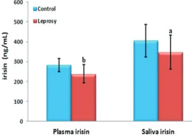
There were 11 males (55%) and 9 females (45%) in the patient group (age range 35-76, mean 65.8 ± 10.01 years); 10 males (50%) and 10 females (50%) in the control group (age range 33-75, mean 56.5 ± 13.08 years). There was no statistically significant difference between the two groups in the distribution of gender (P = 0.752). The mean BMI was 23.53 ± 1.94 and 24.06 ± 2.77 kg/m2 in the patient and control groups, respectively (P = 0.351) (Table 1). The plasma and salivary irisin levels were significantly lower in the patient group than those in the control group (P = 0.001 and P = 0.033, respectively) (Figure 1).
The serum and salivary irisin levels showed a positive correlation in the patient and control groups (r: 576, p: 0.008 and r: 764, P: <0.001, respectively). Additionally, the plasma irisin levels had a negative correlation with triglyceride (r: −481, p: 0.032) and CRP (r: −468, p: 0.037) and a positive correlation with HDL levels (r: 535, p: 0.015) in the patient group.
a: P = 0.033 (When compared with the control group) b: P = 0.001 (When compared with the control group)
Figure 1.Serum and salivary irisin levels of the patient and
control groups
DISCUSSION
Since the irisin is secreted from peripheral nerves besides the other tissues, has been associated with several inflammatory diseases, and may have an immunomodulatory activity in leprosy, we conducted the current study. Although irisin is a novel and popular protein examined in recent studies, there is no study investigating the role of irisin in skin diseases other than acne vulgaris and psoriasis. We found the serum and salivary irisin levels decreased in the patients with leprosy than in the healthy controls. In addition, we demonstrated a negative correlation between the irisin and CRP levels.
The main involvement sites of leprosy are peripheral nerves, skin, mucous membrane, bone, and visceral tissue (2). Because Mycobacterium leprae grows at 35°C gradient, it infects cold regions of the body including nose, testicles, and earlobes where the peripheral nerves are close to the skin (2). Leprosy bacillus infects macrophages and peripheral nerves, particularly Schwann cells. When the leprosy bacillus enters the body, two alternative situations occur. In the first case, immunity and resistance develop, leprosy bacillus is phagocytized by the Schwann cells, and the primary infection is terminated without the disease. In the second case, the leprosy disease develops, the bacillus escapes from the phagocytosis in macrophages, and foamy leprosy bacillus cells develop (2).
It has been showed that mutations in the HLA-DR2 and HLA-R3 genes are responsible for the tuberculoid
Table 1. Sociodemographic characteristics and biochemical parameters of the patient and control groups
Leprosy group n=20
mean ± SD Control group n=20mean ± SD P
Age (years) 65.8 ± 10.01 56.5 ± 13.08 0.013
Sex (female/male) 9/11 10/10 0.752
Body mass index (kg/m2) 23.53 ± 1.94 24.06 ± 2.77 0.351
C-reactive protein (mg/dl) 1.14 ± 0.8 0.52 ± 0.25 0.02 Glucose (mg/dl) 89.35± 6.43 86.55±7.66 0.218 Hemoglobin A1c (%) 5.2±0.24 5.21±0.52 0.907 Total Cholesterol (mg/dl) 194.35±37.16 193.05±31.53 0.906 HDL (mg/dl) 45.15±10.23 48.55±8.83 0.268 LDL (mg/dl) 119.54±29.89 116.15±28.37 0.714 Triglyceride (mg/dl) 148.15±53.25 142.73±9.48 0.715 Plasma irisin (ng/ml) 238.48±45.76 282.38±32.30 0.001 Salivary irisin (ng/ml) 346.87±85.70 405.10±80.73 0.033
form and mutations in HLA-DQ1 are responsible for the lepromatous form (11). The Th1 CD4 T-cell response is dominant in tuberculoid leprosy, whereas the Th2 response is dominant in lepromatous leprosy. The Th2 response in lepromatous leprosy leads to the release of interleukin (IL)-4, IL-5, IL-10, and IL-13 to suppress macrophages. In addition, phenolic glycolipid-1 in the basal cell wall reduces T-cell response and interferon (IFN)-γ production (11).
Irisin is a novel protein that exists in many tissues including peripheral nerves. It was firstly demonstrated in muscle tissue and interestingly, the post-exercise concentrations of irisin have been found increased in both mice and humans (11). In some animal models, irisin has been shown to prolong the life span, reduce overall body weight by increasing total energy consumption, and reduce dietary-induced insulin resistance (4). In several studies, the increased irisin levels have been associated with obesity, insulin resistance, metabolic diseases, and hepatic steatosis (12-15). Therefore, irisin has been suggested to be a predictive marker of diabetes mellitus, carotid arteriosclerosis, chronic renal failure, and polycystic ovarian syndrome (16-19).
In addition to the association of irisin with the inflammatory diseases, its anti-inflammatory activity has also been reported (20,21). A negative correlation between the irisin levels and C-reactive protein has been demonstrated in several studies (22,23). Gannon et al. have reported that irisin shows antitumor activity by inducing apoptosis with the reduction of the activation of NFκB in breast cancer cells (24). Also, Mazur-Bialy et al. showed that irisin works as a regulator on macrophages and improves phagocytosis ability in mice (25). These findings suggest that irisin is effective on the immune system and may play a potential modulatory role in immune response or macrophage activity. In the current study, we found lower irisin levels in patients with leprosy compared with those in the control group. Our results may be related to the fact that releasing of cytokines related to Th2 response such as IL-4, IL-5, IL-10, and IL-13 suppress macrophages and IFN-γ production in the cell wall of the bacillus and thus, the irisin expression may be inhibited by this inflammatory cytokines. In addition, we found a negative correlation between the irisin and CRP levels similar to the literature data which also supports the hypothesis of the inhibition of irisin by increased inflammatory markers.
Peripheral nerve involvement in leprosy results in the loss of sensation of temperature, mild tingling, pain, loss of deep pressure sensation, and motor complications, respectively. Motor complications such as muscle weakness and muscle atrophy may also develop in patients with leprosy (10,26). It is known that the secretion of irisin from skeletal muscles increases after exercise. In a study conducted by Chang et al. the levels of irisin in patients with muscle weakness and atrophy were lower than those in the control group (27). Based on this information, the reduced levels of irisin in leprosy patients noted in our study is not an unexpended result because of muscular involvement due to leprosy.
Whereas M. leprae requires a 35°C gradient to grow, the irisin contributes to keeping the body temperature at 36.5°C (11,28). The irisin increases the synthesis of uncoupling protein-1 (UCP-1) in the mitochondrial membrane. Because of the increased synthesis of UCP-1, the protons leak back to the mitochondrial membrane and heat generation takes place instead of energy generation (28). The decreased irisin levels in leprosy patients that we found may be related to the temperature difference they prefer or contribute. Further studies should be needed to elucidate that whether the bacillus suppresses the synthesis of irisin to reach the temperature required for bacillus reproduction or this finding is just a coincidence. The limitations of our study were; relatively small number sample size and that it included the leprosy patients who had completed the treatment.
CONCLUSION
In conclusion, we found the plasma and salivary irisin levels lower in patients with lepromatous leprosy than those in the healthy controls and a negative correlation between the irisin and CRP levels. To the best of our knowledge, this is the first study evaluating plasma and salivary irisin levels in the patients with lepromatous leprosy. The irisin may play a role in the pathogenesis of lepromatous leprosy as an immunomodulatory agent. However, it should be deepened that whether the lower irisin levels in leprosy patients is related to the inhibition of irisin by increased inflammatory markers, to the development of muscular atrophy in the patients with leprosy, or to the suppression of irisin synthesis by the bacillus to reach the temperature required for bacillus reproduction. Further, the irisin levels should be examined before and after the treatments to show its role in the leprosy pathogenesis more clearly and whether it can be useful as a prognostic marker in leprosy.
Competing interests: The authors declare that they have no competing interest.
Financial Disclosure: There are no financial supports.
Ethical approval: The Ethics Committee approval was obtained from the Muğla Sıtkı Koçman University Clinical Research Ethics Committee prior to the study.
Emine Tugba Alatas ORCID: 0000-0002-5727-9474 Mehmet Kalayci ORCID: 0000-0001-9122-9289 Asude Kara Polat ORCID: 0000-0002-5040-6901 Gursoy Dogan ORCID: 0000-0001-6103-9417 Asli Akin Belli ORCID: 0000-0002-4197-9716
REFERENCES
1. Pereira RM, Calegari-Silva TC, Hernandez MO, et al. Mycobacterium leprae induces NF-kappaB-dependent transcription repression in human Schwann cells. Biochem Biophys Res Commun 2005;335:20-6. 2. Lockwood DNJ. Leprosy. In: Burns DA, Breatnach SM,
Cox N, Griffits CE, et al, editors. Rook’s text book of dermatology. 7th edition. Oxford: Blackwell Publishing 2004;1-21.
3. Rea TH, Modlin RL. Leprosy. In: Wolf K, Goldsmith LA, Karz S, Gilchrest BA, Paller AS, Leffell DJ, et al, editors. Dermatology in General Medicine. 7th edition. New York: McGraw Hill Professional 2008;1786-96.
4. Bostrom P, Wu J, Jedrychowski MP, et al. A PGC1-alpha-dependent myokine that drives brown-fat-like development of white fat and thermogenesis. Nature 2012;481:463-8.
5. Norheim F, Langleite TM, Hjorth M, et al. The effects of acute and chronic exercise on PGC-1alpha, irisin and browning of subcutaneous adipose tissue in humans. FEBS J 2014;281:739-49.
6. Alatas ET, Kalayci M, Kara A, et al. Association between insülin resistance and serum and salivary irisin levels in patients with psoriasis vulgaris. Dermatologica Sinica 2017;35:12-5.
7. Hojlund K, Boström P. Irisin in obesity and type 2 diabetes. J Diabetes Complications 2013;27:303-4. 8. Gagnini M, Cabiati M, Del Turco S, et al. Increased
FNDC5/Irisin expression in human hepatocellular carcinoma. Peptides 2017;88;62-6.
9. Aydin S, Kuloglu T, Aydin S, et al. Cardiac and skeletal muscle serum irisin responses to with or without water exercise in young and old male rats: cardiac muscle produces more irisin than skeletal muscle. Peptides 2014;52:68-73.
10. Aydin S, Aydin S, Kobat MA, et al. Decreased saliva/ serum irisin concentrations in the acute myocardial infarction promising for being a new candidate biomarker for diagnosis of this pathology. Peptides 2014:56;141-5.
11. Ramos-e-Silva M, Ribeiro de Castro MC. Dermatology. In: Bolognia JL, Jorizzo JL, Rapini RP, et al, editors. Leprosy. 3rd edition. Philadelphia: Elsevier Saunders 2012;1221-42.
12. Lopez-Legarrea P, de la Iglesia R, Crujeiras AB, et al. Higher baseline irisin concentrations are associated with greater reductions in glycemia and insulinemia after weight loss in obese subjects. Nutr Diabetes 2014;4:110.
13. Shi X, Lin M, Liu C, et al. Elevated circulating irisin is associated with lower risk of insulin resistance: Association and path analyses of obese Chinese adults. BMC Endocr Disord 2016;16:44.
14. Chen JQ, Huang YY, Gusdon AM, et al. Irisin: a new molecular marker and target in metabolic disorder. Lipids Health Dis 2015;14:2.
15. Park MJ, Kim DI, Choi JH, et al. New role of irisin in hepatocytes: The protective effect of hepatic steatosis
in vitro. Cell Signal 2015;27:1831-9.
16. Liu JJ, Wong MD, Toy WC, et al. Lower circulating irisin is associated with type 2 diabetes mellitus. J Diabetes Complications 2013;27:365-9.
17. Lee MJ, Lee SA, Nam BY, et al. Irisin, a novel myokine is an independent predictor for sarcopenia and carotid atherosclerosis in dialysis patients. Atherosclerosis 2015; 242:476-82.
18. Wen MS, Wang CY, Lin SL, et al. Decrease in irisin in patients with chronic kidney disease. PLoS ONE 2013; 8:64025.
19. Polak K, Czyzyk A, Simoncini T, et al. New markers of insulin resistance in polycystic ovary syndrome. J Endocrinol Investig 2017;40:1-8.
20. Polyzos SA, Kountouras J, Anastasilakis AD, et al. Irisin in patients with nonalcoholic fatty liver disease. Metabolism 2014;63:207-17.
21. Dulian K, Laskowski R, Grzywacz T, et al. The whole body cryostimulation modifies irisin concentration and reduces inflammation in middle aged, obese men. Cryobiology 2015;71:398-404.
22. Hou N, Han F. Sun X. The relationship between circulating irisin levels and endothelial function in lean and obese subjects. Clin Endocrinol 2015;88:339-43. 23. Xiang L, Xiang G, Yue L, et al. Circulating irisin levels
are positively associated with endothelium-dependent vasodilation in newly diagnosed type 2 diabetic patients without clinical angiopathy. Atherosclerosis 2014;235:328-33.
24. Gannon NP, Vaughan RA, Garcia-Smith R, et al. Effects of the exercise- inducible myokine irisin on malignant and non-malignant breast epithelial cell behavior in vitro. Int J Cancer 2015;136:197-202.
25. Mazur-Bialy AI. Irisin acts as a regulator of macrophages host defense. Life Sci 2017;176:21. 26. Ernst JD. Leprosy (HansenDisease). In: Goldman L,
Schafer AI, et al, editors. Goldman’s Cecil Medicine. 25th edition. Philadelphia: ElsevierSaunders; 2016;326:2042-6.
27. Chang JS, Kim TH, Nguyen TT, et al. Circulating irisin levels as a predictive biomarker for sarcopenia: A cross-sectional community-based study. Geriatr Gerontol Int 2017;17:2266-73.
28. Albayrak S, Atci IB, Kalayci M, et al. Effect of carnosine, methylprednisolone and their combined application on irisin levels in the plasma and brain of rats with acute spinal cord injury. Neuropeptides 2015;52:47-54.