Ankara Üniv Vet Fak Derg, 61, 107-110, 2014
The effects of pre-milking and post-milking teat disinfection in goats
on udder health and milk quality
*Ali DURALIOĞLU1, Ayhan BAŞTAN1, Seçkin SALAR1, Mehmet CENGİZ3, Mehmet AKAN2 1 Department of Obstetrics and Gynaecology, 2Department of Microbiology, Faculty of Veterinary Medicine, Ankara University,
Ankara; 3Department of Obstetrics and Gynaecology, Faculty of Veterinary Medicine, Atatürk University, Erzurum -Turkey.
Summary: In this study, the effect of pre-milking and post-milking teat disinfection on the intramammary infection and milk quality in goats was examined. In the study, 65 healthy Saanen goats were used. Before sampling and in the 1st, 2nd and 3rd months of
the study, individual mixed milk samples were taken from animals used in the study. Somatic cell count and bacteriological examination were carried out on these milk samples. When the initial sampling and the 1st month sampling were compared, a
decrease at somatic cell count was noticed in the sample taken in the 1st month. In contrast, a slight increase at somatic cell count was
noticed in the samples taken in the 2nd and 3rd months. The average somatic cell counts by months were 878.450 cell/ml, 757.230
cell/ml, 853.770 cell/ml, and 1.143.420 cell/ml, respectively. In the bacteriological examination, the mostly isolated bacteria species among the samples were coagulase negative staphylococcus (CNS), S. aureus and Enterococcus. It was concluded that pre-milking and post-milking teat disinfection cause a decrease in milk somatic cell count and also decrease the rate of a new intramammary infection.
Key words: teat disinfection, udder health, milking hygiene, goat.
Keçilerde sağım öncesi ve sonrası meme başı dezenfeksiyonunun meme sağlığı ve süt kalitesi üzerine etkisi
Özet: Bu çalışmada, keçilerde sağım öncesi ve sonrası uygulanan meme başı dezenfeksiyonunun meme içi enfeksiyon ve süt kalitesi üzerine etkisi araştırıldı. Çalışmada 65 adet sağlıklı Saanen ırkı keçi kullanıldı. Çalışmada kullanılan hayvanlardan çalışmaya başlamadan önce ve çalışmanın 1., 2., ve 3. aylarında karma süt örnekleri alındı. Alınan süt örneklerinden somatik hücre sayımı ve bakteriyolojik muayene yapıldı. Çalışmada başlangıç örneklemesi ile 1. ay örneklemesi karşılaştırıldığında 1. ayda alınan örnekte somatik hücre sayısında bir düşme vardı. Oysa 2. ve 3. aylardaki örneklemelerde ise somatik hücre sayısında hafif bir artış görüldü. Aylara göre ortalama somatik hücre sayıları 878.450, 757.230, 853.770, 1.143.420 hücre/ml idi. Bakteriyolojik muayenede örneklerden en sık izole edilen bakteri türleri koagulaz negatif stafilokok (KNS), S. aureus ve Enterococcus türleri idi. Sonuç olarak sağım öncesi ve sonrası meme başı dezenfeksiyonunun süt somatik hücre sayısında azalmaya neden olduğu ve yeni meme içi enfeksiyon oranını azalttığı tespit edildi.
Anahtar söcükler: meme başı dezenfeksiyonu, meme sağlığı, sağım hijyeni, keçi.
Introduction
Mastitis is a disease shaped depending on various reasons in udder tissue. This disease causes physical and chemical changes in milk and udder tissue (2, 5). Mastitis significantly affects the milk yield and quality. It particularly causes to an increase in somatic cell count (SCC) which is the indicator of milk quality (2). Mastitis is one of the most important problems of dairy goat farms as in the cows (6).
Somatic cell count can be used as a criterion to reveal the health and milk quality. Seventy percent of the somatic cells in the goat milk consists of neutrophil, 21%
consists of lymphocyte, and 0,4% consists of epithelial cells; and somatic cell count of milk is nearly 750.000 per ml (7). Somatic cell count in the goat milk is generally more than the somatic cell count in cow milk, and in the late lactation period, the somatic cell count is over 1.000.000 per ml in milk in healthy dairy goats (4).
In mastitis control programs, treatment of infections present in herd of goats and protect from new infections are the fundamental purposes. Some of the pathogenic microorganisms that cause mastitis can occur through a general circulation, or can also transmit from the teat orifice. Hence, it is essential to prevent the occurrence of
* This study is summarized from the MSc thesis of the first author. Ethics Committee approval was obtained for thesis from Ankara University.
Ali Duralıoğlu - Ayhan Baştan - Seçkin Salar - Mehmet Cengiz - Mehmet Akan 108
new infections decreasing the intensity of bacteria on teat orifice. Disinfection of teat is one of the significant applications carried out for that purpose (1, 3).
The aim of this study is to search for the effect of pre-milking and post-milking teat disinfection in a machine milking goat farm on udder health and milk quality.
Material and Method
In this study, 65 Saanen goats milked once in the mornings in a private farm and varying from the ages of 2 to 4, and between the 100th – 130th days of lactation
were used as animal material. The material was randomly chosen among the goats which had no health problems and were fed similarly.
For the somatic cell count and bacteriological examination, 2 each mixed milk samples were taken from each goat before starting to the study and in the 1st,
2nd and 3rd months during the study period. Within the
specified period, somatic cell count and bacteriological examination were performed from the samples.
Before the sampling, the teats were wiped with 70% sterile cotton swabs, the foremilk was discarded, and then mixed milk samples from the 2 udder halves were taken; one sample for the somatic cell count and the other for bacteriological examination. The same day, milk samples were brought to the laboratory for somatic cell count and bacteriological examination in the cold chain. The somatic cell count was carried out with a device called as Bentley IBC-M Bactoscan ® (Bentley Instruments, USA); and the bacteriological examination was performed according with the conventional method.
During the milking periods after the initial sampling, the teats were dipped into disinfectant solution before and after the milking until the study was completed. Before the milking, a ready-to-use foamy teat disinfectant called Deosan Iomin-D plus ® (Johnson Diversey) which contains iodine against teat disinfection was used, and after the milking, iodine based Deosan Mastimin H ® (Johnson Diversey) which contains glycerin and requires 1/3 dilution was used.
In order to analyze the changes of somatic cell counts by months in statistical calculations, the data
which were not primarily appropriate for normal distribution were made appropriate for the normal distribution through the logarithmic transformation method. The difference between the averages of somatic cell counts by months was analyzed through the help of general linear model for the iterative transformations. All statistical calculations were evaluated with 5% margin of error, and SPSS 14.1 package program was used for the calculation. The results of the bacteriological examination by months were evaluated in percentage.
Results
When the first sample (0th day) and 1st month
sampling were compared in terms of the somatic cell count, a decrease was noticed at somatic cell count in the sample taken 1 month later after disinfection. However, in samples taken 2 and 3 month after the onset of the study, there was a slight increase at somatic cell count. The average somatic cell counts by months were 878.450 cell/ml, 757.230 cell/ml, 853.770 cell/ml, and 1.143.420 cell/ml, respectively. The change of average somatic cell counts according to the order of samples was presented in Figure 1.
Figure 1. Average Somatic Cell Counts by months. Şekil 1. Aylara göre somatik hücre sayısı ortalamaları.
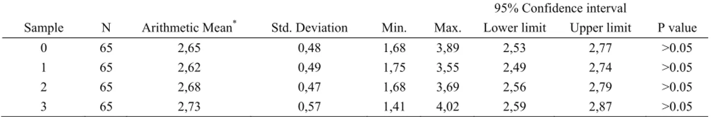
The difference between the average somatic cell counts by months was evaluated by the help of the general linear model for the iterative measurements. As result of the logarithmic transformation; log was determined as log 2,65; log 2,62; log 2,68, and log 2,73; and the difference between the somatic cell counts by
Table 1. Logarithmic view of the average rates of the somatic cell counts. Tablo 1. Somatik hücre sayısı ortalamalarının logaritmik görünümü.
95% Confidence interval
Sample N Arithmetic Mean* Std. Deviation Min. Max. Lower limit Upper limit P value
0 65 2,65 0,48 1,68 3,89 2,53 2,77 >0.05
1 65 2,62 0,49 1,75 3,55 2,49 2,74 >0.05
2 65 2,68 0,47 1,68 3,69 2,56 2,79 >0.05
3 65 2,73 0,57 1,41 4,02 2,59 2,87 >0.05
* Average Somatic Cell Counts were transformed to log 10 to normalize the data.
Ankara Üniv Vet Fak Derg, 61, 2014 109
months was statistically insignificant (p>0.05). The logarithmic view of the average somatic cell counts was shown on Table 1. However, when the average SCC in samples found as infected and negative as result of the culture was compared, a significant difference was obtained in somatic cell counts. In the sampling of the 0th
day, the average SCC of the infected and healthy goats was 972.833 cell/ml and 857.075 cell/ml, respectively. The average SCC in the 1st month sampling was 853.000
cell/ml and 747.491 cell/ml, in the 2nd month it was
1.139.666 cell/ml and 824.694 cell/ml, in the 3rd month it
was 977.000 cell/ml and 1.146.020 cell/ml, respectively. The graphic view of the obtained data was shown in Table 2.
Table 2. The average SCC of infected and healthy goats. Tablo 2. Enfekte ve sağlıklı keçilere ait somatic hücre sayısı ortalamaları. Sample Average SCC Infected Healthy 0 972833 857075 1 853000 747491 2 1139666 824694 3 977000 1146020
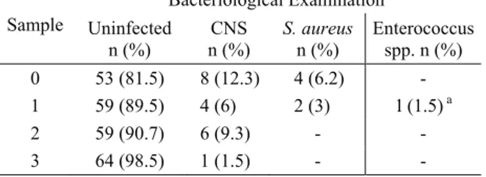
Table 3. The findings of the isolated bacteria. Tablo 3. İzole edilen bakteriler.
Sample Bacteriological Examination Uninfected n (%) n (%) CNS S. aureus n (%) Enterococcus spp. n (%) 0 53 (81.5) 8 (12.3) 4 (6.2) - 1 59 (89.5) 4 (6) 2 (3) 1 (1.5)a 2 59 (90.7) 6 (9.3) - - 3 64 (98.5) 1 (1.5) - -
a Mix infection due to S. aureus and Enterococcus spp. was
detected in one sample.
a Bir örnekte S. aureus and Enterococcus
In the bacteriological examination, CNS, S. aureus and Enterecoccus species among the samples were isolated. Especially CNS was the most frequently isolated bacteria (12.3 % in the initial sampling). In the first sampling taken at the onset of the study (0th day), 53
samples were bacteriologically negative, CNS was isolated in 8 samples, and S. aureus was isolated in 4 samples. In the mixed milk samples obtained in the 1st
month, 59 samples were bacteriologically negative. CNS was isolated in 4 samples, S. aureus was isolated in 2 samples, and both S. aureus and Enterecoocus spp. were isolated in one sample. In the sampling in the 2nd month
of the study, whereas CNS was isolated in 6 samples, the other samples were bacteriologically negative. In the samples obtained in the 3rd month, whereas CNS was
isolated in 1 sample, 64 samples were bacteriologically negative. The findings of the isolated bacteria related to this study were presented in Table 3.
Discussion and Conclusion
Turkey is a country available for common sheep and goat farming through its natural and economic conditions, agricultural structure, and traditions. In Turkey, there have been approximately 6.7 million goats as of 2004, and 280 000 ton milk has been provided milking 3.4 million of those (15).
Mastitis, which is described as the inflammation of the udder tissue, is the most significant problem of the dairy farming throughout the world; moreover, despite the control precautions adopted by dairy farms mastitis causes major economic losses.
White and Hinckley (17) reported the mastitis prevalence as 36.4% in their study in which they carried out through single sampling in 2911 goats. They isolated Staphylococcus spp. from 38.2% of the milk samples with mastitis, S. aureus from 11%, Streptococcus spp. from 4.1%, E. coli from 1.6%, and Pseudomonas spp. from 1.2%. In another study carried out in Spain, Staphylococcus spp. was determined in 70% of the infected goats, Corynebacterium spp. in 24%, Gram negative bacilli in 3%, and mixed infection in 3% (14). In this study, the prevalence of mastitis in the initial sampling was found as 18,5%, and in the milk sampling, CNS was isolated in 12,3% of the samples, and S. aureus was isolated in 6,1%. When the obtained data and the data in the literature were compared (14, 17), no difference was found in mastitis prevalence. The reason for this can be explained through the epidemiological and managing difference.
In another study that was carried out with goat milk with subclinical mastitis, CNS was determined in 60.8% and S. aureus (15.9%) was determined in 15.9% (8). In another study, mastitis pathogens were examined in 630 goat udder halves and it was reported that 68 udder halves was infected with CNS and 2 udder halves were infected with Staphylococcus spp. (10). In their study Virdis et al. (16) isolated the bacteria from 15,6% of the goat udder halves. It was determined that 88.5% of the isolated udder halves were CNS and 5.3% were S. aureus. In their study they carried out with 305 goats, Morini et al. (9) noticed CNS in 87% of the infected goats, S. aureus in 5%, Streptococcus spp. in 3%, and other bacteria and environmental factors in 5%.
In the study, bacteria was isolated in 18.5% of the initial samples. In the infected samples of post-isolation; CNS was determined in 66,48%, and S. aureus in 32,97%; in the 2nd sampling CNS was determined in
33,24% and S. aureus was determined in 16,48%; in the 3rd sampling CNS was determined in 100%, and in the 4th
sampling CNS was determined in 100%. As it can be understood from this result, CNS is the most frequently isolated bacteria, and this result is compatible with the literature data. This result proves us that CNS has a great importance on goat mastitis.
Ali Duralıoğlu - Ayhan Baştan - Seçkin Salar - Mehmet Cengiz - Mehmet Akan 110
The number of leukocytes and epithelial cells in blood increases when an infection occurs in udder tissue. The somatic cells which consist of udder epithelial cells and leukocytes in milk can be used as a criterion to reveal the udder health. It has been reported that the amount of somatic cells in goat milk is generally higher than the amount in cow milk; and in the late lactation period, there have been over 1.000.000 somatic cells in 1 ml milk even in healthy dairy milk goats (4).
In the USA, SCC ≤1.000.000 cell/ml is the legal standard value in goat milk. It has been explained that this amount has been affected by several factors such as location of farm, farm management, feeding, age, stage and period of lactation, intramammary infections, milking (11).
Milk SCC was tried to be determined in a study; SCC was determined as 70% of <500.000 cell/ml in 70% of the samples, as SCC <1.000.000 cell/ml in 83%, and as SCC <1.500.000 cell/ml in 89%. Moreover; as the lactation period proceeds, SCC was reported as being increased both in infected and uninfected goat milks (9). In this study, SCC was measured as 878.000 cell/ml in the initial sampling, as 757.000 cell/ml in the 1st month,
as 853.000 cell/ml in the 2nd month, and as 1.143.000
cell/ml in the 3rd month. When the obtained data and the
literature data were compared, it was noticed that the changes in the lactation period were similar. As the lactation period proceeded, an increase was noticed in SCC. However, a significant decrease occurred in SCC with implemented milking hygiene protocol, and SCC was noticed as being increased physiologically depending on the progress in lactation period.
Consequently, it was concluded that CNS and S. aureus were the most important pathogens that cause mastitis in goats, and pre-milking and post-milking teat disinfection positively affected the udder health and milk quality.
References
1. Alaçam E (1978): Süt ineklerinin mastitisten korunmasında teat dipping’in etkisi üzerine çalısmalar. Doçentlik Tezi, A. Ü. Vet. Fak. Ankara.
2. Baştan A (2010): Meme Başı Dezenfeksiyonu. 272-280. Alınmıştır: İneklerde Meme Sağlığı ve Sorunları. Kardelen ofset, Ankara.
3. Baştan A, Salar S (2012): Sütçü ineklerde meme başı dezenfeksiyonunun önemi. Hasad Hayvancılık, 323, 40-44.
4. Cedden F, Kor A, Keskin S (2002): Laktasyonun geç döneminde keçi sütünde somatik hücre sayımı; yaş, süt verimi ve bazı meme özellikleri ile olan ilişkileri. YYÜ Tar Bil Derg, 12, 63-67.
5. Contreras A, Luengo C, Sa´nchez A, Corrales JC (2003): The role of intramammary pathogens in dairy goats. Livestock Production Science, 79, 273–283.
6. Doğruer G, Sarıbay MK, Ergün Y, Aslantaş Ö (2010): Treatment of subclinical mastitis in damascus goats during lactation. Small Rum Res, 90, 153-155.
7. Haenlein G (2002): Relationship of somatic cell counts in goat milk to mastitis and productivity. Small Rum Res, 45, 163–178.
8. İlhan Z, Taşal İ, Sağcan S, Solmaz H (2011): Subklinik mastitisli keçi sütlerinden aerobik bakterilerin izolasyonu. YYÜ Vet Fak Derg, 22, 89-91.
9. Morini P, Pisoni G, Ruffo G, Boettcher P (2005): Risk factors for intramammary infections and relationship with somatic cell counts in Italian dairy goats. Preventive Veterinary Medicine, 69, 163–173.
10. Ndegwa E, Mulei C, Mumyua S (2000): Risk factors associated with subclinical subacute mastitis in kenyan dairy goats. Israel J Vet Med, 56, 4-8.
11. Poutrel B, Cremoux R, Ducelliez M, Verneau D (1997): Control of intramammary infections in goats: impact on somatic cell counts. J Anim Sci, 75, 566-570.
12. Raynal-Ljutovak K, Pirisi A, Cremoux R, Gonzalo C (2007): Somatic cells of goat and sheep milk: Analytical, sanitary, productive and technological aspects. Small Rum Res, 68, 126–144.
13. Sabuncuoğlu N, Çoban Ö (2006): Mastitis ekonomisi. Atatürk Üniversitesi Vet Bil Derg, 1, 1-5.
14. Sanchez A, Contreras A, Corrales C (1999): Parity as a risk factor for caprine subclinical intramammary infection. Small Rum Res, 31, 197-201.
15. Şimşek Ü, Bayraktar M, Gürses M (2006): Çiftlik koşullarında kıl keçilerine ait bazı verim özelliklerinin araştırılması. FÜ Sağlık Bil Dergisi, 20, 221-227.
16. Virdis S, Scarano C, Cossu F, Spanu V, Spanu C, Santis E (2010): Antibiotic resistance in staphylococcus aureus and coagulase negative Staphylococci isolated from goats with subclinical mastitis. SAGE-Hindawi Access to Research, 1-6.
17. White E, Hinckley I (1999): Prevalence of mastitis pathogens in goat milk. Small Rum Res, 33, 117-121.
Geliş tarihi: 13.06.2013 / Kabul tarihi: 24.12.2013
Address of correspondence:
Prof. Dr. Ayhan Bastan
Ankara University, Faculty of Veterinary Medicine Department Obstetrics and Gynecology
06110, Dışkapı, Ankara-Turkey. e-mail: [email protected]