Ankara Üniv Vet Fak Derg, 53, 97-102, 2006
Kronik böbrek yetmezliği belirtileri gösteren kedilerde biyokimyasal
ve hematolojik değişiklikler*
Arif ALTINTAŞ1, Nilgün ÜREN1, Mert PEKCAN1, Ali KARADENİZ2, Ali Haydar KIRMIZIGÜL3
1 Ankara Üniversitesi Veteriner Fakültesi, Biyokimya Anabilim Dalı; 2 Fizyoloji Anabilim Dalı; 3 İç Hastalıkları Anabilim Dalı, 06110-Ankara.
Özet: Araştırmada, kronik böbrek yetmezliği (KBY) gözlenen kedilerde hematolojik ve biyokimyasal değişikliklerin incelen-mesi ve bulguların hastalığın derecelendirilincelen-mesinde kullanılması amaçlanmıştır. KBY tespit edilen ve yaşları 3-12 arasında değişen 22 dişi kedi ile aynı yaşlarda ve klinik olarak sağlıklı görünen 10 dişi kedi (kontrol) araştırmanın hayvan materyalini oluşturmuştur. Hayvanlara ait idrar örneklerinde protein varlığı ve dansite; kan örneklerinde hemoglobin, hematokrit değer, alyuvar ve akyuvar sayıları ile akyuvarların % dağılımları; serum örneklerinde ise toplam protein, üre, kreatinin, sodyum, potasyum, kalsiyum, fosfor, osteokalsin (OC), toplam ALP (t-ALP) ve kemik spesifik ALP (k-ALP) değerleri ölçülmüştür. KBY’li kedilerde poliüri, idrar dansitesinde düşüş, azotemi, hipokalemi, hiponatremi, hiperfosfatemi, hiperproteinemi, normositik normokromik anemi ve akyuvar artışı saptanmış; serum osteokalsin değerleri hasta ve kontrol grupta ölçüm sınırlarının altında (<1ng/ml) bulunmuştur. Kan seru-munda üre ve kreatinin değerlerinin KBY’li kedilerde “üremi” düzeylerine (320.7±38.5 mg/dl) yükseldiği (p<0.001); kreatinin değer-lerinin de (6.77±0.75 mg/dl) hastalığın ciddi boyutlarda olduğunu gösterdiği saptanmıştır. Serum Na ve K değerdeğer-lerinin hasta kediler-de önemli kediler-derecekediler-de düştüğü (p<0.05); Ca kediler-değerlerinkediler-de istatistik önemli bir kediler-değişiklik gözlenmezken (p>0.05) Pi kediler-değerlerinin önemli derecede (p<0.001) artış gösterdiği (hiperfosfatemi) saptanmış ve değişiklikler glomeruler filtrasyon hızında düşüş ve tubuler geri emilimde bozukluk ile ilişkilendirilmiştir. İncelenen parametreler kümeleme testi ile değerlendirilerek kedilerde KBY (1+)-(3+) arasında derecelendirilmiş ve 4 hastada şiddetli, 9 hastada orta ve geri kalan 9 hastada ise hafif seyrettiği saptanmıştır. Kan hematokrit yüzdesi ve alyuvar sayısındaki önemli düşüşler (p<0.01) KBY ile uyumlu bulunmuştur (normositik normokromik anemi) ve akyuvar sayısındaki artış olası bir sekonder enfeksiyona bağlanmıştır. Kan hemoglobin değerlerindeki düşüş belirgin olmasına rağmen istatistik önemli bulunmamıştır (p>0.05). Serum t-ALP ve k-ALP düzeylerindeki önemli düşüşler; Ca/P dengesindeki bozuk-luk ve düşük osteokalsin düzeyleri kemik metabolizmasının kronik yetmezlikten henüz belirgin şekilde etkilenmediği yönünde değer-lendirilmiştir.
Anahtar sözcükler: Biyokimyasal belirtiler, hematolojik belirtiler, kedi, kronik böbrek yetmezliği.
Biochemical and hematological changes in cats with chronic renal failure
Summary: In this study, it is aimed to evaluate the bone metabolism, biochemical and haematological changes of cats with chronic renal failure. 22 female cats aging between 3-12 years with Chronic Renal Failure (CRF) and for control group 10 clinicaly healty female cats aging nearly the same with the patients constituted the research’s animal material. Urine samples densities were determined and assayed for the presence of protein. Blood count and total protein, urea, creatinine, sodium, potassium, calcium, phosphorus, osteocalcine, total ALP and bone ALP assays have been made in blood and samples of these animals. Serum osteocalcine values were found under detection limits (<1ng/ml) in patient and control groups. Age, urine findings (proteinuria, density, polyuria), haematological findings (Hb,Ht and erithrocyte count) and biochemical findings (serum urea, creatinin and electrolites) were evaluated in animals with chronic renal failure. When all the parameters of cats in the patient group analysed with grouping test, the degree of CRF resulted from 1+ to 3+ and it was determined in 4 patients severe, 9 patients intermediate and the remaining 9 patients as mild. Significant decrease (p<0.01) in blood haematocrit values and the number of erythrocyte (normositic normochromic anemia) was found pertinent with CRF. Although the blood haemoglobine markedly low, it was not found statistically significant (p>0.05). Blood serum urea values were increased in uremia with chronic renal failure, and also creatinine values proved that failure was found to be significant (p<0.01). Values in control group respectively 40.8±3.42 mg/dl and 1.33±0.10 mg/dl; in sick group respectively 320.7±38.5 mg/dl and 6.77±0.75 mg/dl was found. In patient cats serum Na and K values were determined that they were decreased and this was related with tubuler reabsorbtion disorder. In cats with chronic renal failure, while serum Ca values were not significant compared to control group, serum Pi values were significantly increased in patients and this finding was related with tubuler disorder. Serum t-ALP and b-ALP levels were decreased importantly related with CRF (respectively control group 68.8±15.89 and 13.17±3.82 U/L; in patient 30.04±3.71 and 4.24±0.53 U/L); Ca/P non-equilibrium and low osteocalcin levels were interpreted as bone metabolism yet not affected.
Key words: Biochemical findings, cat, chronic renal failure, haematological findings.
Arif Altıntaş - Nilgün Üren - Mert Pekcan - Ali Karadeniz - Ali Haydar Kırmızıgül 98
Giriş
Kronik böbrek yetmezliği (KBY) yaşla birlikte böb-reklerde görülen fonksiyon kaybı nedeniyle, başta orta ve ileri yaşlı kediler olmak üzere tüm evcil hayvanlarda önemli bir sorun oluşturan; poliüri, polidipsi ve zayıfla-ma gibi şikayetlerle seyreden bir hastalıktır. Böbreklerin kompenzasyon yeteneğinin yüksek olması nedeniyle, sağlam nefronların %10’u kalana kadar böbreklerin glomeruler filtrasyona devam etmesi ve özellikle kedile-rin KBY’nin son dönemlekedile-rine kadar idrarı konsantre edebilmeleri nedeniyle klinik belirtiler geç ortaya çıkar (16). Bu nedenle, hastalığın erken tanısında kan ve idrar analizleri büyük önem taşır. KBY; azotemi, polidipsi, poliüri, hipokalemi, idrar dansitesinde azalma ve dehidrasyon ile karakterizedir (9). Böbreklerin kronik yetmezliğine bağlı olarak suyu tutamaması ve idrarı konsantre edememesi nedeniyle KBY’nde idrar miktarı artar. Hayvanlar su ihtiyacını karşılamak için fazla mik-tarda su tüketirler (polidipsi). Yeterli su içemediklerinde ise kanda bazı toksinlerin birikimine bağlı olarak ağırlık kaybı, çevreye ilgisizlik, iştah kaybı gibi belirtiler ortaya çıkar (5). KBY’nde toplam H+ atılımı bozulur. Bu
du-rum, böbreğin NH4 üretimi kapasitesindeki yetmezlikle
ilgilidir. Hastaların çoğunda metabolik asidoz vardır. Burada H+larının nötralizasyonunda böbrek dışı bir
me-kanizma devreye girer ve H+ları muhtemelen kemikte Ca
tuzları ile tamponlanır. Bu durum, kronik böbrek yetmez-liğinde sıkça rastlanan kemik demineralizasyonuna neden olur. Plazma Ca seviyesi düşmeye meyillidir. Bu, kısmen plazma albumin düşüşü ile ilgili olabilir. GFR düşüşü ile ilgili olarak plazma P düzeyi yükselir (18).
Genelde kemik yıkımının ve yapımının biyokimya-sal belirteçleri osteoblastlar ve osteoklastları tanımlayan enzimler ya da kemik matriksinin sentez ve rezorbsiyonu sırasında salınan organik bileşiklerdir (19). Kemik spesi-fik alkalen fosfataz (k-ALP) ve osteokalsin (OC) kemik yapımının önemli belirteçleri içinde yer alır (10). Serum OC düzeyleri genelde osteoblast aktivitesi ve kemik sentezinin yansıması olarak değerlendirilir. Osteokal-in’in yarı ömrü yaklaşık 5 dakika olup hızla böbreklerden atılır. Serum OC düzeyleri, kronik böbrek yetmezliği olan hastalarda, kemik metabolizmasının izlenmesinde kullanılır (8).
Çalışmada, KBY’ne bağlı olarak kedilerde biyo-kimyasal ve hematolojik değişikliklerin saptanması, hastalıkla ilişkili olarak değerlendirilmesi ve hastalığın derecelendirilmesinde kullanılması amaçlanmıştır.
Materyal ve Metot
Araştırmada; Ankara Üniversitesi Veteriner Fakül-tesi İç Hastalıkları Anabilim Dalı Kliniği’ne getirilen, klinik ve labaratuvar olarak KBY belirlenen ve yaşları 3-12 arasında değişen 22 adet dişi kedi ile klinik olarak sağlıklı görünen ve hastalarla aynı yaşlarda 10 dişi kedi (kontrol) kullanılmıştır.
Klinik olarak poliüri, polidipsi ve anemi belirtileri gösteren dişi kedilerden alınan kan örneklerinden bir kısmı 3000 RPM’de 10 dakika santrifüj edilerek serum elde edilmiştir. Serum örneklerinde öncelikli yapılan analizlerden sonra KBY tespit edilen kedi serumları diğer analizler için dondurularak (–80°C) saklanmış, heparinli tüplere alınan kanlar ise kan sayımı ve hemoglobin mik-tarının tespitinde kullanılmıştır.
Serum üre miktarı Modifiye Gentzkow Yöntemi ile (2); kreatinin miktarı Jaffe Yöntemi ile (27); toplam protein miktarı Biüret Yöntemi ile (25); Ca miktarı O-cresolphthalein complexon yöntemi ile (23); Pi miktarı Modifiye Younburg Yöntemi ile (21); Na ve K miktarı Alev fotometrik yöntem ile (15); toplam ALP miktarı ticari kit kullanılarak otoanalizör ile (8); kemik ALP ve serum osteokalsin düzeyleri immunoradiometrik yöntem (IRMA) ile ölçülmüştür (17). Hemolizli serum ve plaz-malar tercih edilmemiştir. Çünkü, alyuvarlar OC yıkımlayan proteazlar içerir (22).
Kan hemoglobin düzeyleri Sahli Yöntemi ile öl-çülmüştür (14). Hemogram için alınan kandan eritrosit, lökosit sayımları Hemositometrik yöntem ile; hematokrit değerleri mikrohematokrit yöntem ile; akyuvar % oranla-rı May Grunwald-Giemsa kaoranla-rışık boyama yöntemi ile (28) ölçülmüştür.
Kontrol ve hasta gruba ait ortalama değerler arası fark Student t- test ile değerlendirilmiş, hastalar üzerinde ilgili parametrelerle kümeleme analizi yapılarak (20) KBY 1+, 2+ ve 3+ şeklinde hafif, orta ve şiddetli olarak yorumlanmıştır.
Bulgular
KBY şüpheli kedilerde klinik olarak genelde zayıf-lık, iştahsızzayıf-lık, halsizlik, sık idrar yapma ve bir çoğunda kusma ve göz mukozalarında anemik belirtiler gözlen-miştir. Hematolojik ve Biyokimyasal parametrelere ait ortalama değerler ile gruplararası farkın isatatistik önemi Tablo 1 ve 2’de sunulmuş; Hematolojik ve Biyokimyasal değerler arasında istatistik olarak önemli farklılıklar (p<0.05) tespit edilmiştir.
Hasta kedilerde kontrol grubuna göre serum üre, kreatinin, fosfor ortalama düzeylerinde istatistik açıdan önemli artışlar görülürken, Na, K, t-ALP, k-ALP ve Ca/P oranında istatistik önemli düşüşler tespit edilmiştir (Tab-lo 2). Serum osteokalsin (OC) değerleri hasta ve kontrol grupta ölçüm sınırlarının altında (<1ng/ml) bulunmuştur.
Hastalarda idrar dansitesinde düşme, poliüri, azotemi, hiponatremi, hipokalemi, hiperfosfatemi ve hiperproteinemi, hematokrit değerde ve alyuvar sayısında düşüş, anemi ve akyuvar artışı saptanmıştır. Kontrol grubu oluşturan kedilerde idrar dansitesi 1.019-1.030 arasında ve hasta hayvanlarda ise 1.007-1.017 arasında bulunmuş ve ortalama değerleri kontrol grubuna göre istatistik önemli derecede (p<0.01) düşük olduğu sap-tanmıştır (Tablo 2). Kontrol grubuna ait 10 hayvandan 5’inde idrarda hafif proteinüri tesbit edilmiş, buna
karşı-Tablo 1. Kronik böbrek hastası kediler ile kontrollerinde hematolojik değerler ve istatistik analiz sonuçları Table 1. Statistical results and heamotological values of cats in control and patient group
Kontrol grup Hasta grup
Parametre n x Sx n x Sx ± t Hemoglobin (g/dl) 10 11.72 0.67 22 9.97 0.85 1.293 Hematokrit (%) 10 40.50 1.22 22 32.32 2.31 2.301* Eritrosit (x106/mm3) 10 8.15 0.16 22 6.55 0.47 2.207* Lökosit (x104/mm3) 10 6.44 0.43 22 12.54 1.24 3.244*** Nötrofil (%lökosit) 10 62.60 3.39 22 83.41 2.09 5.408*** Lenfosit (%lökosit) 10 28.50 4.06 22 12.95 1.91 3.966*** Monosit (%lökosit) 10 4.50 0.22 22 2.59 0.36 3.379** Eosinofil (%lökosit) 10 2.70 0.26 22 1.18 0.31 3.042** ∗ : p<0.05 ∗∗: p<0.01 ∗∗∗: p<0.001
Tablo 2. Kronik böbrek hastası kediler ile kontrollerinde biyokimyasal değerler ve istatistik analiz sonuçları Table 2. Statistical results and biochemical values of cats in control and patient group
Kontrol grup Hasta grup
Parametre n x Sx n x Sx ± t Üre (mg/dl) 10 40.80 3.42 22 320.77 38.54 4.850∗∗∗ Kreatinin (mg/dl) 10 1.33 0.10 22 6.77 0.75 4.795∗∗∗ Toplam protein 10 7.08 0.20 22 8.34 0.26 3.094** Kalsiyum (mg/dl) 10 10.16 0.21 22 10.57 0.60 0.453 Fosfor (mg/dl) 10 5.41 0.48 22 13.76 1.45 3.801∗∗∗ Sodyum (mg/dl) 10 349.50 4.74 22 306.36 8.21 3.398∗∗ Potasyum (mg/dl) 10 17.98 0.79 22 13.42 1.18 2.471∗ t-ALP (U/L) 10 68.80 15.89 22 30.04 3.71 3.262∗∗ k-ALP (U/L) 10 13.17 3.82 22 4.24 0.53 3.370∗∗ Ca/P 10 1.99 0.15 22 1.04 0.17 3.321∗∗ Serum OC (ng/ml) 10 <1 22 <1 - İdrar Dansitesi 10 1.024 0.005 22 1.013 0.009 10.451*** ∗ : p<0.05 ∗∗: p<0.01 ∗∗∗: p<0.001
lık KBY’li hayvanların tamamında şiddetli proteinüri gözlenmiştir. Kan serumu toplam protein ortalama değer-leri kontrol kedilerde 7.08±0.20 g/dl; hastalarda ise 8.34±0.26 g/dl bulunmuş ve gruplararası farkın istatistik önemli (p<0.01) olduğu saptanmıştır.
Hasta hayvanların tamamında serum üre ve kreatinin değerlerinin kontrollerden önemli derecede yüksek olduğu belirlenmiştir. Kontrol grupta serum üre ve kreatinin ortalama değerleri sırasıyla 40.80±3.42 mg/dl ve 1.33±0.10 mg/dl; hasta kedilerde ise 320.77±38.54 mg/dl ve 6.77±0.75 mg/dl olarak hesap-lanmış ve gruplararası fark istatistik olarak önemli (p<0.01) bulunmuştur (Tablo 2).
Serum Pi değerleri açısından gruplar arasında ista-tistik önemli fark tespit edilmiş (p<0.001), Ca değerleri açısından ise önemli bir fark saptanamamıştır. Serumda t-ALP ve kemik spesifik t-ALP aktiviteleri açısından gruplararası farkın istatistik önemli (p<0.01) olduğu kaydedilmiştir. Serum OC değerleri kontrol ve hasta grupta 1ng/ml’nin altında bulunmuştur. Kullanılan test kiti bu değerin altındaki düzeylerin okunmasına olanak vermediğinden kedilere ait bireysel değerler elde
edile-memiş ve sadece <1 ng/ml şeklinde değerlendirilmiştir (Tablo 2).
Serum Na ve K değerleri açısından gruplararası far-kın istatistik önemli (p<0.05) ve hastalarda düşük (hipo-natremi ve hipokalemi) olduğu saptanmıştır (Tablo 2).
Kan hemoglobin düzeyi kontrol kedilerde 9.0-14.0 g/dl arasında değişirken hasta kedilere ait değerler 5.0-18.4 g/dl arasında hesaplanmıştır. Hemoglobin ortalama değerleri açısından gruplar arasındaki fark istatistik önemli bulunmamıştır (p>0.05). KBY’li kedilerde hematokrit değerde istatistik önemli derecede (p<0.05) düşüş saptanmıştır. Eritrosit sayısı ortalama değerleri kontrol ve hasta grupta sırasıyla 8.15±0.16 ve 6.55±0.47x10⁶/mm3 olarak hesaplanmış ve gruplararası
farkın istatistik önemli olduğu (p<0.05) hesaplanmıştır. KBY belirlenen kedilerde kan alyuvar sayısının önemli derecede düşük (normositik normokromik anemi) olduğu tespit edilmiştir. Kronik böbrek hastası kedilerde kan akyuvar sayılarının kontrol gruba göre önemli düzeyde artış gösterdiği (p<0.01); artışın nötrofillerde görüldüğü; monosit, eozinofil ve lenfositlerde ise önemli düşüş şe-killendiği (p<0.01) saptanmıştır (Tablo 1).
Arif Altıntaş - Nilgün Üren - Mert Pekcan - Ali Karadeniz - Ali Haydar Kırmızıgül 100
Tablo 3. Kronik Böbrek Yetmezliği saptanan kedilerde hastalığın derecelendirilmesi (kümeleme testi sonuçları) Table 3. The degree of disease in cats with chronic renal failure (results of grouping test)
KBY 1 2 3 4 5 6 7 8 9 10 11 12 13 14 15 16 17 18 19 20 21 22 Top
Hafif + + + + + + + + + 9
Orta + + + + + + + + + 9
Şiddetli + + + + 4
Kronik böbrek yetmezliğinin yaşlı kedilerde daha çok görüldüğü ve hastalığın incelenen 4 hayvanda şiddet-li; 9’unda hafif ve diğer 9’unda ise orta derecede seyret-tiği belirlenmiştir (Tablo 3).
Tartışma ve Sonuç
KBY şüpheli kedilerde klinik olarak gözlenen za-yıflık, iştahsızlık, halsizlik, sık idrar yapma ve bir çoğun-da kusma ve göz mukozalarınçoğun-da anemik belirtiler İmren (1998)’in bildirimine benzer klinik belirtiler olarak de-ğerlendirilmiş ve KBY tanısında yararlanılmıştır. Çalış-mada, hasta grupta 7 yaş ve üzeri hayvan sayısı belirgin derecede yüksek bulunmuştur (Tablo 3). Bu bulgu, KBY’nin her yaşta kedide gözlenebilirse de daha çok yaşlı kedilerde görüldüğü bildirimini (4) destekler nite-liktedir.
KBY saptanan kedilerde kan alyuvar ve Hb değerle-rinde düşme tespit edilmiş ve normositik normokromik anemi olarak değerlendirilmiştir. Anemi, muhtemelen KBY ile ilgili olarak böbrekten sentezlenen eritropoietin seviyesinin azalmasından ve eritrositlerin hayatta kalma sürelerinin kısalmasından kaynaklanabilir. Kontrol ve hasta kedilere ait kan hemoglobin, hematokrit ve eritrosit sayısı ortalama değerleri ayrı ayrı sırayla 11.72±0.67 g/dl ve 9.97±0.85 g/dl; %40.50±1.22 ve %32.32±2.31 ve (8.15±0.16)×106 ve (6.55±0.47)×106 olarak hesaplanmış
ve gruplararası farkın hematokrit ve eritrosit sayısı için istatistik önemli olduğu saptanmıştır. Hemoglobin değer-lerinin de hastalarda önemli düşüş göstermesine rağmen istatistik düzeyde önemli bulunamamıştır (Tablo 1). Hemoglobin değerlerinin değişim sınırları 9.2-14.0 g/dl arasında ve Karagül ve ark (2000)’larının bildirdikleri referans değerler ile (8.0-15.0 g/dl) uyumlu bulunmuştur. Hastalarda Hb değişim sınırları 5.0-18.4 arasında olup ortalama değerleri açısından gruplararası fark önemsizdir (Tablo 1). Hematokrit değerlerin kontrol hayvanlarda %31.0-45.0 arasında seyrettiği ve referans değerler ile (%24-45) (18) paralellik arzettiği belirlenmiştir. Çalış-mada kontrol kedilerde lökosit alt gruplarının % dağılım-larının referans sınırlar arasında olduğu gözlenmiştir. KBY’li kedilerde yürütülen benzer bir çalışmada (22) hematokrit değerler bozukluğun derecesi ile ilişkilendi-rilmiştir. Çalışma sonunda hastalara ait hematokrit değer-lerdeki önemli düşüş literatür veriler ile uyumlu bulun-muştur. Kan lökosit sayılarında kontrollere göre önemli artış belirlenmiş ve bir enfeksiyon olasılığı ile birlikte değerlendirilmiştir.
KBY azotemi, polidipsi, poliüri, hipokalemi, idrar dansitesinde azalma ve dehidrasyon ile karakterizedir (9). Çalışmada, bu özelliklere ek olarak hiponatremi, hiperfosfatemi ve hiperproteinemi de tespit edilmiştir. Hastalarda idrar dansitesinin 1.007’lere kadar düştüğü gözlenmiş ve poliüri ve glomeruler filtrasyon hızında düşüş ile ilişkilendirilmiştir. Böbreklerin kronik yetmez-liğe bağlı olarak suyu tutamaması ve idrarı yoğunlaştıra-maması nedeniyle KBY’nde idrar miktarı artar ve dansite düşer. KBY olan kedilerde glomeruler filtrasyon hızının normal kedilerden üç kat daha düşük olabileceği (6); hafif ve orta derece KBY olgularında hastalığı gruplama-da kullanılabilecek düzeyde önemli farklılık gösterdiği bildirilmiştir (22). Kontrol gruba ait idrar dansitesi değer-leri 1.019-1.030 arasında bulunmuş ve Karagül ve ark (2000)’nın bildirimindeki referans değerler ile (1.020-1.040) paralellik göstermiştir.
Klinik olarak sağlıklı görünen kedilerden bazıların-da hafif proteinüri görülmesi böbrek eşiğinin düşük ol-masından ve hayvanların bakım besleme koşullarının iyi olmamasından kaynaklanabilir. Çünkü hafif üşütmelerde bile idrara albumin kaçabilir ve proteinüri gözlenebilir (18). Hasta kedilerde gözlenen şiddetli proteinüri ise kronik böbrek bozukluğu ile ilişkilendirilmiştir.
Serum t-protein değerleri kontrol kedilerde 6.2-8.2 g/dl (ortalama değer: 7.08±0.20 g/dl); böbrek hastası kedilerde 6.5-11.9 g/dl (ortalama değer: 8.34±0.26 g/dl) olarak hesaplanmıştır. Kedilerde serum t-protein referans değerleri 5.8-8.0 g/dl (7) ya da 5.4-7.8 g/dl (1) olarak bildirilmiştir. Kontrol kedilere ait bireysel değerlerin referans değerler ile paralellik arzettiği hastalara ait de-ğerlerin ise önemli derecede yüksek olduğu (p<0.01) saptanmıştır. KBY gözlenen kedilerde serum t-protein değerlerindeki anlamlı artış böbrek fonksiyon bozukluğu ile ilgili olarak gelişen poliüri sonucu kan hacminin azalması ve hematokrit değerde düşüş ile uyumlu bulun-mamıştır. Hematokrit değerdeki düşüş kan hacminin artışı ile ilişkili olup bu durumda serum t-protein değerle-rinde de düşme beklenirken tersine artmış olması muh-temelen kronik hastalığa bağlı olarak immun sistemin uyarılması sonucu serum gama-globulinlerindeki artış ile ilgili olabilir. Nitekim, hasta kedilerde kan akyuvar sayı-larında da önemli artış saptanmıştır (Tablo 1). İmmun sistemin herhangi bir nedenle uyarılması dolaşımda immun globulinlerin artışı ile sonuçlanır (3, 24).
Çalışmada, serum üre ortalama değerleri kontrol grubunda 40.80±3.42 mg/dl, KBY’li grupta 320.77±38.54
mg/dl; serum kreatinin ortalama değerleri ise kontrol grubunda 1.33±0.10 mg/dl, KBY’li grupta 6.77±0.75 mg/dl olarak hesaplanmış ve gruplar arası farklılıkların istatistik önemli (p<0.001) olduğu saptanmıştır (Tablo 2). Üre ve kreatinin değerlerindeki şiddetli artış (azotemi) Dibartola ve ark (11)’nın; Squires (26)’in ve Deguchi ve Akuzawa (9)’nın bildirimleri ile de paralellik göstermiş-tir. Kedilerde serum ya da plazma üre düzeyi için refe-rans aralık 42.8–64.2 mg/dl (1); kreatinin değeri ise 0.5-1.5 mg/dl (7) ya da 0.8-1.8 mg/dl (1) olarak bildirilmiştir. Kontrol hayvanlarda üre değerleri 28.0-60.0 mg/dl kreatinin için 0.9-1.9 mg/dl olarak hesaplanmıştır. Kont-rol hayvanlarda üre ve kreatinin için bulunan değerlerin referans değerler içinde yer aldığı belirlenmiştir. Çalış-mada; hastalarda üre ve kreatinin değerleri anormal dere-cede yüksek (azotemi) ve böbrek hastalığı ile uyumlu bulunmuştur. Özellikle üre değerlerinin “üremik” düzey-de olduğu belirlenmiş ve bununla ilgili olarak hastaların bir çoğunda kusma ya bizzat gözlenmiş veya hasta sahibi tarafından dile getirilmiştir. Serum kreatinin düzeyleri de çok anlamlı olarak değerlendirilmiştir. Çünkü >3.0 mg/dl kreatinin değerlerinin anlamlı olduğu bildirilmiştir (1). KBY’nin şiddeti ile serum üre ve kreatinin değerleri arasında belirgin bir paralellik olduğu rapor edilmiştir (13). Hastalarda serum kreatinin değerlerinin 2.0-14.0 mg/dl arasında (ortalama 6.77±0.75 mg/dl) olduğu ve şiddetli KBY olarak değerlendirilen 4 hayvanda en yük-sek değerlere rastlandığı kaydedilmiştir.
Yüksek serum üre değerleri (>100-120 mg/dl) ekskresyon bozukluğu düzeyinde anlamlıdır. Tek başına >96 mg/dl değerler protein yıkımında artışla; >210 mg/dl değerler yalnız böbrek perfüzyon bozukluğu (kronik böbrek yetersizliği, primer akut böbrek yetmezliği yada idrar yollarının tıkanması veya kopması) ile ilişkili olabi-lir; >400 mg/dl değerler ise prognozun iyi olmadığını gösterir (26). Hastalar kötü prognoza rağmen uzun süre yaşamlarını sürdürebilirler. KBY bulunan hastalarda tam sağaltım mümkün olmamakla birlikte uygun semptoma-tik ve destekleyici sağaltım ile yıkım en aza indirilebilir.
Kontrol grubu ile KBY’li hasta kedilerden oluşan grubun serum Ca ve Pi düzeyleri sırasıyla 10.16±0.21mg/dl, 10.57±0.60 mg/dl; 5.41±0.48 mg/dl ve 13.76±1.45 mg/dl olarak hesaplanmış ve gruplararası fark Pi için istatistik önemli (P<0.001), Ca için önemsiz olduğu saptanmıştır (Tablo 2). Serum Pi değerlerindeki önemli artış başlıca glomeruler filtrasyon hızında düşüş ve idrarla Pi atılamaması ile ilişkilendirilmiştir. Referans değerler serum Ca için 7.2-12.0 (7) ve 6.2-10.2 mg/dl (1); Pi için 4.0-8.0 (7) ve 3.7-9.0 mg/dl (1) olarak veril-miştir. Kontrol hayvanlara ait elde edilen değerler refe-rans değerlere ait sınırlar içinde olduğu belirlenmiştir. Benzer bir çalışmada (22) hafif derecede KBY saptanan kedilerde Ca ve Pi değerlerinin referans değerlerin üze-rinde olduğu ve orta derecede KBY olan kedilerde ise değerlerin daha yüksek olduğu bulunmuştur. KBY’li pek
çok hayvanda kan Ca düzeyi normaldir (18). Çalışmada serum Ca değerlerinin KBY’den etkilenmediği söylene-bilir. Hastalarda paratiroid uyarımı nedeniyle hastalığın patogenezi olarak gelişen hipokalsemi gözlenebilir, aynı zamanda serum Pi değerleri artabilir (17, 18).
ALP kemik döngüsünün yapım aşamasında çok yüksek yoğunlukta üretilir ve genel kemik etkinliği hak-kında iyi fikir verir. Serum toplam ALP aktivitesi karaci-ğer, böbrek, kemik, plasenta ve bağırsakta üretilen izoenzimleri oluşturur. Ancak, en büyük kısmı kemik ve karaciğer izoenzimlerince oluşturulur (17). Serum t-ALP aktivitesi kontrol kedilerde 68.80±15.89 U/L olarak he-saplanmıştır. Referans aralık kediler için 25-93 IU/L; 10-40 IU/L ve <70 IU/L olarak verilmiştir (1). Referans değerler arasında önemli farklılıklar olduğu görülmekte-dir. Bireysel değerler 13-145 U/L arasında dağılmış ve değerlerin önemli varyasyon gösterdiği tesbit edilmiştir. Ortalama değerdeki standart hata’nın yüksek oluşu da bunu destekler özelliktedir. Kemik spesifik ALP değerle-ri de geniş bir değişim sınırlar içinde (1.2-34.4 U/L) yer almıştır. Serum ALP aktivitesindeki artış karaciğer ve kemik dokusundan kaynaklanabilir. Çünkü, diğer dokula-ra ait olan izoenzimlerin yarılanma süreleri çok kısa olduğundan toplam aktivitedeki miktarları çok düşüktür (17).
Kontrol ve hasta gruba ait k-ALP seviyeleri sırasıy-la 13.17±3.82 U/L ile 4.24±0.53 U/L osırasıy-larak hesapsırasıy-lanmış ve gruplararası fark istatistik önemli (p<0.01) bulunmuş-tur (Tablo 2). Tespit edilen bu önemli düşüş muhtemelen KBY’nin kemiklerdeki olumsuz yansımasından kaynak-lanabilir. Benzer düşüş t-ALP değerlerinde de gözlenmiş-tir (Tablo 2). Renal osteodistrofide k-ALP değerlerinin artacağı bildirilirken (8) çalışmada tersine düşüş saptan-mıştır.
Kedilerde kemik spesifik alkalen fosfataz (k-ALP) ve osteokalsin (OC) kemik yapımının belirteçleri içinde yer alır (10). Çalışmada, serum OC düzeylerinin ölçü-münde insan test kiti kullanılmıştır. Türler arasında OC için olan antikorlar çapraz reaksiyon gösterirler. Örneğin sığır OC antikoru, insan ve tek tırnaklı OC düzeyinin radyometrik ölçümünde kullanılabilir (17). Kedilerde serum OC değerlerinin ölçülemeyecek düzeylerde (<1 ng/ml) olduğu anlaşılmış ve bu nedenle istatistik değer-lendirme yapılamamıştır. Serum OC değerlerinin ölçüle-meyecek düzeylerde olmasının kullanılan test kitinin insana özgü olmasından değil, kedi için değerlerin çok düşük olmasından kaynaklandığı görüşündeyiz.
Kronik böbrek yetmezliği tanısı konmuş kedilerde serum Na ve K değerlerinde gözlenen önemli düşüşler (hiponatremi ve hipokalemi) (Tablo 2) dikkat çekici bulunmuş ve KBY sonucu gelişen bir tubuler geriemilim bozukluğu ile ilişkilendirilmiştir. Nitekim, sağlıklı kedi-lerde Na geri emilimi %99’un üzerinde iken KBY olan kedilerde %87.2’den düşük olduğu bildirilmiştir (9, 12). Kedilerde kan K seviyesinin 4.1 mEq/L (15.9 mg/dl)
Arif Altıntaş - Nilgün Üren - Mert Pekcan - Ali Karadeniz - Ali Haydar Kırmızıgül 102
altına düştüğünde hipokalemi şekillendiği KBY olan kedilerde bu değerin 3.0 mEq/L’nin (11.7 mg/dl) altına düştüğü, KBY olan kedilerde K’un idrarla dışarı atılımı-nın artması (kaliüri) nedeniyle hipokalemi gözlendiği, böbrek tubuluslarından K geri emilimi normal kedilerde %89.9 iken KBY olan kedilerde %56.5 olduğu bildiril-miştir. İdrarla aşırı K atılımı idrar miktarında artış, Na geri emilimde azalma ile birlikte görülebilir (9, 12).
KBY’li kedilerden oluşan hasta grup içerisinde ana-liz edilen tüm parametreler kümeleme testi ile değerlen-dirilmiş ve hasta hayvanlar 1+’dan 3+’ya kadar derece-lendirilmiştir. KBY tesbit edilen kedilerde hematolojik değerler, üre ve kreatinin artışı ve idrar bulguları dikkate alınarak yapılan derecelendirme sonunda 4 kedide şiddet-li; 9’unda hafif ve diğer 9’unda ise orta şiddete seyrettiği söylenebilir (Tablo 3).
Sonuç olarak; hasta hayvanların yaşı, idrar ve he-matolojik bulguları, serum üre, kreatinin ve elektrolit düzeyleri kronik böbrek yetmezliği tanısı için yeterli kanıtlar olarak değerlendirilmiş; hastaların çoğunda üre değerleri “üremik” kreatinin değerleri de “anlamlı” bu-lunmuş; serum Pi düzeyleri ile Ca/P oranındaki artışlar böbrekler tarafından Pi atılamaması ile ilişkilendirilmiş; KBY’nin kemik metabolizması üzerine etkisi net olarak ortaya konamamış, serum t-ALP ve k-ALP düzeyleri ile OC değerlerinde beklenen artışlar gözlenememiş ve KBY ile ilişkili olarak kemik metabolizmasının henüz etkilen-mediği kanısına varılmıştır.
Kaynaklar
1. Altıntaş A, Fidancı URF (1993): Evcil hayvanlarda ve
insanda kanın biyokimyasal normal değerleri. Ankara
Üniv Vet Fak Derg, 40, 173-186.
2. Annino JS (1964): Clinical Chemistry. Little, Brown and Co. London.
3. Anonim (2002a): Chronic renal failure. http://www. vetcentric.com/reference/encycEntry.cfm. Erişim tarihi: 01.12.2004
4. Anonim (2002b): Chronic renal failure. http://www.hdw-inc.com/ healthcrf.htm. Erişim tarihi:01.12.2004
5. Anonim (2002c): Chronic renal failure. http://www. marvistavet.com/html/body-chronic-renal-failure.html. Erişim tarihi:11.10.2004.
6. Anonim (2002,e): Kidney disease.http://Ibah.com/ feline/kidney.htm. Erişim tarihi:21.11.2004.
7. Bush BM (1996): Interpretation of Laboratory Results for
Small Animal Clinicians. Blackwell Science. Inc.
Cambridge.
8. Carl AB, Edward RA (1999): Mineral and bone
metabolism. 1427-1434. In: CA Burtis, ER Ashwood (Ed),
Tietz Textbook of Clinical Chemistry. Third Edition. W.B. Saunders Company. U.S.A.
9. Deguchi E, Akuzawa M (1997): Renal clearance of
endogenouse creatinine, urea, sodium and potassium in normal cats and cats with chronic renal failure. J Vet Med
Sci, 59, 509-512.
10. Delaurier A, Jackson B, Pfeiffer D, Ingham K, Horton AM, Price JS (2004): A comparison of methods for
measuring serum and urinary markers of bone metabolism in cats. Res Vet Sci, 77, 29-39.
11. Dibartola SP, Rutgers HC, Zack PM, Tarr MS (1987):
Clinicopathologic finding associated with chronic renal disease in cats. JAVMA, 190, 1196-1201.
12. Dow SW, Fettman MJ, Curtis CR, LeCouter RA (1989): Hypokalemia in Cats: 186 cases (1984-1987). JAVMA, 194, 1604-1608.
13. English PB (1973): Chronic renal failure in the dog and
cat. Aust Vet J, 49, 74-82.
14. Fairbanks FV, Klee GG (1999): Biochemical aspects of
hematology. In: Tietz Textbook of Clinical Chemistry.
1687-1688. Third Edition. W.B. Saunders Company. U.S.A. 15. Hilgers A (1954): Erfahrungen bei
flammephotometrisc-hen Natrium, Kalium- und Calcium- Bestimmungen in Blutserum. Hoppe- Seyler’s Ztschr Physol Chem, 294,
61-74.
16. İmren, HY (1998): Kedi ve Köpek Hastalıkları. 222-223. Medisan Yayın Serisi. No 32 Medisan Yayınevi- Ankara. 17. Kaneko JJ, Harway JW, Bruss ML (1997):
Calcium-regulating hormones and diseases of abnormal mineral metabolism. 673-674. In: Kaneko JJ (Ed), Clinical
Biochemistry of Domestic Animals. Fifth Edition. Academic Press.U.S.A.
18. Karagül H, Altıntaş A, Fidancı UR, Sel T (2000): Klinik
Biyokimya. Medisan Yayınevi. 74, 225.Ankara.
19. Kargın F, Fidanci UR (2002): Kemik hastalıklarında
biyokimyasal belirteçler. Türk Vet Hek Derg, 14, 52-55.
20. Kutsal A, Alpan O, Arpacık R (1990): İstatistik
Uygula-malar. Ankara.
21. Levinson, AS, McFate, RP (1952): Clinical Laboratory
Diagnosis. 345-349. Lea and Febiger (Ed). Philadephia.
22. Martignoni L (2000). Biochemical picture of chronic
renal insufficiency in cats and dogs. (Proceedings of the IX
Congress of the International Society of Animal Biochemistry, Toulouse, 2000). Revue de Medecine Veterinaire Tome 151 no 7. http://www.medvet. umontreal.ca/biblio/litt/t151n7-00.html. Erişim tarihi: 28.02.2005
23. Morin LG (1974): Colorimetric determination of serum
calcium with O-crezolphthalein complexon. Am J Clin
Path, 61, 114-117.
24. Newman C (2002): Kidney failure.
http://www.kidneydisease.htm. Erişim tarihi:21.11.2004 25. Natelson S (1961): Microtechniques of Clinical
Chemistry. Springfield, Illinois, USA.
26. Squires RA (1996): Uremia. 52-67. In: SJ Ettinger, (ed), Text Book of Veterinary Internal Medicine. W.B. Saunders, Philadelphia
27. White WL, Erickson, MM, Stevens SC (1976):
Chemistry for the Clinical Laboratory. 103-104. The
Mosby comp. Saint Louis.
28. Yılmaz B. (2000): Kan. 45-152. Alınmıştır: Yılmaz B (Ed) Fizyoloji. Feryal Matbaacılık, Ankara.
Geliş tarihi: 22.07.2005 / Kabul tarihi: 12.09.2005
Yazışma adresi:
Prof. Dr. Arif Altıntaş
Ankara Üniversitesi Veteriner Fakültesi Biyokimya AD, 06110-Ankara
Ankara Üniv Vet Fak Derg, 53, 103-109, 2006
Repair of osteochondral defects with autologous chondrocyte
implantation: clinical study on the stifle joint of 9 dogs*
Hasan BİLGİLİ1, Cemil YILDIZ2, Barış KÜRÜM3, Yasemin SOYSAL4, Muhterem BAHÇE4
1 Department of Orthopaedics and Traumatology, Faculty of Veterinary Medicine, University of Ankara, Ankara. 2Department of Orthopaedics and Traumatology, Faculty of Medicine, Military Medical Academy of Gülhane, Ankara.
3Department of Surgery, Faculty of Veterinary Medicine, University of Kırıkkale, Kırıkkale. 4Department of Biology and Genetic, Faculty of Medicine, Military Medical Academy of Gülhane, Ankara.
Summary: This study was based on 9 dogs taken to Ankara University, Faculty of Veterinary Medicine, Department of Orthopaedics and Traumatology. The aim was production of chondrocyte cell culture, implantation of autologous chondrocytes to osteochondral defects (OCD) produced in monolayer cell cultures and repairment of defects under in-vitro conditions. Dogs taken to clinic with lameness were clinically and radiographically examined. Dogs with chronical cruciate ligament rupture, suspected to have OCD on femoral condyle were examined. Nine of them were taken to research with the permission of their owners. By arthroscopic technique, cartilage tissue samples were taken from right shoulder joints under general anesthesia.
Samples were exposed to enzymatical digestion with 1 mg/ml type-II collagenase. The viabilities and numbers of alive cells were obtained by tripsinization and measured. The autologous chondrocyte cell grafts which became injectable on the 15th day were
implanted on the OCD in affected stifle joint of same cases. Ruptured cruciate ligaments were repaired by Modified Utrecht University Technique. Cases were clinically and radiographically examined postoperatively 1st, 3rd, 5th and 7th weeks and
arthroscopically at the end of the 8th week. In clinical examination on the 1st and 3rd weeks there was mild lameness, but after 5th and
7th weeks there was no pain. In radiographical examinations OCD were completely filled on the 5th and 7th week. In the arthroscopical
examination on the 8th week, it was observed that the defect were completely filled, healed, even the borders disappeared, the tissue
filling the defect was the same in colour and as transparent as the surrounding and it was strong enough. As a result; with this project for the first time in Turkey, cells taken from shoulders joints, healthy chondrocytes were isolated and implanted on the OCD in the stifle joints at the end successful results were achieved.
Key words: Chondrocyte, dog, implantation, osteochondral defect, stifle joint.
Otolog kondrosit implantasyonu ile osteokondral defekt onarımı: 9 köpeğin diz ekleminde klinik çalışma
Özet: Bu çalışma Ankara Üniversitesi Veteriner Fakültesi Ortopedi ve Travmatoloji Bilim Dalı Kliniği’ne getirilen 9 köpek üzerinde gerçekleştirildi. İn-vitro koşullar altında köpeklerde kondrosit hücre kültürü yapılması, monolayer kültürde üreyen otolog köpek kondrositlerinin osteokondral defektlere implantasyonu ve tamir olanaklarının araştırılması amaçlandı. Veteriner Fakültesi Ortopedi ve Travmatoloji Bilim Dalı Kliniğine diz eklemi topallık şikayeti ile getirilen köpekler detaylı bir şekilde klinik, radyolojik muayeneye tabi tutuldu. Kronik çapraz bağ kopuğu olan olgular osteokondral defekt olasılığı yönünden tekrar incelenerek, femoral kondiluslarında defekt bulunan 9 olgu, hayvan sahiplerinin izni ile çalışmaya alındı. Genel anestezi altında sağ omuz eklemlerinden artroskopik teknikle kıkırdak doku örnekleri alındı. Örnekler 1 mg/ml tip II kollagenaz ile enzimatik parçalamaya tabi tutuldu. Hücrelerin canlılıkları, sayıları, tripsinizasyon işlemini izleyen 15. günde enjektabl hale gelen otolog kültüre kondrosit hücre greftleri, aynı olgunun diz eklemindeki osteokondral defekte implante edildi. Olguların kopuk ön çapraz bağları Modifiye Utrecht Üniversitesi Tekniği ile onarıldı. Olgular postoperative 1., 3,, 5., ve 7. haftalarda klinik ve radyolojik olarak, 8. hafta sonunda da artroskopik olarak değerlendirildi. Klinik kontrollerde olguların; ekstremitelerini 1. ve 3. haftada kullanırken ağrı duydukları, 5. ve 7. haftalarda ise giderek daha rahat kullandıkları ve ağrı duymadıkları belirlendi. Radyolojik kontrollerde osteokondral defektin 5. haftada tamamen dolduğu (İki olguda ise 7. haftada tam dolum) izlendi. Sekizinci haftada yapılan artroskopik muayenede defektin tamamen iyileştiği, dolduğu, kenarındaki sınırının bile belli olmadığı, defekti dolduran dokunun çevresiyle aynı renkte ve şeffaf olduğu, dayanıma karşı güç verdiği izlendi. Sonuç olarak; bu proje ile Türkiye’de ilk kez köpeklerin omuz ekleminden alınan ufak miktardaki kıkırdak doku örneklerinden hücreler ayrıştırıldı, sağlıklı otolog kondrositler izole edildi ve steril koşullar altında, in-vitro ortamda monolayer kültür sistemi ile hücre sayısı arttırıldı ve olguların diz eklemlerindeki osteokondral defektlere eklem şekil ve fonksiyonunu restore etmek için transplante edildi ve başarılı sonuçlar alındı.
Anahtar sözcükler: Diz eklemi, implantasyon, kondrosit, köpek, osteokondral defekt.
Hasan Bilgili - Cemil Yıldız - Barış Kürüm - Yasemin Soysal - Muhterem Bahçe 104
Introduction
In contrast to other tissues originated from epithelial and mesenchymal, highly differentiated mature joint cartilage does not have the capability of replacing new cells and tissues damaged by synthesized extracellular matrix because it lacks vascular blood support and have limited regeneration ability. Thus, it is insufficient in restoring its normal functions. Cartilaginous injuries, which are not penetrated to subchondral bone, cannot improve and develop and as a result this leads to the degeneration of articular cartilage. Therefore, developed therapy methods are needed in cartilaginous tissue damages (1, 4). Insufficiency (loss of function) and pain induced by joint cartilage damages led researchers to find ways to facilitate and increase cartilage repair (12, 13, 17, 18, 20). In order to have repaired or regenerated cartilage to function as joint tissue sufficiently, normal painless motion synovial joint should be restored. Realization of this requires that; structure, composition, mechanical properties and strength of the repairing tissue should be similar to the surface of natural joint (11, 19, 20). Under the light of the latest developments in understanding of biology (5, 6, 7), composition, metabolism, and biomechanical properties of joint cartilage, new researchers on joint cartilage offer hope about biologically, based procedures as alternative to conventional methods (subchondral drilisation, abrasion and spongiolisation) in the treatment of joint injuries and degenerative joint diseases (8, 9, 14, 15, 21). Since many different results are reported, the most suitable method in treatment of focal defects is not exactly known (11, 16).
In this study, we aimed at repairing osteochondral defects induced by us in the dog stifles, by using autologous chondrocytes reproduced through monolayer system and transformed into injectable form to be used in transplantation.
Materials and Methods
This project was based on 9 dogs in different breeds, age and sex taken to Ankara University, Faculty of Veterinary, Department of Orthopaedics and Traumatology.
Taking cartilage tissue sample
Anaesthesia of the cases was induced by 10 mg/kg Ketamine HCl intramuscularly after premedication by injecting 0.1 ml/kg Xylazine HCl. After the anaesthesia, right shoulder areas of the subjects were shaved and disinfected. Subjects were fastened to the operation table so as to keep their right shoulders upward, and the regions to which operation will be applied were sterilized and covered. With lateral triangular arthroscopic technique, were reached to the right shoulder and from 2-3 non-weigh bearing areas there were 2-2-3 tissue samples
taken with biopsy knife. After that operation area was closed with sutures.
Transfer of tissue samples to laboratory
Biopsy material was extracted from donor region under sterile conditions, and then it was taken to the laboratory in a sterile tube containing 10 cc transfer solution. Samples were washed 3 times with PBS or serum physiologic and then transferred to new tube. Until the experiment, it was kept in a refrigerator under +4°C (10). Mechanical segmentation process
In a cabinet containing laminar air flow, cartilaginous tissue samples taken were firstly transferred to 35 mm petri dishes. Thereafter, they were neatly segmented into pieces smaller than 0,5 mm with the lancet numbered 15.
Decomposition of chondrocyte with enzymatic digestion process
Type II collagenase (Biochrom. Germany) and tissue culture medium were added onto the 35 mm petri dishes so as to have 1 mg/ml final concentration. It was kept in incubator at 37 oC, in an atmosphere of 5% CO
2
and 90% humidity for 12 hours. Then it was controlled using a Leica (Germany) inverted light microscope. If the decomposition was not completed totally, it was incubated for two or four more hours. When it was observed that all cells were decomposed, they were filtered using nylon filter and then after adding 10 ml culture medium centrifuged for 10 minutes at 1000 rpm. After disposing supernatant, 10 ml culture medium was added to the pellet part remaining at the bottom of the tube and it was carefully homogenized with a glass pipette and then mixed for a minute in vortex.
Determining viability of chondrocytes and the number of the living cells
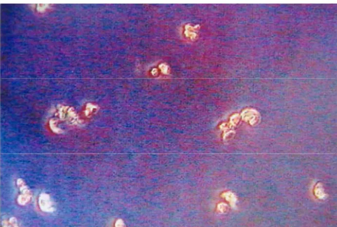
Trypan blue viability test was employed to estimate the number of the living chondrocytes (Fig. 1).
Figure 1. Trypan blue viability test was employed fort he living chondrocytes.
Şekil 1. Canlı kondrosit hücrelerine Tripan Mavisi Canlılık Testi uygulanması
Incubation of the cells
Amount of the living cells were counted and; if there were less then 2 million cells, they were put into 25 cm2 tissue culture flasks (Falcon, USA), if there were
more than 2 million cells, and then they were put into 75 cm2 tissue culture flasks. Thereafter, they were placed in
the incubator at 37°C, in an atmosphere of 5% CO2 and
90% humidity with its cover half-closed.
Providing subculture through trypsinisation process Aim of this process was to decompose dead cells and to transfer produced living cells to bigger flasks. The material was first washed 7 times using sterilized PBS or RPMI 1640. Then, 3 ml Trypsin/EDTA (0,25/0,02) (Sigma USA) solution was added onto the material in order to cover flask base. It was incubated at 37 °C, in an atmosphere of 5% CO2 in autoclave for
a minute. Later on, 5 ml culture was added to it, and it
was centrifuged at 1000 rpm for 10 minutes. Liquid left in the tube was disposed, and after adding 5 ml culture medium, it was homogenized with a glass pipette. This process was repeated 10 times and then suspension was put in the flask and placed in a 5% CO2
incubator.
Providing injectable chondrocytes to the transplanted
In monolayer system, after 15 days chondrocytes started to cover the entire surface, and depending on the initial number of the cells, they became confluent in about 20 days (Fig. 2). After the top cover of the flask was removed. Chondrocytes adhered on the base of the flask were scraped using a cell scraper (Falcon. USA), and when it became jelly, it was aspirated with a pipette and collected in a 5 cc injector.
Transplantation of autologous chondrocytes to osteochondral defect
The stifle joints of the cases which have osteochondral defects were reached with lateral para patellar incision under the same anesthesia protocol. Following the skin incision, subcutaneous connective tissue was passed while dissecting as retinaculum bluntly, fascia latea was cut and after incision, through 0.5 cm lateral to ligamentum patellare inner part of the stifle joint was reached while ligamentum collaterale laterale was secured, through vastus lateralis of m. quadriceps and m. biceps femoris, patella was deviated to medial, so the joint was totally opened up. Defects in the stifle joint of the subjects were washed using hyaluronidase (Sigma, UK) for 5 minutes until its final concentration became 1 U/ml. Purpose of this process was to remove anti-adhesive character of the region by depolymerising GAGs. Osteochondral defects were filled in injectable chondrocyte grafts (Fig. 3). 15x15 mm, periosteal grafts taken from proximal of the tibia or the lateral condyle of the femur were sutured with 4/0
polyglactin 910 (Vicryl, Ethicon), as the cambium layer lying on the defect. After controlling the position of the defect, ruptured cranial cruciate ligaments of the cases were repaired with Modified Utrecht Technique and then stifle joint was washed with solution consist of physiologic saline (Serum Fizyolojik, Eczacibasi) and
Figure 2. After 15 days, chondrocytes started to cover the entire surface in monolayer system and they became confluent in about 20 days.
Şekil 2. 15 gün sonunda, kondrositlerin tüm monolayer sistemin yüzeyini kaplamaya başlaması ve 20 gün içinde tamamen yayılması
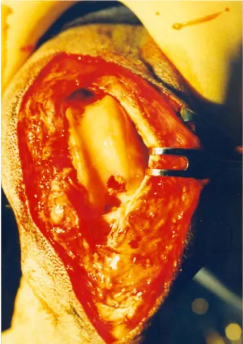
Figure 3. In the stifle joint, the osteochondral defect was filled in injectable chondrocyte grafts.
Şekil 3. Diz eklemi içindeki osteokondral defektli bölgeye enjektabl kondrositlerin aktarılmış şekli.
Hasan Bilgili - Cemil Yıldız - Barış Kürüm - Yasemin Soysal - Muhterem Bahçe 106
Figure 4 At the monolayer cell cultures after 5th days, dog
chondrocytes started to take polygonal and rhomboidal shapes. Şekil 4. Monolayer hücre kültüründeki köpek kondrosit hücrelerinin 5. gün sonunda poligonal ve romboidal şekil almaya başlaması.
Figure 5. At the 8th week, the arthroscopic view was evaluated.
Şekil 5. 8. haftadaki artroskopik değerlendirme.
Table 1. All data of 9 dogs with osteochondral defects, treated with autologous chondrocyte implantation. Tablo 1. Otolog kondrosit implantasyonu ile sağaltılan osteokondral defektli 9 köpeğe ait toplu veriler.
Cases Signalment Orthopedic problems Operative treatment Follow-up
period, results 1 Anatolian Shepherd,
5 years old, Male
Rupture of right ligamentum cruciate anterior,
OCD lesion on lateral femoral condyle
Autologous chondrocyte implantation, Repair of anterior cruciate ligament with modified Utrecht University Technique.
3 months Good
2 German Shepherd, 3.5 years old, Female
Rupture of left ligamentum cruciate anterior,
OCD lesion on lateral femoral condyle
Autologous chondrocyte implantation, Repair of anterior cruciate ligament with modified Utrecht University Technique.
6 months Good
3 Boxer, 2 years old, Male
Rupture of right ligamentum cruciate anterior,
OCD lesion on medial femoral condyle
Autologous chondrocyte implantation, Repair of anterior cruciate ligament with modified Utrecht University Technique.
2,5 months Good 4 German Shepherd mixed, 8 years old, Male
Rupture of right ligamentum cruciate anterior,
OCD lesion on lateral femoral condyle
Autologous chondrocyte implantation, Repair of anterior cruciate ligament with modified Utrecht University Technique.
9 months Good
5 Collie, 6,5 years old, Female
Rupture of left ligamentum cruciate anterior,
OCD lesion on medial femoral condyle Tears of medial meniscus
Autologous chondrocyte implantation, Repair of anterior cruciate ligament with modified Utrecht University Technique. Partial menisectomie. 10 months Satisfactory 6 Pointer, 4 years old, Male
Rupture of right ligamentum cruciate anterior,
OCD lesion on medial femoral condyle
Autologous chondrocyte implantation, Repair of anterior cruciate ligament with modified Utrecht University Technique.
4 months Good
7 Anatolian Shepherd, 5 years old,
Male
Rupture of right ligamentum cruciate anterior,
OCD lesion on lateral and medial femoral condyle
Autologous chondrocyte implantation, Repair of anterior cruciate ligament with modified Utrecht University Technique.
3,5 months Good
8 Golden Retriever, 4,5 years old, Male
Rupture of left ligamentum cruciate anterior,
OCD lesion on lateral femoral condyle
Autologous chondrocyte implantation, Repair of anterior cruciate ligament with modified Utrecht University Technique.
3 months Good 9 Anatolian Shepherd Mixed, 5,5 years old, Male
Rupture of right ligamentum cruciate anterior,
OCD lesion on medial femoral condyle, Tears of medial meniscus
Autologous chondrocyte implantation, Repair of anterior cruciate ligament with modified Utrecht University Technique. Partial menisectomie.
7 months Satisfactory
rifamicine (Rifosin, Hoescht). After that, operation area was closed with suitable techniques. No bandage was done for the extremities. After the operation a 5 day antibiotheraphy was done. At the 1st, 3rd, 5th and 7th
weeks cases were radiographically and at the 8th week
arthroscopically examined. At the clinical examination using of the extremities and pain relief; at the radiographical examination repairment of the osteochondral defects, and at the arthroscopic examination the observation of the osteochondral defect was evaluated. Clinical and radiographical evaluation was done with O’Driscoll Scoring System, and arthroscopical evaluation was done with Modified Lukoschesk Scoring System (2).
In order to decide whether the formed tissue is hyaline cartilage or not, O'Drisscoll, et al. criteria was employed (3, 16). If the surface of the regenerated tissue was smooth, if its surface was the same with that of the surrounding joint and if it was covered with hyaline cartilage, then the graft was considered successful. Furthermore, if infiltration of inflammatory cells were low, and if regeneration of the tissue surrounding subchondral bone was realized until osteochondral junction level, it was also considered successful.
Results
During the culturization, chondrocytes were examined under invert light microscope. The widths of the cells were a little differentiated and have small cytoplasmas which contains multiple thin granules. At the monolayer cell cultures after 3rd and 5th days, dog
chondrocytes started to take polygonal or rhomboidal shapes (Fig. 4). After 18 days it was observed that the cells got enlarged. The proliferation speed was maximum between 7th - 11th days and minimum at the 15th day. In
clinical examinations on the 1st and 3rd weeks there was
pain when using the extremities. But at the 5th and 7th
weeks it was observed that there was good function and no pain. In the radiologic examination osteochondral defects were depressed at the 1st and 3rd weeks and the 5th
week it was fully filled. In two cases the defects was fully filled at the 7th week. Because of the surface of the
regenerated tissue was smooth, the surface was the same with that of the surrounding joint cartilage and weight bearing, the transplantation of the chondrocytes was succesfull due to the criteria of the O’Driscoll et, all. In the arthroscopical examination done at the 8th week (Fig.
5), the synovial inflamation due to Modified Lukoschesk Scoring System was degreed as (0) normal, (1) mild inflamation, (2) moderate inflamation, (3) severe inflamation. At the end of the study 2 cases (0), 4 cases (1) and 2 cases had (3) degrees inflamation. As a result 2 cases were satisfactory, and 7 cases were good (Table 1).
Discussion and Conclusion
The most appropriate method to recover chondral and osteochondral defects, which cause an important degree of pain, loss of function and human power, and high medical treatment costs by inducing osteoarthritis in old ages, is still not known (14). Having an increasing interest, autologous chondrocytes transplantation (ACT) is a hope offering method in repairing osteochondral defects on femorotibial joint surface. In this method, cells isolated from the patient can be reproduced in vitro and autologous chondrocytes can be retransferred to the defected area in order to repair damaged cartilage (8, 21).
One of the advantages of this technique is that although the number of cells obtained from small sample pieces of cartilage is little, it can be increased in a short period of time. At this stage great care should be taken about the sterilization conditions otherwise culture flasks can easily be infected, as we experienced in our study (16, 17, 19-21).
We took cartilage samples from the shoulder joints of the dogs in order not to damage their stifles on which the study was performed. Their stifles are very small, thinner layers of articular cartilage and vulnerable to damage compared to human knees (This is a challenge because there is no ideal animal model that duplicates completely human situation). However, we should consider morbidity risk of donor in humans also (8, 16). In this study, we used autologous chondrocytes for transplantation because of their advantages like lower risk of transferring organisms and absence of immunological reaction.
However, when we look at literature, we can see that after 24 weeks of observation, Wakitani et al. (14), recovered osteochondral defects in rabbit’s stifle joint using allogenic chondrocytes which were embedded into collagen gel, and they indicated that it recovered through hyaline cartilage formation. In their study, although defects were shown as filled with hyaline cartilage, a clear subchondral bone formation was not seen when histological photomicrographs were evaluated. At the same time, they reported that histological monolayer cell infiltration was not observed and no immunological reaction by transplanted chondrocytes or collagen was seen (14). On the other hand, in their study, Noguchi et al. (12), repaired osteochondral defects using culture chondrocyte grafts and compared allogenic and isogenic grafts. They reported that in early stages of defects (in the second and fourth weeks), which were treated using isogenic or allogenic chondrocytes, they were filled with hyaline cartilage, and in peripheral sections there was a light inflammatoric
Hasan Bilgili - Cemil Yıldız - Barış Kürüm - Yasemin Soysal - Muhterem Bahçe 108
cell inflammation which was more clear in allogenic defects. However they increase subchondral bone formation and do not have any risk to transfer organisms like HIV from donor to receptor, autografts are relatively more superior than allografts. Besides, its donor maturity is automatically constant in clinical practices and the amount of cartilaginous tissue to be extracted is limited without causing any insufficiency in material (8). Furthermore, isogenic chondrocytes have more definite regeneration in subchondral bone tissue than allogenics.
One of the problems we encountered was that we did not know the number of optimal cells that we had to place into the defected area, so we filled the defect until it was full (8, 17). Further research is needed to solve this problem.
The second problem we had was the difficulty of inducing a defect in the dog stifles joint cartilage which is very thin. Moreover there are many factors that need to be standardized, for instance; geometric shape and volume of the lesions (the results in large lesions are less predictable), the depth of injury (penetration of the subchondral bone alters the response), the age of the animal and individual animal's biologic response and activity level (9).
We used hyaluronidase to wash the defects. Pre-treatment of the articular cartilage lesion with enzymes such as hyaluronidase and chondroitinase has been advocated to enhance these cell based repair strategies. Such treatment is intended to improve cell attachment and possibly promote the integration of the regenerated repair tissue with the native cartilage (9).
Another problem in our study was closing the defects. We used periosteal graft since it is easy to get. With recent studies it has been understood that using periosteal graft is not effective on its own. In their study on rabbit stifles, Brittberg et al. (3), reported that, compared to the group in which chondrocytes were used, repairing tissues formed in the group periosteal grafting was employed, is histologically of lower quality. This led the studies carried with ACT to come into agenda. Therefore, we did not use periosteal graft in the dog’s stifles were damaged less and rehabilitation period was shorter. Because of their advantages like no donor morbidity, less invasive surgery and shorter operation time, biologic and synthetic polymers and tissue engineering products need to be used in closing and holding the cells in the defects instead of periosteal grafts (15, 18).
Brittberg et al. (2), carried out a research for the first time on the usage of ACT in the treatment of localized cartilage defects of patella or femur condyle in 23 patients, cultured for 14 and 21 days and they
were injected to the defected region and covered with periosteal flab. Two years after the transplantation, good or perfect clinical results were reported about 14 of 16 patients (who had condyle defect) and 2 of 7 patients (who had patellar defect). Hyaline cartilage was shown in the biopsies taken from one patellar defect and 11 femoral defects. Finally, they published results of a bigger group of patients. After the treatment of chondral and osteochondral defects in knee through chondrocyte transplantation, subjects were monitored for 2 or more months and in 47 of 66 subjects there was an increase in function. These results show that chondrocyte transplantation together with periosteal grafts increase restoration of joint surface in humans. But there is still a need for research to define newly formed tissue and to determine its functions (2).
To summarize, there are a lot of parameters that need to be explained, for instance; differences in the species, the age of the animals, extent of the lesion, depth of the defect, the lack of retention of the cells in the defect due to possible displacement of the periosteal flap, location of the defect (the patella compared with the trochlea), the numbers of the cells that can be injected, and immobilization of the animals.
Consequently, in order to prevent joint surface damages and degenerative joint defects to occur in the future, autologous chondrocytes transplantation to repair osteochondral defects is a new alternative treatment method. There is a need for further studies to develop this method.
To sum up; with this project for the first time in Turkey, cells taken from shoulder joints of the dogs, healthy chondrocytes were isolated and implanted on the OCD in the stifle joints at the end successful results were achieved.
Aknowledgements
The researchers would like to thank the Research Foundation of Ankara University for their financial support. We also express appreciation to Professor Dogan Aslanbey for his valuable comments and contributions.
References
1. Bilgili H, Orhun S (2002): The research of the changes on
joint tissues radiological and histopathologically on an experimental osteoarthritis performed on rabbit stifle joint.
Turk J Vet Surg, 8, 8-12.
2. Brittberg M, Lindahl A, Nilsson A, Ohlsson C, Isaksson O, Peterson L (1994): Treatment of deep cartilage defects
in the knee with autologous chondrocyte transplantation.
3. Brittberg M, Lindahl A, Nilsson A, Ohlsson C, Isaksson O, Peterson L (1996): Rabbit articular
cartilage defects treated with autologous cultured chondrocytes. Clin Orthop, 326, 270-283.
4. Buckwalter JA, Mankin HJ (1997): Articular
cartilage. Part II: Degeneration and osteoarthritis, repair, regeneration, and transplantation. J Bone Joint
Surg, 79A, 612-627.
5. Cook JL, Kreeger JM, Payne JT, Tomlinson JL (2002):
Effects of EGF, FGF, and PDGF on canine chondrocytes in three-dimensional culture. Vet Comp Orthop Traumatol,
10, 210-213.
6. Fortier LA, Nixon AJ, Mohammed HO, Lust G (1997):
Altered biological activity of equine chondrocytes cultured in a three-dimensional fibrin matrix and supplemented
with transforming growth factor β–1. Am J Vet Res, 58,
66-70.
7. Fortier LA, Nixon AJ, Williams J, Cable CS (1998):
Isolation and chondrocytic differentiation of equine bone marrow-derived mesenchymal stem cells. Am J Vet Res,
59, 1182-1187.
8. Gillogly SD, Voight M, Blackburn T (1998):
Treatment of articular cartilage defects of the knee with autologous chondrocyte implantation. J O S P T, 28,
241-251.
9. Jackson DW, Simon TM (1999): Tissue engineering
principles in orthopaedic surgery. Clin Orthop, 376,
31-45.
10. Kim W, Mooney D, Vacanti JP, Upton J, Ibarra C, Vacanti CA (1996): Functional viability of chondrocytes stored at 4°C. Tissue Eng, 2, 75-81.
11. Messner K, Gillquist J (1996): Cartilage repair: A
critical review. Acta Orthop Scand, 67, 523-529.
12. Noguchi T, Oka M, Fujino M, Neo M, Yamamuro T (1994): Repair of osteochondral defects with grafts or
cultured chondrocytes: comparison of allografts and isografts. Clin Orthop Rel Res, 302, 251-258.
13. Tunay S, Bilgili H, Yıldız C, Yanmış İ, Solakoğlu C, Gür E (2002): The use of intraarticular hyaluronic acid
on the treatment of experimental osteoarthritis: Radiological and histopathological study on rabbit stifle joint. Turk J Vet Anim Sci, 26, 939-947.
14. Wakitani S, Kimura T, Hirooka A, Ochl T, Yoneda M, Yasui N, Owaki H, Ono K (1989): Repair of rabbit
articular surfaces with allograft chondrocytes embedded in collage gel. J Bone Joint Surg, 71B, 74-80.
15. Yaylaoğlu MB, Yıldız C, Korkusuz F, Hasırcı V (1999): A novel osteochondral implant. Biomaterials, 20, 1513-1520.
16. Yıldız C, Bahçe M, Başbozkurt M, Uğur G, Oğuz E, Deveci S, Gür E (1997): Cultured chondrocytes in
monolayer system. Turk J Bone Joint Surg, 3, 88-93.
17. Yıldız C, Bahçe M, Şehirlioglu A, Tunay S, Erler K, Başbozkurt M, Gür E (1998): Injectable Chondrocyte. Acta Orthop Trauma Turc, 2, 78-85.
18. Yıldız C, Yaylaoglu MB, Korkusuz F, Başbozkurt M, Hasırcı V (1999): Calcium phosphate loaded lyophilized
collagen graft material for induction of chondrocytes. J
Bone Joint Surg, 81A, 64.
19. Yıldız C, Bilgili H, Bahçe M, Başbozkurt M, Aslanbey D, Gür E (1999): An experimental study of chondrocyte
cell culture from cartilage tissue of rabbit shoulder joint.
Turk J Vet Surg, 5, 20-23.
20. Yıldız C, Bilgili H, Bahçe M, Başbozkurt M, Aslanbey D, Gür E (2000): Two different in-vitro methods for
chondrocyte proliferation: An experimental study on rabbit stifle joint cartilage. Turk J Vet Surg, 6, 47-51.
21. Yıldız C, Bahçe M, Deveci S, Bilgili H, İde T, Tunay S, Başbozkurt M, Gür E (2001): Autologous chondrocyte
transplantation (An experimental study in rabbit knee joints). Turk J Arthrop Arthros Surg, 12, 75-82.
Geliş tarihi: 14.06.2005 / Kabul tarihi: 22.07.2005
Yazışma adresi:
Doç. Dr. Hasan Bilgili,
Ankara Üniversitesi Veteriner Fakültesi Cerrahi Anabilim Dalı
06110 Dışkapı, Ankara.
e-posta: [email protected] [email protected]