ABSTRACT
Although diverticular disease of the colon is common, the occurrence of rectal diverticula is extremely rare with only sporadic reports in the literature since 1911. Symptomatic rectal diverticula are seen even less frequently, and surgical intervention is needed for only complicated cases. Here we report the case of a 63-year-old woman pre-senting with rectal diverticulitis mimicking rectal carcinoma with intestinal obstruction.
Keywords: Rectal diverticulitis, rectal carcinoma, intestinal obstruction
INTRODUCTION
Diverticular disease is common in the Western world, manifesting as a sac of an abnormally protruding bowel wall and usually occurring in the distal colon; 90% of patients have sigmoid colon involvement (1) and only 15% have right-sided diverticula (1). Diverticular disease is an acquired disease with herniations of the mucosa and part of the submucosa through the muscularis pro-pria due to a lack of dietary fiber, but the pathogenesis of colonic diverticula remains poorly understood. The occurrence of rectal diverticula is extremely rare, with only a few reports published in the literature since 1911 (2). The cause of rectal diverticula is unknown; however, most are asymptomatic and do not require treatment. Surgical intervention is generally only nec-essary for complicated cases. In this report, we describe a rectal diverticulum that mimicked rectal carcinoma with intestinal obstruction. Low anterior resection and temporary colostomy were performed via emergent laparotomy because of complete intestinal obstruction. CASE PRESENTATION
A 63-year-old woman was admitted into the hospital with abdominal pain, nausea, and vomiting. She had experi-enced chronic constipation for 10 years, but had stopped
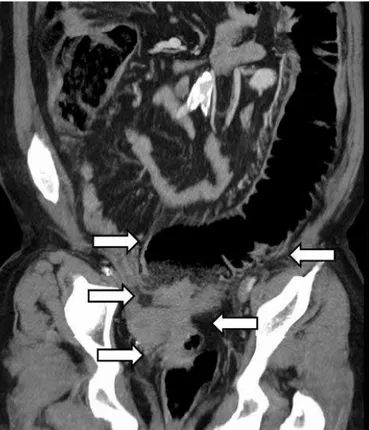
producing gas and gaita discharge 4 days prior to admis-sion. The patient’s medical history included an appen-dectomy 30 years previously, coronary bypass surgery 10 years earlier, and an explorative laparotomy for a suspect-ed left ovarian mass that resultsuspect-ed in a left salpingo-oo-phorectomy 3 years earlier. Upon physical examination, scars from a median incision and the appendectomy inci-sion were seen. She had abdominal disteninci-sion with ten-derness, but did not have rebound tenten-derness, and her bowel sounds were increased and obstructive. During a digital rectal examination, her ampulla recti was empty. A perianal fistula suggestive of inflammatory bowel disease was not apparent, and her laboratory results were within normal ranges. A computed tomography (CT) scan was suggestive of rectal cancer with a diffuse, irregular wall thickening that was 9-cm long and located in the supe-rior rectum, approximately 10 cm above the anal verge (Figure 1); a rectal contrast transition above the mass was not observed (Figure 2). The proximal colon segments were dilated (maximum, 8 cm) with fecal retention due to a mechanical obstruction. Multiple pericolic lymph nodes (<10 mm in size) were detected around the mass and at the obturator chains.
With these findings, an emergent explorative laparot-omy was performed for mechanical ileus and severe Turk J Gastroenterol 2015; 26: 60-2
Address for Correspondence: Ümit Özçelik, Department of General Surgery, Başkent University Faculty of Medicine, İstanbul, Turkey E-mail: [email protected]
Received: November 13, 2012 Accepted: June 30, 2013
© Copyright 2015 by The Turkish Society of Gastroenterology • Available online at www.turkjgastroenterol.org • DOI: 10.5152/tjg.2015.4521
Rectal diverticulitis mimicking rectal carcinoma with intestinal
obstruction: Case report
Ümit Özçelik1, Hüseyin Yüce Bircan1, Eryiğit Eren1, Ebru Demiralay2, İclal Işıklar3, Alp Demirağ1, Gökhan Moray4
1Department of General Surgery, Başkent University Faculty of Medicine, İstanbul, Turkey 2Department of Pathology, Başkent University Faculty of Medicine, İstanbul, Turkey 3Department of Radiology, Başkent University Faculty of Medicine, , İstanbul, Turkey 4Department of General Surgery, Başkent University Faculty of Medicine, Ankara, Turkey
60
Cas
e R
epor
bowel distension due to a rectal obstructing tumoral lesion. During surgery, a 5-cm infiltrative mass was detected adherent to the bladder and uterus, as well as neighboring severe fibro-sis. A small bladder perforation occurred during the isolation of the bladder from the mass, and was sutured primarily. Because of uterine infiltration, a gynecologist participated into the op-eration and hysterectomy; a right salpingo-oophorectomy was also performed. After this, low anterior resection and terminal
colostomy were performed. Frozen section examination re-vealed a 2-cm tumor-free zone to the distal surgical border. A pathologic examination revealed a 4-cm, off white, infiltra-tive lesion around the rectum. Upon microscopic examination, mucosal ulceration, diffuse active chronic inflammation with abscess formation reaching to the serosal surface and removing the submucosa, and multiple diverticuli were detected; there was no evidence of rectal cancer. The microscopic examination was compatible with diverticulitis with micro-perforations (Figure 3). Ten lymph nodes were detected; all were reactional. The patient was discharged on postoperative day 5 without complications. Written informed consent obtained from the patient for the publication of this case report.
DISCUSSION
Diverticular disease usually involves the sigmoid colon (65%), followed by the ascending colon and cecum. Involvement of the entire colon occurs in up to 10% of cases; however, rectal involvement is extremely rare, with an estimated incidence of <0.1% of the cases (3,4). Two theories have been described to explain the low incidence of rectal diverticula. The first theo-ry suggests that the tenia coli muscle fibers, which surround the rectum, protect it from intraluminal pressures. The second theory suggests that the rectum is protected due to reduced internal pressure from accumulated feces and the reduced peristaltic activity associated with the rectum compared with that associated with the sigmoid colon. Although the cause of rectal diverticula remains unknown, possible predisposing factors have been described, including weakness in the cir-cumferential muscles surrounding the rectum, primary muscle atrophy, absence of supporting structures such as the coccyx, a relaxed rectovaginal septum, constipation or recurrent impac-tion causing rectal distension, and rectal trauma or infecimpac-tion that leads to a weakened rectal wall (5,6).
61
Cas
e R
epor
t
Özçelik et al. Rectal diverticulitis mimicking rectal carcinoma
Turk J Gastroenterol 2015; 26: 60-2
Figure 1. Reconstructed coronal computed tomography image showing diffuse, irregular rectal wall thickening and dilated proximal colon seg-ments (arrows).
Figure 2. There was no rectal contrast transition above the mass (arrows).
Figure 3. Multiple diverticula (arrows) are shown as sacs that spread with atrophic mucosa and the surrounding, thin muscularis propria layer (he-matoxylin and eosin stain, ×100).
Most patients with rectal diverticula are asymptomatic and di-agnosed incidentally, and do not require treatment. However, rectal diverticula may become inflamed with impacted feces resulting in abscess formation and perforation. Other compli-cations associated with rectal diverticula include rectal stric-tures, rectovesical fistulas, rectal prolapses, and misdiagnosis of rectal carcinoma (2). Surgical intervention is necessary only in complicated patients, and operative treatment includes drain-ing the abscess, a divertdrain-ing colostomy, resection of the diver-ticular mass, or rectal resection.
Radiologically, differentiating diverticulitis from colon cancer is difficult. Although they are completely different pathologic entities, they show similar segmental colonic wall thickenings, with varying degrees of pericolic infiltration on CT. Pericolic infiltration, lymph node visibility and size, inflamed and non-inflamed diverticula, degree and pattern of enhancement, intestinal obstruction, involved wall thickness and length, fas-cial thickening, and free air and abscess formation have been determined to be significant parameters for differentiating di-verticulitis from colon cancer (7). Diverticula tend to be lon-ger and thinner, with more pericolic inflammatory changes, and are minimally enhanced lesions that show a target or homogenous enhancement pattern (7). Usually, the lymph nodes associated with colon cancer are visible and >10 mm in diameter (7). Sometimes the identification of diverticulosis is challenging because of severe colonic structure destruction or the presence of fluid-filled intramuscular diverticula, and the inflammatory process appears as an inflammatory mass (7). Inflamed and non-inflamed diverticula are the most reliable signs for the diagnosis of diverticulitis, whereas coincidental di-verticula have been reported in 14-29% of colon cancer cases (7). Colonic obstruction is another significant factor for differ-entiating diverticulitis from colon cancer, with a reported inci-dence of 3.8-23% in colon cancer patients (8). In diverticulitis, the chronic inflammatory process results in the intramuscular fibrosis that is responsible for the lumen rigidity, and colonic obstruction occurs in 10-20% of cases (7).
In conclusion, rectal diverticula are extremely rare, and usually do not require treatment; surgical intervention is only neces-sary in complicated cases. The final diagnosis, without post-surgical pathologic confirmation, is very difficult. Although CT
enables accurate diagnosis of diverticulitis, colon cancer re-mains difficult to exclude. The presence of diverticula, absence of lymph nodes, and minimal or target pattern enhancement are the major CT findings used for discriminating between di-verticulitis and colon cancer. Rectal didi-verticulitis should also be considered in the differential diagnosis of patients admitted with rectal masses and mechanical ileus.
Ethics Committee Approval: Ethics committee approval was received
for this study
Informed Consent: Written informed consent obtained from the
pa-tient.
Peer-review: Externally peer-reviewed.
Author contributions: Concept -Ü.Ö., H.Y.B.; Design - Ü.Ö. H.Y.B., E.E.;
Supervision - A.D.; Resource - Ü.Ö. H.Y.B., E.E.; Materials - Ü.Ö., E.D., İ.I.; Data Collection&/or Processing - Ü.Ö., E.D., İ.I.; Analysis&/or Interpreta-tion - Ü.Ö., E.D., İ.I.; Literature Search - Ü.Ö.; Writing - Ü.Ö., H.Y.B.; Critical Reviews - A.D., G.M.
Conflict of Interest: No conflict of interest was declared by the
au-thors.
Financial Disclosure: The authors declared that this study has
re-ceived no financial support.
REFERENCES
1. Jung SH, Kim JH. A case of solitary rectal diverticulum presenting
with a retrorectal mass. Gut Liver 2010; 4: 394-7. [CrossRef]
2. Giffin HZ. VII. Diverticulitis of the rectum: a report of two cases operated upon, one of them with carcinomatous degeneration.
Ann Surg 1911; 53: 533-7. [CrossRef]
3. Kwon JH, Han KH, Chang WS, et al. A case of a mucinous adeno-carcinoma arising from a rectal diverticulum. J Korean Soc
Colo-proctol 2012; 28: 222-4. [CrossRef]
4. Chen CH, Jao SW, Lai HJ, Chiu YC, Kang JC. Isolated rectal divertic-ulum complicating with rectal prolapse and outlet obstruction: case report. World J Gastroenterol 2005; 11: 7697-9.
5. Weston SD, Sclachter IS. Diverticulum of the rectum. Dis Colon
Rectum 1959; 2: 458-64. [CrossRef]
6. Damron JR, Lieber A, Simmons T. Rectal diverticula. Radiology
1975; 115: 599-601. [CrossRef]
7. Shen SH, Chen JD, Tiu CM, et al. Differentiating colonic diverticuli-tis from colon cancer: the value of computed tomography in the
emergency setting. J Chin Med Assoc 2005; 68: 411-8. [CrossRef]
8. Glenn F, McSherry CK. Obstruction and perforation in colorectal
cancer. Ann Surg 1971; 173: 983-92. [CrossRef]
62
Cas
e R
epor
t