UHOD doi: 10.4999/uhod.183074
The Feasibility of Sparing the Hippocampus and
Hypothalamic -Hypophysial Axis During Whole
Brain Radiotherapy: A Dosimetric Study
Yurday OZDEMIR, Ibrahim ACIBUCI, Erkan TOPKAN
Baskent University Faculty of Medicine, Department of Radiation Oncology, Adana, TURKEY
ABSTRACT
We aimed to investigate the feasibility of simultaneously sparing the hippocampus and hypothalamic-hypophysial axis (HHA) during whole brain radiotherapy (WBRT) using helical tomotherapy. Images of totally 12 patients were analyzed. Hippocampi and HHA were delineated according to available guidelines. First, radiotherapy plans were analyzed according to dose constraints of the Radiation Therapy Oncology Group-0933 for 30 Gy (3 Gy/fr). Second, dose constraints were decreased by 1/6 to simulate the 25 Gy prophy-lactic cranial irradiation practice. The mean D98% and D2% was 25.9±0.85 Gy and 35.0±1.32 Gy, and 21.4±0.40 Gy and 30.5±1.33 Gy for planning target volumes in the original and modified protocol plans, respectively. The D100% and Dmax for the hippocampus was 8.1±0.47 Gy and 15.7±0.84 Gy in the original and 7.1±0.68 Gy and 13.2±1.08 Gy in the modified protocol, respectively. The mean doses for the hypothalamus and hypophysis was 12.6±0.76 Gy and 11.4±1.22 Gy in the original and 10.8±1.38 Gy and 9.8±1.50 Gy in the modified plans, respectively. This study demonstrated the feasibility of sparing the hippocampus and HHA simultaneously during WBRT with helical tomotherapy regarding target dose coverage and dose constraints suggested by the literature.
Keywords: Hippocampus, Hypothalamic-hypophysis axis sparing whole brain radiotherapy, Prophylactic cranial radiotherapy
ÖZET
Tüm Beyin Radyoterapisi Esnasında Hipokampüs ve Hipotalamo-Hipofizer Aksın Korunabilirliği: Dozimetrik Çalışma
Tüm beyin radyoterapisi (TBR) esnasında hipokampüs ve hipotalamo-hipofizer aksın (HHA) eşzamanlı olarak helikal tomoterapi yardımıyla korunabilirliğini araştırmayı amaçladık. Toplam 12 hastanın görüntüleri analiz edilmiştir. Hipokampüs ve HHA mevcut re-hberlere göre konturlanmıştır. İlk olarak hastalar 30 Gy (3 Gy/fr) için Radiation Therapy Oncology Group-0933 doz sınırlamalarına göre analiz edilmiştir. İkinci olarak ise doz kısıtlamaları 1/6 oranında azaltılarak 25 Gy’lik proflaktik kranial radyoterapi pratiği simüle edilmiştir. Orjinal ve modifiye edilen protokollerde ortalama D%98 ve D%2 sırasıyla 25.9±0.85 Gy ve 35.0±1.32 Gy; 21.4±0.40 Gy ve
30.5±1.33 Gy olarak bulunmuştur. Orjinal planda hipokampüs için D100% ve Dmax sırasıyla 8.1±0.47 Gy ve 15.7±0.84 Gy iken
modi-fiye planda 7.1±0.68 Gy ve 13.2±1.08 Gy olarak tespit edilmiştir. Orjinal plandaki ortalama hipotalamus ve hipofiz dozları 12.6±0.76 Gy ve 11.4±1.22 Gy; modifiye planda ise 10.8±1.38 Gy ve 9.8±1.50 Gy olarak bulunmuştur. Bu çalışma literatür tarafından önerilen doz sınırlamaları ve hedef doz sarımı açısından helical tomoterapiyle hipokampüs ve HHA’ın aynı anda korunabilir olduğunu gösterir niteliktedir.
UHOD
INTRODUCTION
Prophylactic cranial irradiation (PCI) is an inte-gral part of standard treatment algorithms in some malignancies including small-cell lung carcinoma and acute lymphoblastic leukemia.1-4 The main rationale underlying the use of PCI in patients is the well-recognized limited permeability of the blood-brain-barrier to many chemotherapeutics, rendering the brain a sanctuary site for malignant cells.5 In this setting, PCI decreases brain relapses by eradication of occult tumor cells, which cannot be determined by current diagnostic tools. Thus, PCI enhances the overall survival rates with rela-tively lower radiotherapy doses compared the dose utilized for palliation of brain metastases.6 Despite these advantages of PCI, there is a significant risk of deterioration of neurocognitive functions (NCF) according to the objective baseline measurements, which is the major limitation against its usage.7 Radiation-induced hippocampal injury is the most established cause of neurocognitive dysfunction in patients treated with PCI and/or palliative whole brain radiotherapy (WBRT); this is related to the injury of neural progenitor cells in the subventricu-lar zone and hippocampus in addition to damage of the vascular structure and glial precursors.8-13 In this regard, hippocampus-sparing WBRT has repeatedly been demonstrated to be safe and use-ful in sparing NCF with no sacrifice of potentially microscopically involved brain sites.14-16
Given the separated parts of the brain with sophis-ticated interconnections, it seems reasonable to as-sume that other parts of the brain can contribute to the NCF as well as the master neurocognitive organ, the hippocampus.17 Recent studies report that a limited post-development neurogenesis can occur in other sides of the brain including stria-tum, spinal cord, and neurocortex as well as the hypothalamus.18 The hypothalamus is responsible for the maintenance of homeostatic functions and quality of life (QOL). Therefore the preservation of the hypothalamic-hypophysial axis (HHA), known as the master of the neuroendocrine system, can re-duce the risks of both neuroendocrine dysfunction and NCF particularly in groups of patients with longer survival expectations.19-22
Radiation-induced HHA dysfunction has been de-termined to be a dose- and time- dependent adverse
event, but the radiation doses causing impaired QoL as a result of HHA-related damage has not yet been determined.23,24 On the other hand, some sug-gestions about the dose constraints of hippocam-pus were offered: <7.3 Gy to 40% of bilateral hip-pocampi 14 and a maximum dose (Dmax) of 9 Gy to 100% of the hippocampi.25 Therefore, it is logi-cal to anticipate that sparing of both the hippocam-pi and HHA can be a more effective approach for maximum maintenance of the NCF, rather than the hippocampus-sparing-WBRT alone in patients un-dergoing PCI. Enlightened by the available litera-ture, we evaluated the feasibility of simultaneously sparing hippocampus + hypothalamus + hypophy-sis (3HS) which can be abbreviated as 3HS-WBRT in patients undergoing WBRT.
PATIENTS AND METHODS Contouring
Datasets including planning computerized tomog-raphy (CT) fused with T1-weighted post-contrast axial MRI images (slices 1.5 mm thick) of 12 patients accessible on the Picture Archiving and Communication System were selected. The CT images were obtained 120 kVp, 400 mA, and 1.25 mm thickness. Hippocampi were delineated ac-cording to the Radiation Therapy Oncology Group (RTOG)-093326, and hippocampal avoidance zones were uniformly defined by 5 mm expansion in all directions from the hippocampi.
The hypothalamus was delineated by utilizing the method described by Elson et al27 and additional MRI atlases.28 In brief, the most cranial contouring section corresponded to the anterior commissure, which is located at the medial sides of the third ventricle (Figure 1). The most inferior section of the contour was 1.5 mm (1 axial slice) superior to the slice where the optic chiasm emerges first. The anterior side of the third ventricle and interpedun-cular fossa constituted the anterior and posterior boundaries, respectively. Lastly, either the optic white matter or internal capsule was defined as the lateral border of the hypothalamus. The hypophy-sis was delineated as the concavity within the sphe-noid bone, sella tursica, which is located laterally between the cavernous sinuses, the hypothalamus and optic chiasm cranially. As recommended, the bone window was utilized for this purpose as the
UHOD
most appropriate CT mode for delineation of the hypophysis. A uniform additional 5 mm was added to both the hypothalamus and hypophysis in all directions to constitute the planning organ at risk volumes. The organs at risk constituted the lenses, globes, optic nerves, and chiasms were delineated according to the accessible contouring guidelines.29 The clinical target volume was the whole brain to second cervical vertebra and 2 mm was added to create the planning target volume (PTV). However, the final PTV was defined by subtracting the plan-ning organ at risk volumes of the hippocampus and HHA from the PTV.
Treatment Planning and Procedure
Although our institutional PCI protocol mandates a total dose of 25 Gy in 10 fractions, we also per-formed alternative plans with 30 Gy in 10 fractions for each patient utilizing helical tomotherapy (HT)
Accuray planning system (Tomo HDA, version 2.1.2) to simulate palliative WBRT studies. The dose limitations of the PTV and organs at risk have been identified in accordance with the RTOG 0933 protocol 26 and a modified dose limitation was de-scribed by reducing all the recommended doses in the trial by 5/6, in the view of the 25 Gy of PCI. Planning was performed using the 1.048-cm wide field with a pitch of 0.221 and a modulation fac-tor of 5 and dose calculation grid of 0.203 cm x 0.203 cm. All the dose specifications are shown in Table 1. Although the present study was not a comparison study, the mean homogeneity index (HI) defined as the measure of the steepness of the target, was also calculated to compare our re-sults with similar studies. The HI was defined as [(D2% - D98%)/Dmedian], where a value closer to
0 is associated with optimal plan according to the International Commission on Radiation Units and Measurements.30
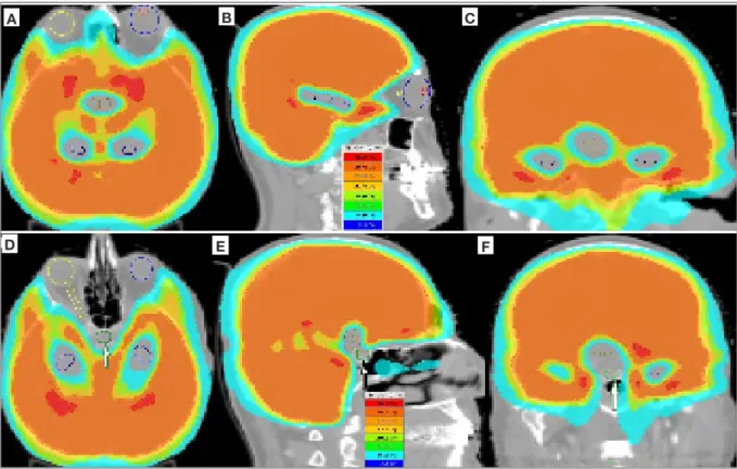
Figure 1. Dose color wash images of a patient receiving 25 Gy of prophylactic cranial irradiation dose utilizing by helical tomotherapy; A. axial section of helical tomotherapy plan with the delineations of hippocampus (dark blue) and hypothalamus (brown); B.
sagit-tal section demonstrating the hippocampus; C. coronal section radiotherapy plan demonstrating the hippocampus (dark blue) and
hypothalamus (brown); D. axial section plan demonstrating the hippocampus bilaterally and hypophysis (white arrow) in the midline; E. sagittal section plan demonstrating the preservation of hypothalamus (brown) and hypophysis (green, white arrow); F. coronal
sec-tion plan demonstrating the hypophysis (white arrow) in the midline A F E D C B
UHOD
Statistical Analysis
All calculations and statistical tests were per-formed utilizing IBM SPSS 17.0 software (SPSS, Inc., Chicago, IL, USA). With respect to dosimetric evaluation, continuous variables were expressed as mean (± SD)
RESULTS
The compliance criteria, limitations of organs at risk which were based on RTOG-0933 and the modified protocol are demonstrated in Table 1. The average dosimetric values in 12 patients are shown in Table 2.
PTV, Homogeneity Index, and Delivery Time
All the plans met the acceptance criteria. The mean D98% and D2% was 25.9 ± 0.85 Gy and 35.0 ± 1.32 Gy, and 21.4 ± 0.40 Gy and 30.5 ± 1.33 Gy for PTV in the protocol and modified protocol plans, respectively (Table 2). The HI was 0.30 ± 0.07 in both of the protocols. The mean delivery time was 31.8 minutes ± 1.81.
Volumes and Dosimetry of the Hippocampus, Hypothalamus and Hypophysis
The mean volumes of the hippocampus, hypothala-mus, and hypophysis were 5.6 cm3 ± 1.82, 0.7 cm3 ± 0.30 and 0.5 cm3 ± 0.16, respectively. The mini-mum (D100%) and maximum doses (Dmax) for the hippocampus were 8.1 ± 0.47 Gy and 15.7 ± 0.84
Gy in the protocol and 7.1 ± 0.68 Gy and 13.2 ± 1.08 Gy in the modified protocol, respectively. The mean doses for the hypothalamus and hypophysis were 12.6 ± 0.76 Gy and 11.4 ± 1.22 Gy in the pro-tocol plan and 10.8 ± 1.38 Gy and 9.8 ± 1.50 Gy, in the modified plan, respectively.
DISCUSSION
To the best of our knowledge, this dosimetric study confirmed the feasibility of simultaneous protec-tion of the hippocampus and HHA without sacri-ficing PTV coverage by utilizing HT; the only cost was relatively longer treatment times (31.8 min-utes ± 1.81) than that for the standard WBRT. Maintenance of NCF as a major component of QoL has gradually gained increasing consideration in patients with cancer undergoing PCI, because of the significantly increased WBRT-related toxicity rates in long-term survivors of PCI in the pres-ence of more effective systemic and loco-regional treatment options.31,32 Studies indicate that unilat-eral or bilatunilat-eral hippocampal damage is related to neurocognitive disorders, particularly in memory and learning, and hippocampal-sparing techniques have been suggested for either WBRT or partial ra-diotherapy of the brain in order to spare NCF.26,33 Although hippocampus-sparing WBRT has been the most utilized approach for this purpose, further complications such as fatigue, sleeping disorders, reduced muscle mass, low energy, and hormonal deficiencies can still alter the NCF, and therefore, the patients’ overall QoL in a negative manner.32
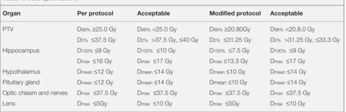
Table 1. Dose specifications
Organ Per protocol Acceptable Modified protocol Acceptable
PTV D98% ≥25.0 Gy D98% <25.0 Gy D98% ≥20.80Gy D98% <20.8.0 Gy
D2% ≤37.5 Gy D2% >37.5 Gy, ≤40 Gy D2% ≤31.25 Gy D2% >31.25 Gy, ≤33.3 Gy
Hippocampus D100% ≤9 Gy D100% ≤10 Gy D100% ≤7.5 Gy D100% ≤9 Gy
Dmax ≤16 Gy Dmax ≤17 Gy Dmax ≤13.3 Gy Dmax ≤17 Gy
Hypothalamus Dmean ≤12 Gy Dmean ≤14 Gy Dmean ≤10 Gy Dmean ≤14 Gy
Pituitary gland Dmean ≤12 Gy Dmean ≤14 Gy Dmean ≤10 Gy Dmean ≤14 Gy
Optic chiasm and nerves Dmax ≤37.5 Gy Dmax ≤37.5 Gy Dmax ≤37.5 Gy Dmax ≤37.5 Gy
Lens Dmax ≤5Gy Dmax ≤10 Gy Dmax ≤5Gy Dmax ≤10 Gy
Abbreviations: PTV= planning target volume; D98%= the dose which %98 of the target volume receives; D2%= the dose which 2% of the target volume
UHOD
In this context, considering the neuroendocrine and homeostatic functions of HHA, as a master organ, 3HS-WBRT can be superior for further sparing of the NCF than the hippocampus-sparing WBRT alone.34
Hippocampus sparing-WBRT techniques includ-ing the volumetric modulated arc technique and HT are the most frequently used methods to de-crease the radiation doses of the hippocampus and HHA.35-37 In the study of Fan et al, which was the first report of simultaneous hippocampus and HHA sparing during PCI, the authors showed that the mean hippocampus, hypothalamus, and hypophy-sis doses might be reduced to 9.6 Gy, 11.06 Gy, and 10.66 Gy, respectively, lending support to our re-spective mean doses in our modified plan: 7.1 Gy, 10.38 Gy and 9.8 Gy.34 In a phase II randomized trial, a mean hippocampal dose of ≤ 10 Gy was re-lated to a 23% improvement in the Hopkins Verbal Learning Test compared to previous hippocampus sparing-WBRT studies.26 Accordingly, in another study, emphasizing its radio-sensitivity, a dose-dependent hippocampal atrophy (>40 Gy vs <10 Gy, p= 0.03) was shown to occur on first year MRI scans.38 Rong et al analyzed the dosimetric differ-ences between HT, step and shot intensity modulat-ed radiotherapy, and rapidarc plans for hippocam-pus-sparing WBRT. They reported that the mean hippocampal doses of Dmax 15.1 Gy and D100% 8.0 with HT were notably lower than their counterparts achieved with other plans which resembled our
re-spective values of 15.7 Gy and 8.1 Gy, achieved in 30 Gy of the WBRT plan.39 Although the mean Dmax values of the bilateral lens (3.3 Gy vs 3.3 Gy) and optic chiasm (33.9 Gy vs 28 Gy) were similar between the study by Rong and our study, those of the optic nerves were considerably lower in our study (33.9 Gy vs 13.3 Gy). Regarding the mean PTV, D98%, and D2% in the present investiga-tion (25.9 Gy and 35.0 Gy) were comparable to the study of Rong et al (27.5 Gy and 32.2 Gy) despite relatively higher HI in our study (0.30 ± 0.07 vs 0.15 ± 0.03), which indicated inferior dose homo-geneity due to the incorporation of HHA sparing. In a study investigating the effect of inclined and neutral head positions on HS-WBRT, the mean D100% (10.45 Gy vs 12.07 Gy, p= 0.007) and Dmax (13.7 Gy vs 15.7 Gy, p= 0.003) of the hippocampus were significantly lower in the inclined position of the head.40 Although the D100% values of the hip-pocampus in both of the positions were higher than the value we found (8.1 Gy), the Dmax in the in-clined position appeared to be more favorable than ours (13.7 Gy ± 0.25 vs 15.7 Gy ± 0.8).
In addition to the apparent hormonal deficiency risk with > 50 Gy of radiotherapy dose, even 12 Gy of hypothalamic dose can induce neuro-endo-crine abnormalities.41,42 The deficiency of growth hormone, the most radio-sensitive hypophysial hormone, can occur after mean radiotherapy doses of 15 Gy43-45, and up to 100% of the patients may suffer from endocrine insufficiencies when the
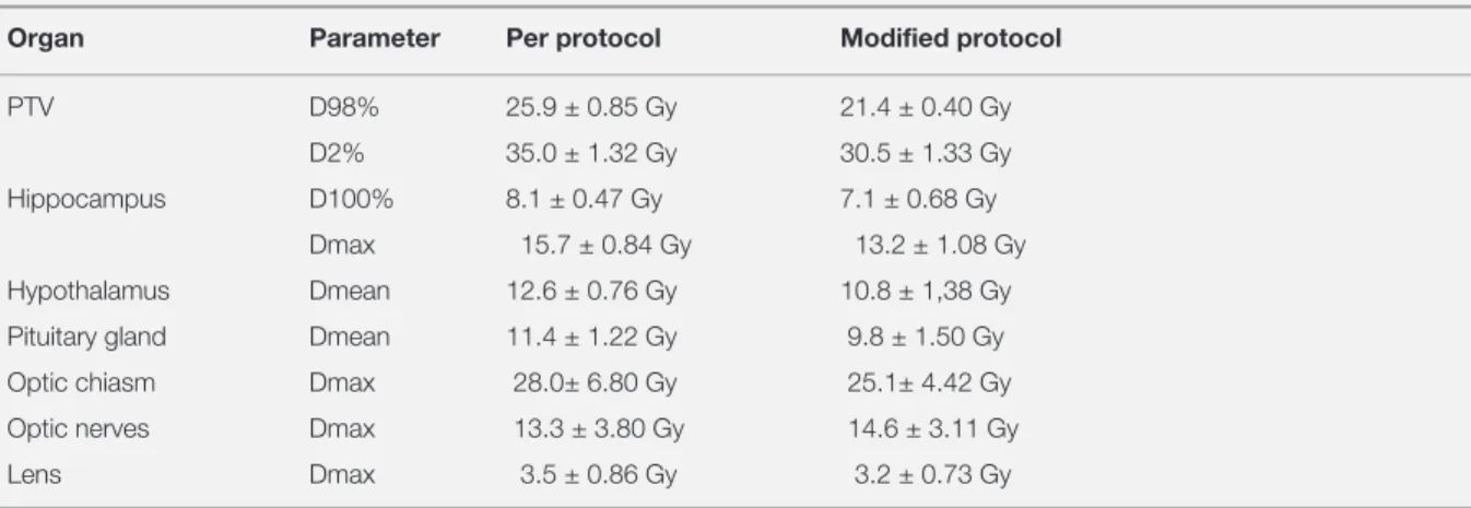
ra-Table 2. Mean doses in 12 patients
Organ Parameter Per protocol Modified protocol
PTV D98% 25.9 ± 0.85 Gy 21.4 ± 0.40 Gy D2% 35.0 ± 1.32 Gy 30.5 ± 1.33 Gy Hippocampus D100% 8.1 ± 0.47 Gy 7.1 ± 0.68 Gy
Dmax 15.7 ± 0.84 Gy 13.2 ± 1.08 Gy Hypothalamus Dmean 12.6 ± 0.76 Gy 10.8 ± 1,38 Gy Pituitary gland Dmean 11.4 ± 1.22 Gy 9.8 ± 1.50 Gy Optic chiasm Dmax 28.0± 6.80 Gy 25.1± 4.42 Gy Optic nerves Dmax 13.3 ± 3.80 Gy 14.6 ± 3.11 Gy Lens Dmax 3.5 ± 0.86 Gy 3.2 ± 0.73 Gy
Abbreviations: PTV= planning target volume; D98%= the dose which %98 of the target volume receives; D2%= the dose which 2% of the target volume
UHOD
diotherapy doses exceed 30 Gy.46 While growth hormone insufficiency risk is reported to be about 50% following 16 Gy of radiotherapy47, 12 Gy for HHA was estimated to be safe.48 Still, the actual in-cidence of treatment-related hormone insufficiency can vary as a result of heterogeneities including the presence or extent of surgery, follow-up intervals, cut-off laboratory values, definitions of hormone-insufficiency, and radiotherapy techniques.49-51 In this respect, the mean doses of the hypothalamus (12.6 Gy) and hypophysis (11.4 Gy) achieved in our study were concordant with the recommended safer range.
The hippocampal dose constraints for 25 Gy of PCI were D100%≤ 7.5 Gy and Dmax≤ 13.3 Gy which
appeared to be significantly lower than the values of D100%≤ 9 Gy and Dmax≤ 16 Gy, recommended in the RTOG 0933 guidelines for the protocol of 30 Gy of WBRT.26 The rationale to perform further modifications for hippocampal dose constraints can be the potential intrinsic radiosensitivity vari-ations of the patients which can also enhance the risk of NCF decline even for the cohort receiving a relatively lower dose schedule. Furthermore, be-cause of the lack of the long-term NCF outcomes of RTOG 0933, it can be rational to reduce the threshold radiotherapy doses for hippocampi in ac-cordance with the results of available studies re-porting better NCF with lower constraints, such as <7.3 Gy to 40% of bilateral hippocampi14, a Dmax of 9 Gy to 100% of the hippocampi25 and a Dmax of 5.83 Gy to 100% of the bilateral hippocampi.52 In the view of the reasons aforementioned, the use of 3HS-WBRT should not be restricted to only the adult patients with small-cell lung carcinoma pa-tients undergoing PCI but also applied to the pedi-atric cohort who are relatively more vulnerable to the effects of radiation compared to the adults.53 Additionally, the length of survival in patients with childhood brain tumors at 70% at 10 years renders the further minimization of radiotherapy-related neurotoxicity more meaningful than adults with a relatively shorter expectation of survival.54 For instance, there is an increased dementia effect af-ter craniospinal irradiation, which was described about a half century ago in patients with medul-loblastoma aged < 2 years.55 This can necessitate further maneuvers besides the efforts of decreasing
the radiotherapy doses or delaying the craniospi-nal irradiation until 3 years of age. Additiocraniospi-nally, underlining the importance of sparing of the in-tracranial endocrine organs the 20-year follow-up series by Institute Curie demonstrated that the endocrine complications were as common as 52% in medulloblastoma survivors after craniospinal ir-radiation.56 Accordingly, 3HS-WBRT appears to be rational for conserving NCF.
Radiation-induced HHA damage can also be a source of severe metabolic, social, financial, psy-chiatric, and educational problems which alto-gether can negatively influence psychosocial and medical status of the patients.57 For instance, HHA-associated endocrine abnormalities or obesity can cause diabetes mellitus, hypertension, dyslipidem-ia, cardiovascular problems, fertility, and pubertal disorders with resultant diminishments in the QoL measures and even shortened life spans.57 Consid-ering the educational outcomes, compared to those children treated with intravenous/intrathecal meth-otrexate alone, the combination of chemotherapy with PCI has been shown to cause significantly more severe declines in the achievement, intelli-gence and attention tasks, and therefore the school success in survivors of acute lymphoblastic leuke-mia.58,59 Additionally, recent data indicated that un-employment was reported to be 5 times higher in brain tumor survivors, associated with the primary tumor and related treatments.57 All these adverse effects of whole- or partial brain radiotherapy men-tioned above support our current efforts to achieve 3HS-WBRT or 3HS-partial brain radiotherapy and appear to be an important step in minimizing the social, financial, psychiatric, and educational bur-den due to impaired HHA.
The present study is the second in the literature to investigate 3HS-WBRT to the best of our knowl-edge. In addition to available dose constraints for 30 Gy of WBRT, we also recommend an alterna-tive reduced dose schedule of 1 in 6 of the original which can provide a further protection for the co-hort who can be more sensitive to radiation. On the other hand, although the delineation of hippocam-pus and HHA was performed according to the ac-cessible guidelines and atlases, the lack of a neuro-radiologist in the study may potentially weaken the accuracy of the 3H contouring process. Despite the
UHOD
main goal of the study to research the feasibility of HHA as well as the hippocampus, the interconnec-tions between hypothalamus and hypophysis were not included in the sparing volume, which can de-crease the reliability of the 3HS-WBRT. Although the mean treatment time was reported to be 15.93 and 20.18 minutes in a dosimetric study evaluat-ing the feasibility of hippocampus sparevaluat-ing-PCI and -WBRT, respectively, the relatively longer treat-ment time of 31.8 minutes elapsed during 3HS-WBRT can be questionable in cases requiring an-esthesia during the treatment process.60
In conclusion, as a consequence of improved sur-vival rates of the patients with cancer requiring whole brain or partial brain radiotherapy during the course of disease, the maintenance of neurocogni-tive and neuroendocrine functions, as the predic-tors of QoL, has become one of the main goals as well as disease control in the brain. In this context, this study demonstrated the feasibility of 3HS-WBRT with HT taking into account the suggested dose constraints in the literature. Considering the fact that a technique investigation can be more valuable for what it offers in practice, prospective studies evaluating the baseline and follow-up ob-jective measurements for 3H can be required for precise deductions.
REFERENCES
1. Govindan R, Page N, Morgensztern D, et al. Changing epide-miology of small-cell lung cancer in the United States over the last 30 years: analysis of the surveillance, epidemiologic, and end results database. J Clin Oncol 24: 4539-4544, 2006. 2. Cortes J. Central nervous system involvement in adult acute
lymphocytic leukemia. Hematol Oncol Clin North Am 15: 145-162, 2001.
3. Pui CH, Evans WE. Treatment of acute lymphoblastic leuke-mia. N Engl J Med 354: 166-178, 2006.
4. Bassan R, Hoelzer D. Modern therapy of acute lymphoblastic leukemia. J Clin Oncol 29: 532-543, 2011.
5. Larson RA. Managing CNS disease in adults with acute lymphoblastic leukemia. Leuk Lymphoma 59: 3-13, 2018. 6. Auperin A, Arriagada R, Pignon JP, et al. Prophylactic cranial
irradiation for patients with small-cell lung cancer in complete remission. Prophylactic Cranial Irradiation Overview Collabo-rative Group. N Engl J Med 341: 476-484, 1999.
7. Gondi V, Paulus R, Bruner DW, et al. Decline in tested and self-reported cognitive functioning after prophylactic cranial irradiation for lung cancer: pooled secondary analysis of
Ra-diation Therapy Oncology Group randomized trials 0212 and 0214. Int J Radiat Oncol Biol Phys 86: 656-664, 2013. 8. Roman DD, Sperduto PW. Neuropsychological effects of
cra-nial radiation: current knowledge and future directions. Int J Radiat Oncol Biol Phys 31: 983-998, 1995.
9. Rola R, Raber J, Rizk A, et al. Radiation-induced impairment of hippocampal neurogenesis is associated with cognitive deficits in young mice. Exp Neurol 188: 316-330, 2004. 10. Saxe MD, Battaglia F, Wang JW, et al. Ablation of
hippocam-pal neurogenesis impairs contextual fear conditioning and synaptic plasticity in the dentate gyrus. Proc Natl Acad Sci U S A 103: 17501-17506, 2006.
11. Madsen TM, Kristjansen PE, Bolwig TG, et al. Arrested neu-ronal proliferation and impaired hippocampal function follow-ing fractionated brain irradiation in the adult rat. Neuroscience 119: 635-642, 2003.
12. Winocur G, Wojtowicz JM, Sekeres M, et al. Inhibition of neu-rogenesis interferes with hippocampus-dependent memory function. Hippocampus 16: 296-304, 2006.
13. Gondi V, Tome WA, Mehta MP. Why avoid the hippocam-pus? A comprehensive review. Radiother Oncol 97: 370-376, 2010.
14. Gondi V, Hermann BP, Mehta MP, et al. Hippocampal dosim-etry predicts neurocognitive function impairment after frac-tionated stereotactic radiotherapy for benign or low-grade adult brain tumors. Int J Radiat Oncol Biol Phys 85: 348-354, 2013.
15. Redmond KJ, Hales RK, Anderson-Keightly H, et al. Pro-spective Study of Hippocampal-Sparing Prophylactic Cranial Irradiation in Limited-Stage Small Cell Lung Cancer. Int J Ra-diat Oncol Biol Phys 98: 603-611, 2017.
16. Wolfson AH, Bae K, Komaki R, et al. Primary analysis of a phase II randomized trial Radiation Therapy Oncology Group (RTOG) 0212: impact of different total doses and schedules of prophylactic cranial irradiation on chronic neurotoxicity and quality of life for patients with limited-disease small-cell lung cancer. Int J Radiat Oncol Biol Phys 81: 77-84, 2011. 17. Bressler SL. Large-scale cortical networks and cognition.
Brain Res Brain Res Rev 20: 288-304, 1995.
18. Qin Y, Zhang W, Yang P. Current states of endogenous stem cells in adult spinal cord. J Neurosci Res 93: 391-398, 2015. 19. Yoo S, Blackshaw S. Regulation and function of neurogen-esis in the adult mammalian hypothalamus. Prog Neurobiol 170: 53-66, 2018.
20. Quik EH, Valk GD, Drent ML, et al. Reduced growth hormone secretion after cranial irradiation contributes to neurocogni-tive dysfunction. Growth Horm IGF Res 22: 42-47, 2012. 21. Sinha A, Hollingsworth KG, Ball S, et al. Impaired quality of
life in growth hormone-deficient adults is independent of the altered skeletal muscle oxidative metabolism found in condi-tions with peripheral fatigue. Clin Endocrinol (Oxf) 80: 107-114, 2014.
UHOD
22. Prodam F, Caputo M, Belcastro S, et al. Quality of life, mood disturbances and psychological parameters in adult patients with GH deficiency. Panminerva Med 54: 323-331, 2012. 23. Lam KS, Tse VK, Wang C, et al. Early effects of cranial
ir-radiation on hypothalamic-pituitary function. J Clin Endocrinol Metab 64: 418-424, 1987.
24. Rosen T, Eden S, Larson G, et al. Cardiovascular risk factors in adult patients with growth hormone deficiency. Acta Endo-crinol (Copenh) 129: 195-200, 1993.
25. Redmond KJ, Mahone EM, Horska A. Association between radiation dose to neuronal progenitor cell niches and tempo-ral lobes and performance on neuropsychological testing in children: a prospective study. Neuro Oncol 15: 1455, 2013. 26. Gondi V, Pugh SL, Tome WA, et al. Preservation of memory
with conformal avoidance of the hippocampal neural stem-cell compartment during whole-brain radiotherapy for brain metastases (RTOG 0933): a phase II multi-institutional trial. J Clin Oncol 32: 3810-3816, 2014.
27. Elson A, Bovi J, Kaur K, et al. Effect of treatment modality on the hypothalamic-pituitary function of patients treated with radiation therapy for pituitary adenomas: hypothalamic dose and endocrine outcomes. Front Oncol 4: 73, 2014. 28. Baroncini M, Jissendi P, Balland E, et al. MRI atlas of the
hu-man hypothalamus. Neuroimage 59: 168-180, 2012. 29. Scoccianti S, Detti B, Gadda D, et al. Organs at risk in the
brain and their dose-constraints in adults and in children: a radiation oncologist’s guide for delineation in everyday prac-tice. Radiother Oncol 114: 230-238, 2015.
30. Srivastava SP, Cheng CW, Das IJ. The effect of slice thick-ness on target and organs at risk volumes, dosimetric cover-age and radiobiological impact in IMRT planning. Clin Transl Oncol 18: 469-479, 2016.
31. Li J, Bentzen SM, Renschler M, et al. Regression after whole-brain radiation therapy for whole-brain metastases correlates with survival and improved neurocognitive function. J Clin Oncol 25: 1260-1266, 2007.
32. Higham CE, Johannsson G, Shalet SM. Hypopituitarism. Lancet 388: 2403-2415, 2016.
33. Raber J, Rola R, LeFevour A, et al. Radiation-induced cogni-tive impairments are associated with changes in indicators of hippocampal neurogenesis. Radiat Res 162: 39-47, 2004. 34. Fan XW, Wang JQ, Wu JL, et al. Simultaneously
avoid-ing the hippocampus and hypothalamic-pituitary axis dur-ing whole brain radiotherapy: A planndur-ing study. Med Dosim S0958-3947(18)30068-2, 2018. doi: 10.1016/j.med-dos.2018.04.004.
35. Gondi V, Tolakanahalli R, Mehta MP, et al. Hippocampal-sparing whole-brain radiotherapy: a “how-to” technique us-ing helical tomotherapy and linear accelerator-based intensi-ty-modulated radiotherapy. Int J Radiat Oncol Biol Phys 78: 1244-1252, 2010.
36. Oskan F, Ganswindt U, Schwarz SB, et al. Hippocampus sparing in whole-brain radiotherapy. A review. Strahlenther Onkol 190: 337-341, 2014.
37. Prokic V, Wiedenmann N, Fels F, et al. Whole brain irradiation with hippocampal sparing and dose escalation on multiple brain metastases: a planning study on treatment concepts. Int J Radiat Oncol Biol Phys 85: 264-270, 2013.
38. Seibert TM, Karunamuni R, Bartsch H, et al. Radiation Dose-Dependent Hippocampal Atrophy Detected With Longitudinal Volumetric Magnetic Resonance Imaging. Int J Radiat Oncol Biol Phys 97: 263-269, 2017.
39. Rong Y, Evans J, Xu-Welliver M, et al. Dosimetric evaluation of intensity-modulated radiotherapy, volumetric modulated arc therapy, and helical tomotherapy for hippocampal-avoid-ance whole brain radiotherapy. PLoS One 10:e0126222, 2015.
40. Moon SY, Yoon M, Chung M, et al. Comparison of the ex-tent of hippocampal sparing according to the tilt of a patient’s head during WBRT using linear accelerator-based IMRT and VMAT. Phys Med 32: 657-663, 2016.
41. Merchant TE, Goloubeva O, Pritchard DL, et al. Radiation dose-volume effects on growth hormone secretion. Int J Ra-diat Oncol Biol Phys 52: 1264-1270, 2002.
42. Azcona C, Preece MA, Rose SJ, et al. Growth response to rhIGF-I 80 microg/kg twice daily in children with growth hor-mone insensitivity syndrome: relationship to severity of clinical phenotype. Clin Endocrinol (Oxf) 51: 787-792, 1999. 43. Costin G. Effects of low-dose cranial radiation on growth
hor-mone secretory dynamics and hypothalamic-pituitary func-tion. Am J Dis Child 142: 847-852, 1988.
44. Kirk JA, Raghupathy P, Stevens MM, et al. Growth failure and growth-hormone deficiency after treatment for acute lympho-blastic leukaemia. Lancet 1: 190-193, 1987.
45. Vladyka V, Liscak R, Novotny J Jr,et al. Radiation tolerance of functioning pituitary tissue in gamma knife surgery for pitui-tary adenomas. Neurosurgery 52: 309-316, 2003.
46. Clayton PE, Shalet SM. Dose dependency of time of onset of radiation-induced growth hormone deficiency. J Pediatr 118: 226-228, 1991.
47. Merchant TE, Rose SR, Bosley C, et al. Growth hormone se-cretion after conformal radiation therapy in pediatric patients with localized brain tumors. J Clin Oncol 29: 4776-4780, 2011.
48. Rappaport R, Brauner R. Growth and endocrine disorders secondary to cranial irradiation. Pediatr Res 25: 561-567, 1989.
49. Feigl GC, Pistracher K, Berghold A, et al. Pituitary insufficien-cy as a side effect after radiosurgery for pituitary adenomas: the role of the hypothalamus. J Neurosurg 113 Suppl: 153-59, 2010.
UHOD
50. Agha A, Sherlock M, Brennan S, et al. Hypothalamic-pituitary dysfunction after irradiation of nonpituitary brain tumors in adults. J Clin Endocrinol Metab 90:6355-60, 2005.
51. Rieken S, Habermehl D, Welzel T, et al. Long term toxicity and prognostic factors of radiation therapy for secreting and non-secreting pituitary adenomas. Radiat Oncol 8: 18, 2013. 52. Tsai PF, Yang CC, Chuang CC, et al. Hippocampal dosimetry
correlates with the change in neurocognitive function after hippocampal sparing during whole brain radiotherapy: a pro-spective study. Radiat Oncol 10: 253, 2015.
53. Leung RS. Radiation Protection of the Child from Diagnostic Imaging. Curr Pediatr Rev 11: 235-242, 2015.
54. Armstrong GT, Chen Y, Yasui Y, et al. Reduction in Late Mor-tality among 5-Year Survivors of Childhood Cancer. N Engl J Med 374: 833-842, 2016.
55. Bloom HJ, Wallace EN, Henk JM. The treatment and prog-nosis of medulloblastoma in children. A study of 82 verified cases. Am J Roentgenol Radium Ther Nucl Med 105: 43-62, 1969.
56. Frange P, Alapetite C, Gaboriaud G, et al. From childhood to adulthood: long-term outcome of medulloblastoma patients. The Institut Curie experience (1980-2000). J Neurooncol 95: 271-279, 2009.
57. Gupta P, Jalali R. Long-term Survivors of Childhood Brain Tumors: Impact on General Health and Quality of Life. Curr Neurol Neurosci Rep 17: 99, 2017.
58. Hill JM, Kornblith AB, Jones D, et al. A comparative study of the long term psychosocial functioning of childhood acute lymphoblastic leukemia survivors treated by intrathecal meth-otrexate with or without cranial radiation. Cancer 82: 208-218, 1998.
59. Moore BD, 3rd, Copeland DR, Ried H, et al. Neurophysiologi-cal basis of cognitive deficits in long-term survivors of child-hood cancer. Arch Neurol 49: 809-817, 1992.
60. Marsh JC, Godbole RH, Herskovic AM, et al. Sparing of the neural stem cell compartment during whole-brain radiation therapy: a dosimetric study using helical tomotherapy. Int J Radiat Oncol Biol Phys 78: 946-954, 2010.
Correspondence:
Dr. Yurday ÖZDEMİR
Başkent Üniversitesi Tıp Fakültesi Radyasyon Onkolojisi Anabilim Dalı Kisla Saglik Yerleskesi,
01120, ADANA / TURKEY Tel: (+90-505) 594 21 69 Fax: (+90-322) 344 44 52 e-mail: [email protected]