..
..
..
..
..
..
..
..
..
..
..
..
..
..
..
..
..
..
..
..
..
..
..
..
..
..
..
..
..
..
..
..
..
..
..
EuroEcho 2019: highlights
Julien Magne
1,2*, Tara Bharucha
3, Maya Cikes
4, Maurizio Galderisi
5,
Suzanna Price
6, Leyla Elif Sade
7, Bogdan A. Popescu
8, Bernard Cosyns
9, and
Thor Edvardsen
10,11,121
CHU Limoges, Hoˆpital Dupuytren, Service Cardiologie, Limoges F-87042, France;2
INSERM U1094, Univ. Limoges, CHU Limoges, IRD, U1094, GEIST, 2, rue Marcland, 87000
Limoges, France;3
Department of Congenital Cardiology, University Hospital Southampton NHS Foundation Trust, Tremona Road, Southampton SO16 6YD, UK;4
Department
of Cardiovascular Diseases, University of Zagreb School of Medicine, University Hospital Center Zagreb, Kispaticeva 12, 10000 Zagreb, Croatia;5
Department of Advanced
Biomedical Sciences, Federico II University Hospital, via S. Pansini 5, 80131 Naples, Italy;6Unit of Critical Care Medicine, Imperial College School of Medicine, Royal Brompton
Hospital, Dovehouse Street, London SW3 6LY, UK;7
Department of Cardiology, University of Baskent, E Blok, 54. Sokak, Bahcelievler 06490, Ankara, Turkey;8
University of
Medicine and Pharmacy “Carol Davila”—Euroecolab, Institute of Cardiovascular Diseases “Prof. Dr. C. C. Iliescu”, Bucharest, Romania;9
Centrum voor Hart en Vaatziekten
(CHVZ), Universitair Ziekenhuis Brussel, Vrij Universiteit van Brussel, Brussels, Belgium;10
Department of Cardiology, Center for Cardiological Innovation, Oslo University
Hospital, Rikshospitalet, Oslo, Norway;11University of Oslo, Oslo, Norway; and12Faculty of Medicine, University of Oslo, Oslo, Norway
Received 12 February 2020; editorial decision 13 February 2020; accepted 19 February 2020; online publish-ahead-of-print 17 March 2020
The annual meeting of the European Association of Cardiovascular Imaging, EuroEcho 2019, was held in Vienna, Austria, in December 2019. In this article, we present a summary of the ‘Highlights’ session.
... Keywords heart failure
•
heart valve disease•
new technologies•
congenital heart disease•
acute cardiaccare
•
preventionIntroduction
The 23rd annual scientific meeting of the European Association of Cardiovascular Imaging (EACVI), EuroEcho 2019, was held from 4 to 7 December in Vienna, Austria. The main themes were ‘cardio-myopathies’ and ‘imaging in acute cardiac care’.
This year, the world leading congress on echocardiography, reached a total number of 4095 health care professionals coming from 95 countries.
Over 1639 abstracts and clinical cases were submitted and the scientific programme involved 387 international faculties from 44 countries in 233 scientific sessions. Furthermore, five studies were presented in the Late Breaking Trial session, newly launched this year.
Overall, 15 oral abstract sessions and 34 poster sessions were presented. The preferred topics regarding abstracts submission were ‘Tissue Doppler, Speckle Tracking and Strain Imaging’, ‘Valve Diseases’, ‘Systolic and Diastolic Function’, and ‘Stress Echocardiography’.
The ‘Highlights’ session wrapped up the event with a summary of the most relevant abstracts presented throughout the congress on the last day of the meeting. A short report of this session is presented below.
Heart failure
The best abstracts dealing with heart failure (HF) included novelties such as artificial intelligence (AI), myocardial work (MW), shear wave propagation, vortex formation but also strain imaging, standard echo-Doppler, and multimodality imaging approach.
The AI was tested to assess both left ventricular (LV) systolic and diastolic function. Uejima et al.1performed a multiparametric assess-ment of diastolic function in 279 consecutive HF patients. The diastol-ic function was assessed according to a cluster analysis [based on unsupervised machine learning (ML) algorithm] compared to diastolic grading of current recommendations. The hospitalization for LV ejection fraction (EF) worsening was the primary endpoint and the combination cardiovascular (CV) death þ HF hospitalization the secondary endpoint. The comparison of diastolic parameters [E/A, e0 velocity, E/e0, left atrial (LA) volume index (LAVi), and tricuspid regur-gitation (TR) velocity] showed similar trend between grading and clusters and both grading and cluster classification correlated with brain natriuretic peptide levels. However, during a follow-up of 2.6 ± 2.0 years (primary endpoint reached by 62 patients), cluster-based classification showed a superiority to predict HF-free survival
and the combined endpoint of CV death þ HF-free survival
(Figure1). The combination of MAGGIC score (an HF risk calculator
* Corresponding author. Tel:þ33 5 19 76 19 18. E-mail: [email protected]
Published on behalf of the European Society of Cardiology. All rights reserved.VCThe Author(s) 2020. For permissions, please email: [email protected].
European Heart Journal - Cardiovascular Imaging (2020) 21, 469–478
REVIEW
doi:10.1093/ehjci/jeaa037
..
..
..
..
..
..
..
..
..
..
..
..
..
..
..
..
.
including 13 clinical variables, risk factors, and treatment) and cluster analysis showed an additional value in comparison with MAGGIC score þ grading classification in predicting primary and secondary endpoints (Figure1). The authors conclude that AI presents an incre-mental value in comparison with current recommendations for pre-dicting HF decompensation. Popoff et al.2evaluated accuracy and reproducibility of an AI-based semi-automated tool to compute LV volumes and EF, in comparison with manual contouring, using cardiac magnetic resonance (CMR) as reference in a two-centre retrospect-ive study (n = 114 patients in sinus rhythm). LV segmentation using U-Net convolutional network on the frames selected by threeobservers. The authors showed improved accuracy (vs. CMR) and higher reproducibility of AI.
The impact of MW acute redistribution after cardiac resynch-ronization therapy (CRT) on LV reverse remodelling (D% LV end-systolic volume) was evaluated by Duchenne et al.3 in 135 HF patients referred for CRT (four European centres). Before CRT, MW showed clear differences among LV walls. The CRT caused an acute redistribution of MW, with most increase in the septum and most decrease laterally and induced a more homogeneous MW distribution. After 1-year follow-up, acute MW redistribu-tion (D% changes) was positively related to D% LV end-systolic Figure 1Clinical outcomes by grading and cluster (top) and Cox proportional hazard models (bottom, A, primary point and B, secondary end-point). Modified from Sanchez-Martinez et al.35LVEDV, left ventricular end-diastolic volume.
..
..
..
..
..
..
..
..
..
..
..
..
..
..
..
..
..
..
..
..
..
..
..
..
..
..
..
..
..
..
..
..
..
..
..
..
..
..
..
..
..
..
..
..
..
..
..
..
..
..
..
..
..
..
..
..
..
..
..
..
..
..
..
..
..
..
..
..
..
..
..
..
..
..
..
..
..
..
..
..
..
..
..
..
..
..
.
volume. The authors conclude that regional loading modification is the mode of action of CRT.Hubert et al.4evaluated a novel semi-automatic tool for the assess-ment of volume–strain loops in 17 patients with cardiac amyloidosis compared with 18 healthy controls and 18 patients with HF with pre-served EF. The volume–strain area better correlated with average e0of the mitral annulus (r = 0.65, P < 0.001) than all the other indices, thus demonstrating to be a very promising tool to assess semi-automatically diastolic function.
Novel sudden cardiac death (= sudden cardiac arrest or an appropriate intracardiac defibrillator (ICD) intervention) predictors such as global longitudinal strain (GLS), LV average strain, and LAVi (in-stead of LA dimension) were tested in 252 patients with hypertrophic cardiomyopathy by Adamczak et al.5By using Cox’s proportional haz-ard analysis, the models with the novel parameters was highly signifi-cant (P = 0.00094), whereas the model including the standard parameters was marginally significant (P = 0.04385) in predicting ICD implantation during an average follow-up of 3.8 years. The new predic-tors could be taken into account when considering ICD implantation.
Prevention
Clerc et al.6 investigated the predictive value of coronary artery calcium score (CACS) for predicting myocardial ischaemia in 927 consecutive patients without known coronary artery disease, who underwent 82Rb positron emission tomography myocardial perfu-sion imaging and multi-slice computerized tomography. The propor-tion of ischaemia, expressed as summed difference score >_4, increased in relation with CACS. The CACS was a better associated with ischaemia than combined clinical and electrocardiogram data (area under curve = 0.82 vs. 0.69; P < 0.001). A CACS <100 AU had high sensitivity (81%) and negative predictive value (94%) to exclude ischaemia, whereas CACS >1000 AU had high specificity (94%) but moderate positive predictive value (60%) to identify ischaemia, underlining the need for additional testing after positive CACS.
Regarding atherosclerosis, Matangi et al.7performed screening by carotid ultrasound in 1683 consecutive patients to estimate the pro-portion and severity of carotid disease in patients referred for stress echocardiography. Patients had no diabetes, statin treatment, or prior history of any CV disease. Plaque scoring was performed according to Rotterdam method.8 A brief carotid using echocardiographic screening prior to stress echocardiography identified a significant proportion of patients (68% males, 52% females) having high risk for CV events who might benefit from guidelines based CV disease pre-vention therapies including aspirin and statins, regardless of induced wall motion abnormalities and/or ischaemia.
Important data came from the ARIC-NCS (Atherosclerosis Risk in Communities Neurocognitive Study) sub-study regarding stroke prevention. Bianco et al.9 demonstrated the association between age-related LA remodelling and ischaemic stroke in the elderly with normal sinus rhythm, preserved LV ejection fraction (LVEF) and no valvular heart disease. Three subgroups were studied as follow: (i) no cerebral infarcts on CMR (n = 315), (ii) CMR-diagnosed subclinical infarcts (n = 58), and (iii) clinically diagnosed stroke (n = 34). Among investigated echocardiographic parameters, they found that E/e0 div-ided by LA GLS, as a surrogate of LA stiffness, was an independent
risk factor for overall cerebral infarcts [odds ratio (OR) 1.92, P = 0.005] and stroke (OR 2.30, P = 0.009), while LA GLS, was an in-dependent marker for subclinical cerebral infarct (OR 1.0, P = 0.014). These results have important clinical implications for empiric anticoa-gulation to prevent both recurrent subclinical cerebral infarcts and stroke before the development of atrial fibrillation among elderly.
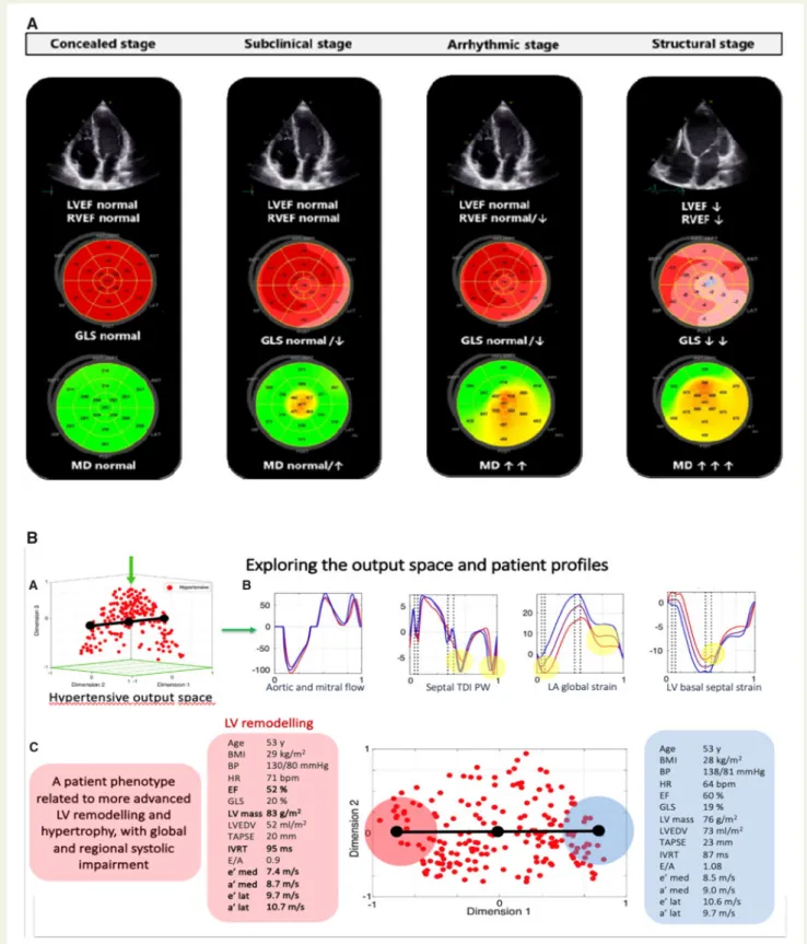
Taha et al.10explored echocardiographic mechanical dysfunction in phospholamban R14del mutation carriers. In all, 120 mutation car-riers and 60 control subjects were studied. Phospholamban (PLN) R14del carriers were classified as: pre-symptomatic (no ventricular arrhythmias, LVEF >_50%), arrhythmic with LVEF >_50%, and structur-ally abnormal (LVEF <50%). GLS was reduced in pre-symptomatic stage that could be further differentiated from concealed stage by means of GLS (Figure 2A). Patients with arrhythmias or structural abnormalities had both increased mechanical dispersion and low GLS. Future studies should aim to investigate the therapeutic and prognostic value of these measurements.
Loncaric et al.11used ML tool to explore hypertensive patient pro-files based on full myocardial deformation and Doppler traces. ML goes beyond the limited number of standard echocardiographic parameters by capturing and integrating the complex and complete information from blood pool and tissue characteristics throughout the cardiac cycle and represents the individual on an output space where patients are grouped based on the similarity of their cardiac function. Individual clinical data, velocity, and deformation patterns can be extracted and compared between positions in the output space (Figure2B). New patients can be projected into the existing space to aid in clinical interpretation. This futuristic model is promis-ing to refine risk assessment and patient management in a cohort with well-regulated blood pressure.
Heart valve diseases
In contrast with past years, aortic stenosis is no longer the most frequently investigated heart valve diseases and tricuspid valve is no longer the ‘forgotten valve’.
Previtero et al.12 studied the prognostic value threshold of semi-quantitative (i.e. vena contracta) and quantitative [i.e. tant volume, effective regurgitant orifice area (EROA), and regurgi-tant fraction (RF)] echocardiographic parameters of functional TR assessment. In this single centre, retrospective study involving 296 patients, they identified new cut-off values, slightly lower than those reported in current guidelines. They also underlined the crucial value of RF in risk stratification of functional mitral regurgitation (MR), related to its less dependency to loading conditions.
In a prospective multicentre study,13 the right ventricular (RV) geometry and function, as well as tricuspid annulus size, of 100 patients with various TR severity (from mild to torrential) were ana-lysed. The EROA of TR was independently associated with right atrial size and tricuspid tethering area but not with RV function parameters. Of note, the vast majority of patients, regardless of TR severity, exhibited tricuspid annulus dilatation (>40 mm).
The prognostic implication of bivalvular functional regurgitation (i.e. concomitant presence of both functional MR and TR) has been investigated by Heitzinger et al.14This context remains challenging to treat and manage, and only limited data are available. Among the
EuroEcho 2019
471
Figure 2(A) Classification of PLN (Phospholamban) mutation (R14del) carriers on the basis of deformation characteristics. Pre-symptomatic stage is differentiated from concealed stage by means of decreased GLS and mechanical dispersion. LVEF, left ventricular ejection fraction; RVEF, right ven-tricular ejection fraction. (B) Exploration of the hypertensive output space and patient profiles from the output space. (A) Hypertensive output space, (B) extraction of individual measurements, and (C) exploration of individual patient profiles. Modified from Sanchez-Martinez et al.35LA, left atrial; LV, left ventricular; PW, pulsed wave; TDI, tissue Doppler imaging.
..
..
..
..
..
..
..
..
..
..
..
..
..
..
..
..
..
..
..
..
..
..
..
..
..
..
..
..
..
..
..
..
..
..
..
..
..
..
..
.
1021 patients with HF and reduced EF studied, 32% have bivalvular function regurgitation. During a maximal follow-up of 5 years, 433 patients died, and as expected, bivalvular function regurgitation was associated with markedly reduced survival, significantly lower than isolated severe functional regurgitation. Furthermore, the authors also found that moderate bivalvular functional regurgitation is associ-ated with higher risk of mortality than isolassoci-ated severe functional MR or TR, or even similar risk that severe functional MR and moderate TR or vice-versa.Mitral annular disjunction (MAD) associated with mitral valve pro-lapse has been studied using CMR by Pavon et al.15They purposely aimed to investigate the relationship between MAD and the presence of interstitial fibrosis, in the basal inferior LV wall (Figure3). A group of 33 patients with both MAD and regurgitant mitral valve prolapse was compared to 22 patients with regurgitant prolapse without MAD. The MAD size and interstitial fibrosis, as assessed as extracellu-lar volume (ECV) derived from T1 mapping, were measured using CMR. The ECV was statistically higher in MAD patients than in those without MAD, in anterior, septal, inferior, and lateral wall. It was also reported a significant correlation between ECV of the basal inferior wall and MAD size. This finding emphasize the potential role of inter-stitial LV fibrosis in the arrhythmogenic abnormalities frequently associated with MAD.
Similarly, the prognostic values of the presence of late gadolinium enhancement (LGE) derived from CMR were tested by Bouttier et al.16in 237 patients with mitral valve prolapse with or without MR.
The presence of LGE was observed in 41% of patients and 32% had LGE in the myocardial wall, 4% in the papillary muscle tip and 5% on both locations. The LGE was only associated with age and LV mass in multivariate analysis. Nevertheless, moderate to severe MR and the presence of LGE were both independently associated with event-free survival (i.e. composite endpoint of CV death, HF, new onset of atrial fibrillation or arterial embolism). These results suggest that CMR may be an additional prognostic tool in patients with mitral valve prolapse.
Acute cardiac care
A major focus of the congress related to the acute and critical care setting, with numerous abstracts relating to acute myocardial infarc-tion (MI) and risk stratificainfarc-tion, and evaluating novel applicainfarc-tions of existing/emerging techniques to optimize and individualize patient care.
Trifunovic et al.17reported part of the PREDICT-VT study, aiming to test whether deformation imaging based on speckle tracking was superior to conventional echocardiography when predicting outcome (i.e. total mortality, hospitalization for HF and New York Heart Association >III) in patients who had undergone percutaneous coronary intervention (PCI) post-ST-elevation acute myocardial infarction (STEMI). A total of 264 patients were included, with follow-up at 1 year. With well-defined cut-off values, peak systolic Figure 3Myocardial ECV by T1 mapping-CMR in mitral valve prolapse with mitral-annulus disjunction. The three-chamber view (A) show MAD (tween 7.1 and 12.3 mm); pre contrast T1 mapping (B) and after contrast T1 mapping (C) divided into four segments to evaluate the ECV. Relation be-tween mitro-annular disjunction and pre contrast T1 mapping (D). Relationship bebe-tween MAD and ECV (E). Modified from Sanchez-Martinez et al.35
EuroEcho 2019
473
..
..
..
..
..
..
..
..
..
..
..
..
..
..
..
..
..
..
..
..
..
..
..
..
..
..
..
..
..
..
..
..
..
..
..
..
..
..
..
..
longitudinal strain, radial strain rate during atrial contraction, and peak systolic circumferential strain (endocardial layer) were superior to standard parameters (including EF and tricuspid annular plane sys-tolic excursion) and predicting outcome.In a study presented by Backhaus et al.,18the interplay of infarct territory-related myocardial mechanics and prognosis post-MI were explored. Here, 795 STEMI and 440 non-STEMI patients underwent CMR 3 days (interquartile range 2–4 days) post-PCI. Primary clin-ical endpoint was the occurrence of major adverse cardiovascular events (MACE) within 1 year. The LV and LA deformation were analysed using standard parameters, including those of LA reser-voir, conduit, and booster pump function. Unsurprisingly, lesion related to left anterior descending (LAD) and left circumflex were associated with higher rate of MACE than right coronary artery lesion. However, LAD risk assessment was most significant using LA strain, suggesting the importance of LA function post-MI. In a separate study with the same group of patients, the impact of right atrial function was also analysed. Schuster et al.19presented data demonstrating that with RVEF <61.3%, impairment of all three elements of right atrial function were associated with a sig-nificant increase in MACE. De Campos et al.20presented further data relating to the impact of right heart function on outcomes post-MI. Numerous variables (physiological, biological, and echo-cardiographic) were studied post-MI in 303 patients with follow-up at short- and long-term (i.e. 1 year and 73 months). Even modest evaluation of pulmonary artery pressure was associated
with a significantly increased mortality, again highlighting the importance of the right heart in outcome prediction.
A study presented by Karagiannidis et al.,21addressed imaging and quantitative estimation of thrombus burden in patients with STEMI with the use of micro-computed tomography. In this methodological study, micro-computed tomography of aspirated thrombus was stud-ies in 3D models, looking at volume, density, and calcification. A strik-ing findstrik-ing was that those with a family history of ischaemic heart disease had higher density thrombus, suggesting a potential genetic contribution to the type of thrombus in different patient populations.
Rodean et al.,22 presented results from the ATHERODENT
study, aiming to evaluate the association between coronary plaque vulnerability and periodontal disease in patients with unstable angina. In this study, 52 patients with unstable angina were underwent (in addition to standard investigations and interventions) complete dental examination (peridontal index), coronary computed tomog-raphy angiogtomog-raphy, carotid ultrasound, and measurement of abdo-men and neck circumference (Figure4). Findings demonstrated that a high periodontal index was associated with an increased calcium score, plaque volume (calcified, non-calcified and lipid-rich), and increased plaque vulnerability.
Congenital heart disease
In congenital heart disease, the right ventricle is often abnormal, ei-ther anatomically or because it is subjected to abnormal pressure or
Figure 4Illustrative case from the ATHERODENT clinical trial. Case of a 47-year-old male patient without any significant history of cardiovascular disease, hospitalized for unstable angina. The complex dental examination revealed severe periodontitis. The coronary computed tomography per-formed highlights the presence of the vulnerability features on the coronary arteries. The severity of periodontal index is directly associated with the plaque vulnerability. The higher the periodontal index, the higher the vulnerability features are. Modified from Sanchez-Martinez et al.35
..
..
..
..
..
..
..
..
..
..
..
..
..
..
..
..
..
..
..
..
..
..
..
..
..
..
..
..
..
..
..
..
..
..
..
..
..
..
..
.
volume loading conditions. Many of our current clinical conundrums relate to assessment of the pathological RV, and imaging parameters which relate to functional/clinical status are increasingly useful.Avesani et al.23used echocardiography and CMR to investigate which parameters correlate best with functional exercise capacity in children with repaired tetralogy of Fallot (TOF), who very commonly have significant pulmonary regurgitation (PR). CMR is the gold stand-ard method to assess PR and RV size and function, but CMR has a number of drawbacks in children, such as the requirement for general anaesthesia in younger patients; echocardiography is a much more at-tractive tool in this population. A number of echocardiographic parameters were studied, but most showed disappointing correlation with CMR, although RV end-diastolic area indexed correlated well with CMR volumes. Right atrial strain, as an indicator of RV diastolic function, was the best determinant of peak VO2in young patients
and could be a useful addition to the routine follow-up echocardio-gram in young patients with repaired TOF.
Akazawa et al.24assessed the effect of electromechanical dyssyn-chrony (EMD) on RV dysfunction and exercise capacity in unoperated patients with Ebstein’s malformation. The EMD within the functional RV was determined by the presence of a septal flash or difference in time to peak shortening between the RV basal segment and apical sep-tum (Figure5). They found that intra-functional RV delay correlated with RV GLS and RV fractional area change, and also with CMR-derived measures of RV function/area, as well as peak VO2. Ostenfeld
et al.25 also investigated exercise capacity in relation to imaging
markers in Ebstein patients but focused on adults having undergone tricuspid repair/replacement surgery. Using CMR, they demonstrated an increase in cardiac index which continued at 5 years following sur-gery, as did improvement in VO2. However, a decline in RVEF may
mitigate potential benefits with regards to incidence of MACE, and therefore longer-term follow-up is required to determine whether there is any lasting alteration to the natural history in this condition.
Patients in whom the right ventricle is the systemic ventricle suffer from early HF. Surkova et al.26,27investigated both the prevalence of MR and systolic dysfunction in the subpulmonary LV in a mixed group comprising patients who had undergone atrial switch operations (Mustard/Senning) and patients with congenitally corrected transpos-ition of the great arteries. They found that non-systemic MR was associated with LV dilatation and systolic dysfunction, raised brain natriuretic peptide levels and HF symptoms. Patients with atrial switch procedures were particularly likely to have severe HF if MR was significant. Dysfunction of the subpulmonary LV was inde-pendently associated with HF in these patients, and more likely to predict functional class than systemic RV function. These findings highlight the importance of LV/RV interaction in these patients and remind us that assessment of the function of both ventricles is informative in their follow-up.
Patients with Fontan (single ventricle) circulation also suffer early HF, and diastolic function in this group is especially important but difficult to assess. Cordeiro Rato Mesquita Da Silva et al.28aimed to characterize the relationship between atrial strain and exercise
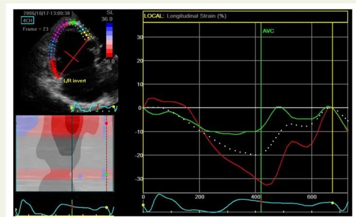
Figure 5 EMD in the functional right ventricle in a patient with Ebstein’s malformation, demonstrating difference in time to peak shortening be-tween the RV basal segment (red line) and apical septum (green line). Modified from Sanchez-Martinez et al.35
EuroEcho 2019
475
..
..
..
..
..
..
..
..
..
..
..
..
..
..
..
..
..
..
capacity. They found that atrial strain parameters are strongly corre-lated with VO2in Fontan patients, and this may be a useful additionto their clinical assessment, especially given the heterogeneity of anatomy in this group, and lack of standardized parameters for assessment of ventricular function.
New technologies
A number of abstracts focused on new technologies and different tasks by which they can support clinical decision-making: from image acquisition,29feature extraction31–34to data interpretation.35
Langet et al.29evaluated AI-assisted TTE: 40 novices performed ap-ical four-chamber acquisitions: initial standard TTE, AI-assisted TTE, and repetition standard TTE. An apical four-chamber acquisition was also performed by an expert. A significant contribution of AI-assisted vs. initial TTE was found in matching reference performance over time, associated with improved clinical use suitability, suggesting that AI-assisted TTE can assess and improve novices’ skills, and support acquisition standardization.
Nordal et al.30aimed to automatize perioperative monitoring of LV function by TOE detection of mitral annular plane systolic excur-sion (MAPSE). Convolutional neural networks were trained in a supervised manner to predict the location of two landmarks on Figure 6ML-based exploration of echocardiographic patterns and clinical parameters in paediatric dilated cardiomyopathy. (A) Three clusters with distinct proportions of the composite outcome of death or heart transplant. (B) Representative traces from Doppler of aortic valve outflow, and 2D strain traces from the basal septal, apical septal and basal lateral wall. (C) Spider plot of clinical characteristics of the studied cohort. (D) The incidence of the composite outcome of death or heart transplant and a summary of clinical and echocardiographic characteristics for the three pa-tient clusters. Modified from Sanchez-Martinez et al.35ACE-I, angiotensin converting enzyme inhibitor; AntiArr, antiarrhythmic therapy; BB, beta blocker; BSA, body surface area; EDD z, LV end-diastolic dimension z score; GLS, global longitudinal strain; LVEF, left ventricular ejection fraction; MRA, mineralocorticoid receptor antagonist; qrsd, QRS interval duration; Wt, weight.
..
..
..
..
..
..
..
..
..
..
..
..
..
..
..
..
..
..
..
..
..
..
..
..
..
..
..
..
..
..
..
..
..
..
..
..
..
..
..
..
..
..
..
..
..
..
..
..
..
..
..
..
..
..
..
..
..
..
..
..
..
..
..
..
..
..
..
..
..
..
..
..
..
..
..
..
..
..
..
..
..
..
..
..
..
..
.
the mitral annulus and automatically track MAPSE in B-mode. The detector was able to adequately track vertical movement of the landmarks, providing a fast calculation of MAPSE and eliminating intraobserver variability.Wheeler et al.31compared the accuracy of a virtual reality (VR) 3D linear measurement system to clinical imaging software, using multiplane reformatting and volume-rendered views. Experts performed VR measurements by selecting two points in the volume rendering on a calibration phantom, and anatomically meaningful measurements, using a VR system and two commercial systems. Distance measurement bias of the VR system was lower than a com-mercial application’s, providing evidence for clinical acceptability of the VR tool.
In a prospective multicentre study (n = 200), Aalen et al.32used re-gional work by pressure–strain analysis before and 7 ± 1 months after CRT to determine whether combined assessment of septal function and viability (by LGE CMR) identified CRT responders. Septal work was improved in CRT responders. The difference between work in the LV lateral wall and septum provided good prediction of CRT response while septal scar predicted non-response; combining these parameters improved the prediction of CRT response.
Petrescu et al.33prospectively studied 22 heart transplant recipi-ents to investigate if natural shear wave velocities increase with the degree of diffuse myocardial fibrosis. Shear wave elastography was performed at 1100 ± 250 frames/s, measuring shear wave propagation velocity at mitral and aortic valve closure from M-mode acceleration maps. Pulmonary capillary wedge pressure was meas-ured invasively, CMR (native T1, ECV) quantified diffuse myocardial fibrosis. End-diastolic and end-systolic shear wave velocities correlated with CMR-defined fibrosis, while end-diastolic shear wave velocities correlated with pulmonary capillary wedge pressure.
Papadacci et al.34 studied semiautomatic evaluation of cardiac Doppler indices within a single heartbeat by 4D ultrafast echocardi-ography (5200 volumes/s). Four-dimensional colour-flow and tissue Doppler imaging were computed from single heartbeats; spectral Dopplers were derived at each voxel, automatically detecting the LV outflow tract, mitral inflow, and basal inferoseptum. Doppler indices from Doppler and tissue velocity spectra were compared to standard 2D echocardiography indices: 4D ultrafast echocardiography was able to quantify major Doppler indices from a single heartbeat acquisition.
Sanchez-Martinez et al.35studied 50 children with dilated cardio-myopathy using unsupervised ML. The input consisted of longitudinal strain traces, LV outflow Doppler patterns, and clinical parameters. According to similarities in these descriptors, three distinct clusters (Figure6) were created with significantly different risk of death or heart transplantation. This suggests that ML-based approaches can be useful to explore the combination of regional and global echocardio-graphic and clinical parameters in association to outcomes.
Conflict of interest: none declared.
References
1. Uejima T, Cho J, Hayama H, Takahashi L, Yajima J, Yamashita T. Multiparametric assessment of diastolic function in heart failure. Eur Heart J Cardiovasc Imaging 2020;21(Suppl. 1):i33.
2. Popoff A, Langet H, Piro P, Ropert C, Allain P, Gauriau R et al. Can we improve the accuracy and reproducibility of left ventricular ejection fraction from 2D
echocardiography using artificial intelligence? Eur Heart J Cardiovasc Imaging 2020; 21(Suppl. 1):i34.
3. Duchenne J, Aalen JM, Cvijic M, Larsen CK, Galli E, Be´zy S et al. Acute re-distribution of regional LV work by CRT determines long-term remodelling. Eur Heart J Cardiovasc Imaging 2020;21(Suppl. 1):i301.
4. Hubert A, Le Rolle V, Galli E, Hernandez A, Donal E. Semi-automated volume-strain loops: a new tool in TTE to assess diastolic dysfunction. Eur Heart J Cardiovasc Imaging 2020;21(Suppl. 1):i21.
5. Adamczak D. New predictors of sudden cardiac death in hypertrophic cardiomy-opathy. Eur Heart J Cardiovasc Imaging 2020;21(Suppl. 1):i730.
6. Clerc OF, Caobelli F, Haaf P, Zellweger MJ. Predictive value of coronary artery calcium score for myocardial ischaemia in positron emission tomography. Eur Heart J Cardiovasc Imaging 2020;21(Suppl. 1):i709.
7. Matangi M, Cases M, Brouillard D, Johri A. Carotid screening prior to stress echocardiography, an opportunity to assess cardiovascular risk? Eur Heart J Cardiovasc Imaging 2020;21(Suppl. 1):i1105.
8. Hollander M, Bots ML, Del Sol AI, Koudstaal PJ, Witteman JC, Grobbee DE et al. Carotid plaques increase the risk of stroke and subtypes of cerebral infarction in asymptomatic elderly: the Rotterdam study. Circulation 2002;105:2872–7. 9. Bianco F, De Caterina R, Chandra A, Goncalves A, Aquila I, Salomon SD et al.
Association of age-related left atrial remodeling with ischemic stroke in patients with normal sinus rhythm. Eur Heart J Cardiovasc Imaging 2020;21(Suppl. 1):i20.
10. Taha K, Te Rijdt WP, De Bruin-Bon H, Cramer MJM, Asselbergs FW, Bouma BJ et al. Early detection of biventricular mechanical dysfunction in PLN R14del mu-tation carriers. Eur Heart J Cardiovasc Imaging 2020;21(Suppl. 1):i669.
11. Loncaric F, Sanchez-Martinez S, Nuno L, Fernandes JF, Mimbrero M, Tirapu L et al. Exploring hypertensive patient profiles based on full myocardial deform-ation and Doppler traces—insights from a machine-learning approach. Eur Heart J Cardiovasc Imaging 2020;21(Suppl. 1):i251.
12. Previtero M, Guta AC, Ochoa-Jimenez RC, Figliozzi S, Palermo C, Baritussio A et al. Prognostic validation of partition values obtained with conventional two-dimensional and doppler echocardiography to grade tricuspid regurgitation se-verity. Eur Heart J Cardiovasc Imaging 2020;21(Suppl. 1):i1.
13. Guerin A, Dreyfus J, Vabret E L, Tourneau T, Lavie-Badie Y, Sportouch C et al. Cardiac remodeling in secondary tricuspid regurgitation: should we just look at the tricuspid annulus diameter? Eur Heart J Cardiovasc Imaging 2020;21(Suppl. 1): i768.
14. Heitzinger G, Bartko PE, Arfsten H, Pavo N, Winter MP, Toma A et al. Natural history of bivalvular functional regurgitation. Eur Heart J Cardiovasc Imaging 2020; 21(Suppl. 1):i300.
15. Pavon A, Hugelshofer S, Rutz T, Pascale P, Pruvot E, Muller O et al. Increased interstitial fibrosis in patients with mitral valve prolapse and mitro-annular dys-junction. Eur Heart J Cardiovasc Imaging 2020;21(Suppl. 1):i676.
16. Bouttier AL, Jobbe-Duval A, Cueff C, Piriou N, Jaafar P, Pattier S et al. Prognostic value of cardiac magnetic resonance imaging in mitral valve prolapse. Eur Heart J Cardiovasc Imaging 2020;21(Suppl. 1):i675.
17. Trifunovic D, Krljanac G, Asanin M, Savic-Spasic L, Aleksandric S, Dudic J et al. Myocardial deformation imaging after STEMI: can we better predict one-year mortality and heart failure development? Eur Heart J Cardiovasc Imaging 2020; 21(Suppl. 1):i319.
18. Backhaus SJ, Kowallick JT, Stiermaier T, Lange T, Koschalka A, Navarra JL et al. Interplay of Infarct-related myocardial mechanics and prognostic implications after acute myocardial infarction. Eur Heart J Cardiovasc Imaging 2020;21(Suppl. 1):i316.
19. Schuster A, Backhaus SJ, Navarra JL, Stiermaier T, Rommel KP, Koschalka A et al. Impact of right atrial physiology on heart failure and adverse events after myo-cardial infarction. Eur Heart J Cardiovasc Imaging 2020;21(Suppl. 1):i314. 20. De Campos D, Saleiro C, Teixeira R, Botelho A, Lopes J, Puga L et al.
Echo-Omics to estimate prognosis after an acute myocardial infarction: which one to pick? Eur Heart J Cardiovasc Imaging 2020;21(Suppl. 1):i320.
21. Karagiannidis E, Konstantinidis NV, Sofidis G, Chatzinikolaou E, Sianos G. Imaging and quantitative estimation of thrombus burden in patients with ST elevation acute myocardial infarction (STEMI) with the use of micro-computed tomog-raphy—a methodological approach. Eur Heart J Cardiovasc Imaging 2020;21(Suppl. 1):i317.
22. Rodean IP, Lazar L, Opincariu DE, Rat N, Hodas RI, Ratiu M et al. Association be-tween periodontal disease, coronary calcium score and markers of subclinical atherosclerosis in patients with unstable angina-a CT-based sub-study from the ATHERODENT clinical trial. Eur Heart J Cardiovasc Imaging 2020;21(Suppl. 1): i318.
23. Avesani M, Borrelli N, Filippini E, Delle Donne G, Krupickova S, Josen M et al. Which parameters do correlate better with functional exercise capacity in chil-dren with repaired Tetralogy of Fallot? An echo and CMR study. Eur Heart J Cardiovasc Imaging 2020;21(Suppl. 1):i702.
24. Akazawa Y, Fujioka T, Kuhn A, Hui W, Slorach C, Roehlig C et al. Right ventricu-lar electromechanical dyssynchrony in relation to right ventricuventricu-lar remodeling,
EuroEcho 2019
477
..
..
..
..
..
..
..
..
..
..
..
..
..
..
..
..
..
..
..
..
..
..
.
dysfunction and exercise capacity in Ebstein anomaly. Eur Heart J Cardiovasc Imaging 2020;21(Suppl. 1):i701.
25. Ostenfeld E, Simard F, Smith G, Ghonim S, Rydman R, Pennell D et al. Improved exercise tolerance and cardiac index at mid-term follow up after tricuspid valve surgery of Ebsteins anomaly. Eur Heart J Cardiovasc Imaging 2020;21(Suppl. 1): i704.
26. Surkova E, Segura T, Dimopoulos K, Flick C, West C, Senior R et al. Prevalence and mechanisms of mitral regurgitation and its association with advanced heart failure in patients with a systemic right ventricle. Eur Heart J Cardiovasc Imaging 2020;21(Suppl. 1):i778.
27. Surkova E, Segura T, Dimopoulos K, Flick C, West C, Senior R et al. Subpulmonary left ventricular dysfunction is associated with severity of heart fail-ure in patients with systemic right ventricle. Eur Heart J Cardiovasc Imaging 2020; 21(Suppl. 1):i380.
28. Cordeiro Rato Mesquita Da Silva JP, Martins D, Cordeiro Mendes S, Anjos R. Atrial strain predicts exercise capacity in patients with the Fontan circulation. Eur Heart J Cardiovasc Imaging 2020;21(Suppl. 1):i708.
29. Langet H, Bonopera M, De Craene M, Popoff A, Denis E, Pizaine G et al. Turning novices into experts: can artificial intelligence transform echocardiography train-ing? Eur Heart J Cardiovasc Imaging 2020;21(Suppl. 1):i293.
30. Nordal T, Berg EAR, Kiss G. Automated detection of mitral annular plane systol-ic excursion in transoesophageal echocardiography based on deep learning. Eur Heart J Cardiovasc Imaging 2020;21(Suppl. 1):i295.
31. Wheeler G, Deng S, Pushparajah K, Schnabel JA, Simpson JM, Gomez A. A virtual reality tool for measurement of 3D echocardiographic images. Eur Heart J Cardiovasc Imaging 2020;21(Suppl. 1):i484.
32. Aalen J, Donal E, Larsen CK, Duchenne J, Cvijic M, Leclercq C et al. Targeting septal work and viability identifies responders to cardiac resynchronization ther-apy. Eur Heart J Cardiovasc Imaging 2020;21(Suppl. 1):i309.
33. Petrescu AM, Bezy S, Cvijic M, Santos P, Orlowska M, Pedrosa J et al. Shear wave imaging using ultra-high frame rate echocardiography for the assessment of structural changes in cardiac transplant recipients. Eur Heart J Cardiovasc Imaging 2020;21(Suppl. 1):i304.
34. Papadacci C, Finel V, Villemain O, Goudot G, Provost J, Messas E et al. Revisiting cardiac Doppler index with single heart beat 4D ultrafast echocardiography. Eur Heart J Cardiovasc Imaging 2020;21(Suppl. 1):i250.
35. Sanchez-Martinez S, Slorach C, Hui W, Mertens L, Bijnens BH, Friedberg M. Machine-learning based exploration of echocardiographic patterns and clinical parameters to understand their relation to death or transplant in pediatric dilated cardiomyopathy. Eur Heart J Cardiovasc Imaging 2020;21(Suppl. 1):i298.