Başlık: MYCOTIC KERATOCONJUNCfIVITIS IN A LAMBYazar(lar):HAZIROĞLU, Rıfkı;ATASEVER, AyhanCilt: 38 Sayı: 1.2 DOI: 10.1501/Vetfak_0000001376 Yayın Tarihi: 1991 PDF
Tam metin
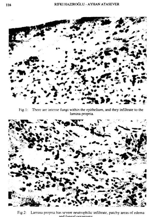
Şekil
Benzer Belgeler
Bunun arkasından İsrail Sina yarımadasını işgal edince, Suriye, Suudi Arabistan, Ürdün Mısır'a askeri destek vereceklerini belirtmelerine rağmen Nasır, Mısır kuvvetlerini
1953 yılında Ankara Üniversitesi Dil ve Tarih-Coğrafya Fakültesi Tarih Bölümü'nden Genel Türk Tarihi, Yeniçağ Tarihi, Yakınçağ Tarihi ve Farsça sertifikaları alarak
Sivas Kongresinden sonra 18 Aralık'ta Sivas'tan ayrılan Mustafa Kemal ve Heyet-i Temsiliye üyeleri Kayseri ve Kırşehir üzerinden dokuz günlük yorucu bir yolculuktan
"1919 Eylül'ünde Mustafa Kemal Rumeli ve Anadolu Müdafaa-i Hukuk Cemiyetinin toplandığı Sivas Kongresinin başkanı olarak seçildiğinde (daha sonra bu kongre Büyük
Öte yandan Yeni Gün gazetesinde çıkan Mustafa Kemal'le ilgili bir iki önemli haber ayrıca dikkati çekmekte ve tarihsel bir belge olan. 29 Yeni Gün, 29 Eylül
Türkiye’de veteriner radyolojinin başlangıcı ve Ankara Üniversitesi Veteriner Fakültesindeki tarihsel gelişimi. The beginning of veterinary radiology in Turkey and the
Akım çevirici stentler anevrizmayı ana damardan hemodinamik olarak ayırarak anevrizmaya akımın yavaşlamasını sağlayıp, boyun defektinde oluşacak neointimal
“İkinci yabancı dil olarak Almanca’ya kendi isteğinizle mi başladınız, yoksa okul gibi mecburi bir nedenle mi?” sorusuna ankete katılan öğrencilerin %85’nin
