Intraabdominal Synovial Sarcoma,
A Rare Tumour and Rare Localisation
İntraabdominal Sinovyal Sarkom,
Nadir Lokalizasyonda Nadir Bir Tümör
Mustafa Yener Uzunoğlu
1, Fatih Altıntoprak
2, Enis Dikicier
2, Zeynep Kahyaoğlu
31 Sakarya University Research and Educational Hospital, Department of General Surgery, Sakarya, Turkey 2 İstinye University, Faculty of Medicine, Department of General Surgery, İstanbul, Turkey 3 Sakarya Research and Educational Hospital, Department of Pathology, Sakarya, Turkey
Yazışma Adresi / Correspondence:
Mustafa Yener Uzunoglu
Sakarya Universitesi Egitim ve Arastırma Hastanesi, Adnan Menderes Cad. Sağlık Sok.No: 195 Adapazarı, 54100, Sakarya / Turkey T: +90 505 650 33 94 E-mail: [email protected]
Geliş Tarihi / Received : 12.03.2018 Kabul Tarihi / Accepted : 07.08.2018
CASE REPORT / Olgu Sunumu
Abstract
Synovial sarcomas are often seen in the soft tissues, intra-abdominal synovial sarcomas are extremely rare. An intra-abdominal mass starting from the umbilicus and extending into the pelvis was detected in a 53-year-old man admitted with abdominal pain and a palpable mass. Total mass excision surgery was performed and the mass was found to be an intra-abdominal synovial sarcoma arise from small bowel wall histopathologically. Unfortunately, this is a malignancy with a poor prognosis and a short survival despite all therapeutic efforts. ( Sakarya Med J, 2018, 8(2):669-673)
Keywords intra-abdominal mass; sarcoma; small bowel wall; synovial sarcoma
Öz
Sinovyal sarkomlar sıklıkla yumuşak dokuda gözlenirler, intra abdominal sinovyal sarkomlar oldukça nadirdir. Karın ağrısı ve palpabl kitle ile başvuran 53 yaşında erkek bir hastada umbilikustan başlayıp, pelvise kadar uzanım gösteren karın içi kitle saptandı. Total kitle eksizyonu yapılan hastada, histopatolojik olarak ince barsak duvarından kaynaklanan intraabdominal sinovyal sarkom bulundu. bu malignite zayıf prognoza sahiptir ve yapılan tüm tedavilere rağmen sağkalımı kısadır.( Sakarya Tıp Dergisi, 2018, 8(2):669-673 ).
Anahtar kelimeler
Introduction
Synovial sarcoma is a rare malignancy accounting for 5~10% of all soft-tissue sarcomas. It has an aggressive course and poor prognosis, with a reported mean 5-year survival of 66%.1 It is typically
encountered in the third to fi fth decades of life with a male preponderance. In 95% of the cases, the tumour is localised to the extremities, most commonly around the knee.2 Rarely, tumours
originating from the retroperitoneum, tongue, neck, oesophagus, and abdominal wall have been reported. The recommended treatment is surgical excision followed by chemotherapy and radiati-on therapy. Here, we report a 53-year-old man who was admitted with a palpable intra-abdominal mass and was ultimately diagnosed with an intra-abdominal synovial sarcoma.
Case report
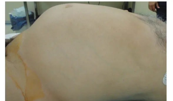
A 53-year-old man presented with abdominal pain for 4 months and a palpable abdominal mass (Fig. 1).
Figure 1. Preoperative patient’s view; apparent palpable intraabdominal mass
Physical examination detected a mass starting from the periumbilical region with indistinct bor-ders. Abdominal ultrasonography (US) revealed a mass extending from the umbilical level to the pelvis, with heterogeneous cystic and degenerated regions and irregular margins. The mass fi lled the entire lower abdominal region (Fig. 2). The symptoms of an intestinal obstruction emerged and we performed surgery. After obtaining written informed consent, surgical exploration showed a 280 × 180 mm mass with lobulated contours and some cystic components, although it was mainly solid in nature, extending from the umbilical level to the pelvis, surrounding a 20-cm ileal segment (Fig. 3A-B). The mass was excised totally and a partial ileal resection and terminal ileostomy were performed. An iatrogenic injury to the urinary bladder during the surgery was repaired primarily. The immunohistochemical staining showed that the major cell types were spindle and epithelial cells revealed a synovial sarcoma with CD99 and EMA positive, but were negative for S-100, pro-tein, desmin, CD34. (Fig. 4,5)
In the postoperative course, the patient had acute renal failure, aspiration pneumonia. The overall
UZUNOĞLU et al. Intraabdominal Synovial Sarcoma, A Rare Tumour and Rare Localisation
Sakarya Med J. 2018;8(2):669-673
UZUNOĞLU et al.
Intraabdominal Synovial Sarcoma, A Rare Tumour and Rare Localisation
Sakarya Med J. 2018;8(2):669-673
671 condition of the patient worsened in the 8th
postoperative day and he died of multi-organ failure on day 10.
Figure 2. Ultrasound image; a mass extending from the umbilical level to the pelvis, with he-terogeneous cystic and degenerated regions and irregular margins
Figure 3A. Operative view; intraabdominal mass is huge, lobulated contour and solid-cystic in nature.
Figure 3B. Operative view; intraabdominal mass is huge, lobulated contour and solid-cystic in nature.
Figure 4. Histological microphotograph of synovial sarcoma, stained with CD99 x20
Figure 5. Histological microphotograph of synovial sarcoma, stained with EMA x20
UZUNOĞLU et al. Intraabdominal Synovial Sarcoma, A Rare Tumour and Rare Localisation
Sakarya Med J. 2018;8(2):669-673
672
Discussion
Synovial sarcomas are rare aggressive tumours that typically arise in the periarticular areas2. Other
rare anatomic locations of this tumour are the thorax, neck, parathyroid, tongue, larynx, medi-astinum, oesophagus, heart, lung, abdominal wall, gastrocolic ligament, small bowel mesentery, prostate, kidney, and retroperitoneum.3,4 In this case the tumor arose from small bowel wall.
Clinically, an intra-abdominal synovial sarcoma often presents as a painful, palpable soft mass, as with tumours in other locations. Non-specifi c gastrointestinal complaints such as abdominal pain, bloating, weight loss, and vomiting may accompany the clinical presentation, depending on the size of the intra-abdominal mass.3 In this case, the abdominal pain had been present for 4 months,
but was overlooked by the patient, until a palpable mass was detected.
Intra-abdominal localisation is an extremely rare presentation of synovial sarcomas. In a retrospec-tive study of 300 cases, Fisher et al. reported that only 11 (3.6%) of the patients had an intra-ab-dominal mass.5 Those 11 cases had a mean age of 49 (range 25–75) years and a mean mass size
of 6 × 47 cm. In our patient, the mass measured 28 × 18 cm and the patient’s age was within the reported age range for synovial sarcomas.
Histopathologically, a synovial sarcoma is a soft-tissue tumour that shows signs of mesenchymal and epithelial differentiation, which can be detected under a light microscope, immunohistochemi-cally, and by electron microscopy.6 Although the diagnosis of biphasic synovial sarcomas is often
straightforward, a monophasic fi brous synovial sarcoma should be differentiated morphologically from fi brosarcoma, malignant peripheral nerve sheet tumour, and solitary fi brous tumour.7 Routine
use of immunohistochemical methods may facilitate the differential diagnosis of these conditions. Changchien et al. reported primary or metastatic intra-abdominal synovial sarcomas stained with SMA, EMA, BCL-2, CD99, and S100, but not with desmin.8 The monophasic and biphasic types were differentiated by theFluorescence In Situ Hybridization (FISH) method using SYT-SSX-1. In a study of 121 cases with a synovial sarcoma, Bergh et al. reported that 60% of the cases were monophasic and 40% the biphasic type.9 Guillou et al. reported that the monophasic type had a
more aggressive course.10 In our case, staining was positive for CD99 and EMA, histopathological
study confi rmed the diagnosis of monophasic synovial sarcoma.
The major determinant of survival is the histological grade of the tumour, and the dimensions and depth of the tumour are other prognostic factors.8 Wide local excision and radiation therapy are
mainstays in the treatment of synovial sarcomas. Chemotherapy has been also suggested in the treatment of some histological subtypes.9 Nevertheless, half of the cases die from distant
metas-tasis despite all treatment modalities.
In conclusion, although synovial sarcomas are rarely seen outside the extremities, they can also present as an intra-abdominal mass, albeit extremely rarely. Wide excision is the fi rst step in the treatment. Unfortunately, this is a malignancy with a poor prognosis and a short survival despite all therapeutic efforts.
UZUNOĞLU et al.
Intraabdominal Synovial Sarcoma, A Rare Tumour and Rare Localisation
Sakarya Med J. 2018;8(2):669-673
673
1. Sultan I, Rodriguez-Galindo C, Saab R, Yasir S, Casanova M, Ferrari A. Comparing children and adults with synovial sarcoma in the Surveillance, Epidemiology, and End Results program, 1983 to 2005. Cancer 2009; 115(15):3537–3547 DOI: 10.1002/cncr.24424
2. Indranil G, Divya M. Synovial sarcoma of the omentum: A rare entity. Indian J Cancer 2015;52:166-7
3. Carrillo R, Rodriguez-Peralto JL, Batsakis JG. Synovial sarcoma of the head and neck. Ann Otol Rhinol Laryngol. 1992 Aprİ; 101(4):367-70. DOI: 10.1177/000348949210100415
4. Fritsch M, Epstein JI, Perman EJ, Watts JC, Argani P. Molecularly confi rmed primary prostatic synovial sarcoma. Hum Pathol 2000; 31:246-250 5. Fisher C, Folpe AL, Hashimoto H, Weiss SW. Intra-abdominal synovial
sarcoma: a clinicopathological study. Histopatology September 2004; 45(3): 245–253
6. Buiga-Potcoav� R, Crişan D, Olinici CD. Primary intraabdominal sarcoma, a case report Romanian Journal Of Gastroenterolojy 2005;14(1): 67-69 7. Harsh KK, Kalwar A, Kapoor A, Jakhar SL, Kumar HS. Giant cell variant of
malignant fi brous histiocytoma of male breast: A rare case report. J Can Res Ther 2015;11:657 DOI: 10.4103/0973-1482.138129
8. Changchien YC, Katalin U, Fillinger J, Fónyad L, Papp G, Salamon F, Sápi Z. A Challenging Case of Metastatic Intra-Abdominal Synovial Sarcoma with Unusual Immunophenotype and Its Differential Diagnosis. Case Rep Pathol. 2012; 2012: 786083. DOI: 10.1155/2012/786083. 9. Eriksen C, Burns L, Bohlke A, Haque S, Slakey DP. Management of
mo-nophasic synovial sarcoma of the small intestine. JSLS. 2010;14(3):421-5. DOI: 10.4293/108680810X12924466006846.
10. Guillou L, Coindre JM, Gallagher G, Terrier P, Gebhard S, Somerhau-sen NSA, et al. Detection of the synovial sarcoma translocation t(X;18) (SYT;SSX) in parafi n-embedded tissues using reverse transcriptasepoly-merase chain reaction: a reliable and powerful diagnostic tool for patho-logist. A molecular analysis of 221 mesenchymal tumors fi xed in different fi xatives. Hum Pathol 2001; 32:105-112. DOI: https://doi.org/10.1053/ hupa.2001.21130