T.C.
ISTANBUL MEDIPOL UNIVERSITY HEALTH SCIENCES INSTITUTE
MASTER THESIS
LOWER RESPIRATORY SYSTEM INFECTIONS BACTERIAL
AGENTS OF ADULTS AND CHILDREN IN ISTANBUL, A CASE
STUDY BY MEDIPOL MEGA HOSPITAL
MOHAMMED MOHAMMED AHMED
DEPARTMANT OF MICROBIOLOGY
SUPERVISOR
Yard.Doç.Dr.AYŞE İSTANBULLUTOSUN
III ACKNOWLEDGEMENT
I would like to thank Almighty Allah for bestowing upon me an opportunity and strength to follow my education. It is His plenty elegance that has brought this research project to a success. I also would like to thank my supervisor Yard.Doç.Dr. Ayşe İSTANBULLU TOSUN who gave me brilliant advise to complete my research project. Without her fully support and encouragement I wouldn’t be the one I am now.
I would like to thank Head Department of Microbiology Prof.Dr.Ahmet Zeki ŞENGİL, Associate Professor Süleyman YILDIRIM and Assistant Professor Deniz DURALI.
I would also like to thank my professors Doç.Dr. Sevgi ERGİN, Yard.Doç.Dr.Özlem GÜVEN, for their support, encouragement and advices.
Especial thanks is due to the Medipol Mega University hospital administrations who have contributed to provide us a very unforgettable and conducive environment for better applied learning in to the modern technology practice. I am deeply indebted to thank Yard Doç.Dr.Canan SEVER for her undivided effort in supporting me to pursue my education of this level. I am deeply indebted to thank to Dr Omar Abdulle Omar for his undivided effort in supporting me to pursue my education at this level.
I would like to thank all my friends Abdulshakur Mohamed, Gözde Inci, Kalif, Hatice, Selcuk, Ahmed, Gözde, Leyla, Müge, Mücahit, Cemile, Ezgi, and especially Fadumo Yusuf.I would like to thank my classmates, friends who have supported me and encouraged me to pursue my education.
Having this opportunity i would like to express my sincere gratitude and thank to my beloved parents for their countless support and also to my brothers and sisters for their encouragement and love.
Finally I would like to thank İHH and Zamzam foundations for providing scholarship and financial support. Thank you all.
IV
CONTENTS
Page number
THESIS APPROVAL FORM ...I DECLARATION ... II ACKNOWLEDGEMENT ... III CONTENTS...IV-VI LIST OF ABBREVIATIONS………....VII-VIII LIST OF TABLES ………...IX-X LIST OF FIGURES ………...………...….XI LIST OF PHOTOGRAPHS...XII 1. ABSTRACT ... 1 2. ÖZET ... 2 3. INTRODUCTION ... 3 4. GENERALINFORMATION ... 9 4.1 Introduction ……….……….…...9 4.2 Acute bronchitis ……….…...9 4.3 Chronic bronchitis ……… …………...13 4.4 Influenza ……….….15 4.5 Pneumonia………..15
4.5.1 Pneumonia in immunocompromised hosts………....…..16
4.6 BAL……..………...18
4.7 Sputum…..……….…….…18
5. MATERIAL AND METHOD………...…....19
5.1 Culture media…...……...……….…..……....19
5.1.1 Blood agar…….………...………..……...19
V
5.1.3 Chrome agar orientation ………...21
5.1.4 Chrome candida medium………...………...…...…....23
5.2 Biochemical tests ………...24
5.2.1 Catalase test…...………...24
5.2.2 Bile esculin agar………..………...25
5.2.3 Coagulase test ………... 26
5.2.4 Cytochrome oxidase test………...26
5.2.5 MIO ( Motility indole ornithine )………..………...27
5.2.6 Citrate test…….……….……...28
5.2.7 Urea test ……….…...29
5.2.8 Triple sugar iron agar ………...30
5.2.9 Molecular hinton agar medium………...32
5.2.10 Antimicrobial susceptibility test………. ...32
5.3 Used instruments Vitek………….………...34
5.4 Specimen collection and handling …...………...34
5.5 Specimen inoculation…...………...35
5.5.1 BAL inoculation: dilution method …….………...36
5.5.2 TA and Sputum inoculation……..………... .37
5.6 Investigation of Gram stain and culture result…...……….... .37
5.7 Bacterial identification … ………....………... 39
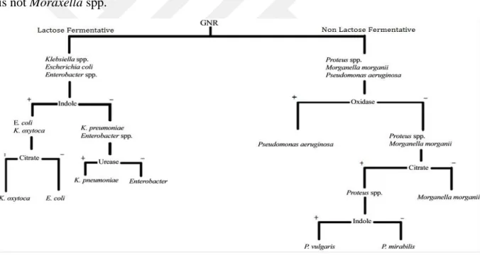
5.7.1 Gram negative identification flowchart...39
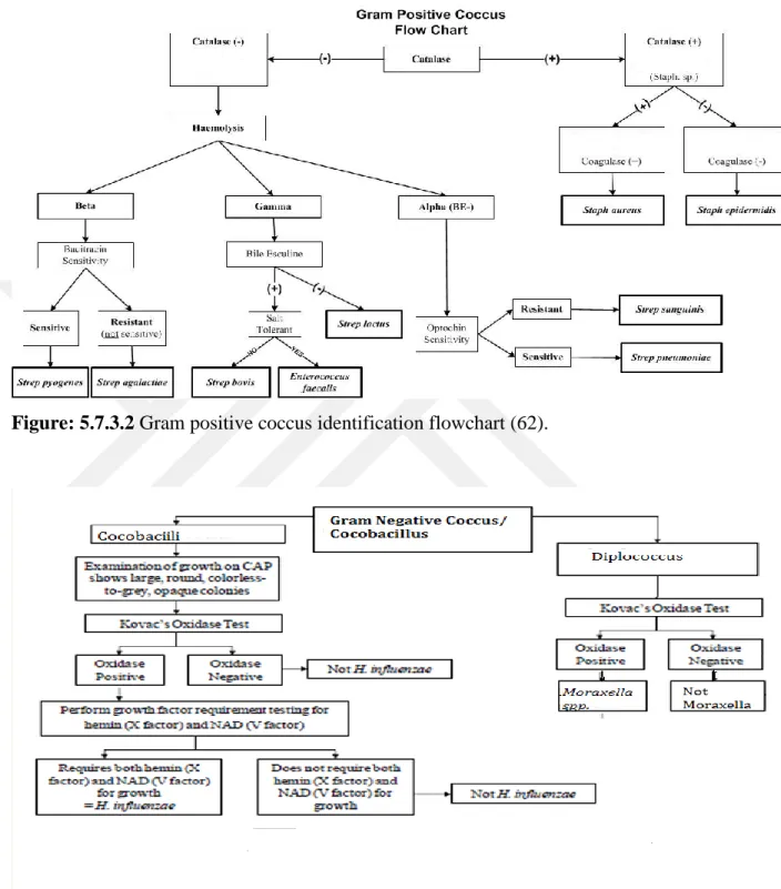
5.7.2 Gram positive coccusidentification flowchart...39
5.7.3 Gram negative coccus/coccobacilliidentification flowchart...40
VI
7. DISCUSSION ... 69
8. CONCLUSION ... 75
9. REFERENCES. ... 76
10. APPROVAL Of ETHİCAL COMMİTTEE ... 84
VII LIST OF ABBREVIATIONS
BAL: Bronchoalveolar Lavage
LRTIs: Lower Respiratory Tract Infections
WHO: World Health Organization
RTIs: Respiratory Tract Infections URI: Upper Respiratory Infections
TB: Tuberculosis
ILDs: Interstitial Lung Diseases
MDRO: Multiple Drug Resistant Organism
ERS: European Respiratory Society
OPD: Outpatient Department
IPD: Inpatient Department ICU: Intensive Care Unit
PSA: Pseudomonas aeruginosa
MIO: Motility Indole Ornithine MHA: Mueller Hinton Agar
VIII CHOC: Chocolate Agar
NaCl: Sodium Chloride
PBB: persistent bacterial bronchitis NAD: Nicotinamide adenine dinucleotide CLSI: Clinical Laboratory Standards Institute
IX LIST OF TABLES
Table 5.1.3.1 Typical colony appearance of Gram negative microorganisms.…... .22
Table 5.1.3.2Typical colony appearance of Gram positive microorganisms...…..22
Table 5.1.4.1Typical colony appearance of Candida species for microorganisms...24
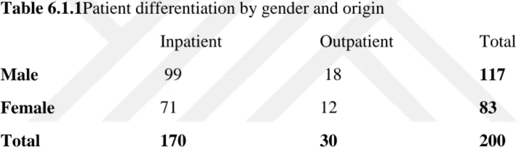
Table 6.1.1 Patient differentiation by gender and origin………....………...…...42
Table 6.2.1BAL, sputum and tracheal samples of adult patients classified as inpatient and outpatient………...43
Table 6.3.1BAL, Sputum and tracheal samples of pediatric patients classified as inpatient and outpatient....………...……...44
Table 6.4.1Respondents distribution by age...………...45
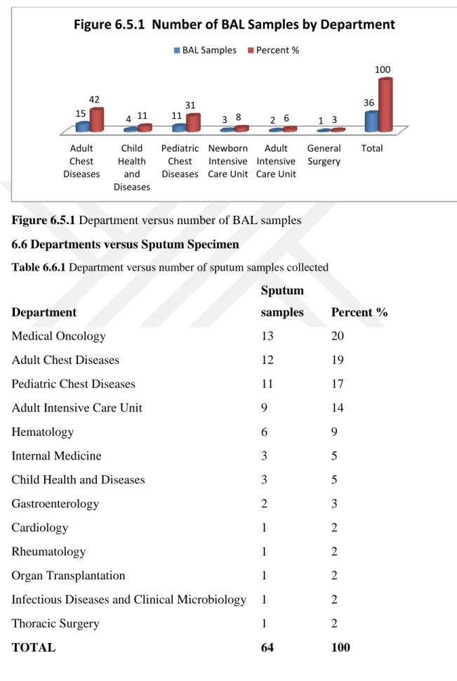
Table 6.5.1Number of BAL samples by department………..46
Table 6.6.1Demonstrates department versus number of sputum cases collected...47
Table 6.7.1This table shows the number of tracheal aspirate samples collected... 49
Table 6.8.1 Distribution of tracheal and sputum samples according to department……50
Table 6.9.1Number of samples collected from BAL, sputum and tracheal aspirate...52
Table 6.10.1A Percentage of bacterial species from BAL, sputum and tracheal aspirate of inpatients with LRTS (n=112),...53
Table 6.10.1.2B Percentage of bacterial species detected from BAL, sputum and tracheal aspirate of outpatients with LRTS (n=18) ...54
Table6.11.1Gram negative bacilli and Gram positive cocci ……...56
Table6.12.1 Antibiotic susceptibility test results of Pseudomonas species…….…...57
Table 6.13Antibiotic susceptibility test results of Klebsiella species………....… ...59
Table 6.14Antibiotic susceptibility test results of E. coli ..………...60
Table 6.15Antibiotic susceptibility test results of Enterobacter species …..………...62
X
Table6.17Antibiotic susceptibility test results of Acinetobacter species.…...64
Table 6.18 Antibiotic susceptibility test results of Haemophilus species…………...65
Table6.19 Antibiotic susceptibility test results of S.pneumoniae …………...67
XI LIST OF FIGURES
Figure 5.7.3.1 Gram negative identification flowchart…...40
Figure 5.7.3.2 Gram positive cocci identification flowchart...41
Figure 5.7.3.3 Gram negative coccobacilli identification flowchart...41
Figure 6.1.1Patient differentiation by gender and origin……….……...…….43
Figure 6.2.1 BAL, sputum and tracheal samples of adult patients cases according to their gender and IPD or OPD ……….……..……...44
Figure 6.3.1BAL, sputum and tracheal specimen cases of pediatric patients classified as inpatient and outpatient:……… ….………..…...45
Figure 6.4.1Participants categorized according to adult- and childhood………...…..46
Figure 6.5.1 Department versus number of BAL samples……….…………47
Figure 6.6.1 Departments versus cases with sputum specimen collected ………..48
Figure 6.7.1 Department versus tracheal specimen collected………...50
Figure 6.8 Outpatient department distributions………...51
Figure 6.9.1 Bacterial species detected from BAL, sputum and tracheal aspirate of patients with LRTS………...52
Figure 6.10.1A& 6.10.1.2 B Percentage of bacterial species detected from BAL, sputum and tracheal aspirate of inpatients and outpatients with LRTS...55
Figure 6.11.1Percentage of Gram negative bacilli and Gram positive cocci…...……...56
XII LIST OF PHOTOGRAPHS
Photo 5.1.1.1Types of Hemolysis………...20
Photo 5.1.3.1Different Bacteria on Chromagar Orientation...21
Photo 5.1.4.1Different Types of Candida on Chromagar Candida...23
Photo:5.2.1.1Catalase Test ………...…..24
Photo:5.2.2.1Bile Esculin Disk ……..………....………...…………...25
Photo:5.2.6.1Citrate Test………...….28
Photo :5.2.7.1Urease Test………...29
Photo:5.2.8.1TSI Media………...………...30
Photo 5.2.10.1Antimicrobial Susceptibility Testing…...………...………..…...33
1
1- ABSTRACT
LOWER RESPIRATORY SYSTEM INFECTIONS BACTERIAL
AGENTS OF ADULTS AND CHILDREN IN ISTANBUL, A CASE
STUDY BY MEDIPOL MEGA HOSPITAL
The general objectives of the study were to explore the incidence of lower respiratory infections due to bacterial pneumonias and to determine antimicrobial susceptibility patterns of infectious agents. The study participants were 200 patients included between January-March, 2017, mainly children and adults which were divided into two age groups 0-17 and those aged >18 years. The samples were collected from both inpatients and outpatients. The studied samples were; sputum, broncheoalveolar lavage (BAL) and tracheal aspirate. After proper incubation time, significant bacteria were identified and antimicrobial susceptibility tests were done.
According to findings of the study, 86% of the samples were Gram-negative bacilli while 14% of the samples showed to be Gram positive. Based on the causative agents of lower respiratory tract infections following findings were discovered 41.1%
Pseudomonas aeruginosa, 29.9% Klebsiella spp., 15% Escherichia coli, 8% Staphylococcus aureus while 5.6% were shown to be Streptococcus pneumoniae.
In conclusion, although some antibiotics were found to be effective, the study findings were indicative of increased resistance to some common antibiotics that are widely prescribed by doctors for treatment of patients with lower respiratory system infections. There were also signs of growing overuse of such antibiotics, which can sadly lead to greater antibiotic resistance in the near future.
Key words: Lower Respiratory System Infections, Bacterial Agents, and Antimicrobial Resistance.
2
2- ÖZET
ERİŞKİN VE ÇOCUK HASTALARDA ALT SOLUNUM YOLU
İNFEKSİYONLARINDA İZOLE EDİLEN
MİKROORGANİZMALAR, MEDİPOL MEGA HASTANESİ VAKA
ÇALIŞMASI
Bu çalışmanın genel hedefleri, alt solunum yolları enfeksiyonlarından pnömonilerin erişkin ve cocuk hastalardaki yoğunluğunu ve infeksiyon etkeni olan mikroorganizmaların antimikrobiyal duyarlılıklarını araştırmaktır. Araştırmamızda Ocak – Mart 2017 arasında hastanemize alt solunum yolu enfeksiyonu şüphesi ile başvuran 200 hastadan alınan alt solunum yolu örnekleri kullanılmıştır. Hastalardan 0-17 yaş arasında olanlar çocuk, 18 yaş ve üstü olanlar erişkin olarak gruplandırılmıştır. Numuneler hem yatan hem de ayaktan hastalardan alınmıştır. İncelemeye alınan numuneler balgam, bronkoalveoler lavaj ve trakeal aspirat olarak ayrı ayrı incelenmiştir. Gerekli inkübasyon dönemi sonrası anlamlı üremeler tiplendirilerek antimikrobiyel duyarlılık testleri yapılmıştır.
Elde edilen örneklerin %86`sı Gram negatif çomak olarak belirlenirken %14`ü Gram pozitif olduğu görüldü. Alt solunum yolu enfeksiyonlarında organizmalar arasında şu etkenler saptandı; Pseudomonas spp. 41,1%, Klebsiella spp. 29,9%, Escherichia coli 15%, Staphylococcus aureus 8%, Streptococcus pneumoniae 5%. Kimi antibiyotiklere karşı duyarlılık görülse de, çalışmamızda hekimler tarafından alt solunum yolları enfeksiyonları tedavisi için çok kullanılan kimi antibiyotiklere karşı da yükselen direnç oranları saptandı. Bu antibiyotiklerin kontrolsüz ve yaygın kullanımından dolayı gelecekte daha da yüksek oranda bir direnç tablosu ile karşı karşıya gelmemiz malesef beklenmektedir.
Anahtar Kelimeler:Alt Solunum Yolu İnfeksiyonları, Bakteriyel Etkenler, Antimikrobiyal Direnç.
3
3. INTRODUCTION AND PURPOSE
Respiratory tract infection is any kind of transmittable disease concerning the respiratory tract. An infection of this sort is generally categorized more detailed as an upper respiratory tract infection (URI or URTI) or a lower respiratory tract infection (LRI or LRTI). Lower respiratory infections, like pneumonia are more likely to be more severe than upper respiratory infections, for instance the common cold.
The upper respiratory tract is normally regarded to be the airway above the glottis or vocal cords together with the nose, sinuses, pharynx, and larynx despite the dispute among scientists on the precise border between the upper and lower respiratory tracts. The lower respiratory tract incorporates the trachea, bronchial tubes, the bronchioles and the lungs. LRIs have been the most important cause of death in the midst of all contagious illnesses; the two widespread LRIs are bronchitis and pneumonia (1).
The upper respiratory tract infection (URI) is a imprecise expression used to explain serious infections relating to the nose, paranasal sinuses, pharynx, larynx, trachea, and bronchi (2). The prototype is the disease identified as the common cold, which is further explained in this research in adding together with pharyngitis, sinusitis and tracheobronchitis. Influenza is a systemic disease that engages the upper respiratory tract and must be distinguished from other URIs (3).
The inflammation of the throat that is brought about by a respiratory virus (Rhinovirus, coronavirus, adenovirus, influenza virus, parainfluenza viruses, respiratory syncytial virus, epstein–barr virus or coxsackievirus is considered to be Pharyngitis (4). Bacterial pharyngitis also is not as common and its only main regular cause is S.
pyogenes, Neisseria meningitidis, Mycoplasma pneumoniae, C.diphtheria and Arcanobacterium haemolyticum are other uncommon bacterial causes of URI or LRI (5).
4 Topmost occurrence is between autumn and spring in moderate climates, and during the rainy season in tropical areas. Nevertheless, the progress of transmission is speedy between societies sharing populated living areas or districts because of the widespread transmission by droplets or direct diffusion. The discovery of S. pyogenes the exploration of most regularly demanded for pharyngitis (4).
This species is identified either by culture on blood agar and subsequent latex agglutination reaction for group-specific polysaccharide, or by direct antigen detection. Thus, there is no method that can differentiate oropharyngeal colonization from actual infection except the culture, which permits antibiotic susceptibility testing. Suspicion of infection with N.gonorrhaea, Mycoplasma spp., Arcanobacterium spp. or
Corynebacterium spp. must be informed to the laboratory so that specialized non-routine
culture media can be utilized. To cure Streptococcal pharyngitis, an oral penicillin or erythromycin is utilized (6).
Medication may not change the course of the primary pharyngeal infection, but it is supposed to decrease the risk of main non-infective sequelae like rheumatic heart disease, post streptococcal glomerulonephritis and Sydenham‟s chorea. The demand for antibiotic medication of streptococcal pharyngitis has been controversial issue in developed countries, since the non-infective sequelae of streptococcal infections are all scarce. However, the current rise in streptococcal infection rates in Europe and North America might advocate this point (7). The other problems of streptococcal pharyngitis comprise of scarlet fever (less common than in the past in developed countries), streptococcal toxic shock syndrome (brought about by toxins) and quinsy (paratonsillar abscess).
5 In quinsy, there might be minor infection with oral anaerobic bacteria, but these are often penicillin susceptible. Moreover, the drainage of purulent foci is demanded. Pathogens (viruses and bacteria) must fight a number of physical and immunologic barriers in order for pathogens to attack the mucus membranes of the upper airways.
General lower RTIs include flu (this can affect both the upper and lower respiratory tract), bronchitis, pneumonia, bronchiolitis and tuberculosis 8). The major sign of a lower RTI is cough; it is typically serious and might create phlegm and mucus. Other probable signs are a tense feeling in the chest, increased rate of breathing, breathlessness and wheezing. RTIs can be expressed in many ways. LRTI is one of the foremost sources of morbidity and death throughout the world (9). In rising countries, the condition is more complex and management is often complicated because of the troubles related to the recognition of the etiological agents and the management of suitable medication in cases antibiotic therapy is needed (10). LRTI is not a group of definite infections with diverse epidemiology‟s, pathogeneses, clinical presentations, and outcomes.
Variation of the etiology and symptomatology of respiratory diseases are attributed to, but not limited to the disparity of the following factors; age, gender, season, the type of population at risk. LRTIs are commonly the first infection to happen after birth and pneumonia is often too the final illness to happen before death (11). The etiological agents of LRTIs cannot be identified clinically and distinguished from zone to zone (12).
6 To identify suitable antimicrobial therapy, the recognition of bacteria bringing about lower-airway infections is significant. Despite the possibility of easy sputum collection in adults, it is complicated to gather sputum from children who usually cannot expectorate. Thus, flexible bronchoscopy with broncheoalveolar lavage (BAL) is frequently utilized to get a lower-airway specimen for culture of microorganisms in children. The European Respiratory Society (ERS) Task Force has made available advices and guiding principles for the performance of BAL in children and processing the return fluid. Typically BAL is conducted in the main affected zone, or the right middle lobe in case of diffuse lung disease (13).
BAL fluid gathered during bronchoscopy is normally chronological with two or more lavages carried out. The ERS suggests that the first lavage (lavage-1) is utilized for bacterial culture and any following lavage is used for cytology and non-cellular studies such as immunology-based work (14). Lavage-1 is regarded to be more reflective of bronchial airways, while the second (lavage-2) is reflective of distal airways (bronchioles and alveoli), and cellularity findings from the two lavages are dissimilar (15). Therefore, it is biologically reasonable that cultivable bacteria also vary between lavage-1 and lavage-2. Gram-positive bacteria, for instance Staphylococcus aureus,
Streptococcus pneumoniae, etc. in addition to Gram negative bacteria like Haemophilus influenzae, Pseudomonas, Acinetobacter, and Klebsiella species are recovered from
LRTIs (16).
Supervising and observing the antimicrobial resistance samples of the etiological agents is required not only to direct the clinician when running cases requiring antibiotic therapy but also to observe the development of these infections. Bacteria are recognized to bring about primary infection or superinfection, and in majority of the cases, they need aimed antimicrobial therapy (17).
7 Pneumonia keeps on to be a main health problem although there are developments in the determination of causal microbes and the accessibility of new antimicrobial drugs. Additionally, there are still many arguments concerning diagnostic methods and medication preferences. Pneumonia is the sixth most important reason of death in the United States and the main general cause of mortality from infectious disease. Moreover, pneumonia is the foremost cause of death in hospital-acquired infections. Left untreated and relying on the causal microbe and population, bacterial pneumonia has a mortality rate that may extend 30%. Every year more than 60,000 Americans die from pneumonia. Pneumococcal pneumonia alone represents probably 40,000 deaths yearly.
The illness is a special worry for older adults and people with chronic diseases or harmed immune system, but it can also strike previously healthy, young people. Worldwide, pneumonia is a main cause of mortality in children, many of whom are younger than 1 year of age. Bronchiectasis is a main problem of bacterial pneumonia where damaged alveoli and bronchioles are replaced by small sacs filled with infected debris. A low-grade, smoldering infection devastates more lung tissue with time. Bacterial pneumonia may also cause destruction of lung tissue with succeeding scarring, a permanent reduction in gas exchange, and a loss of respiratory reserve. The lungs also turn to be less elastic and inflation of the lungs need more energy and work for the duration of the aspiratory phase of respiration.
Bacterial pneumonia can evolve deadly when fluid loads the air sacs and obstructs the capability to swap oxygen and carbon dioxide. Circulating oxygen levels reduces (i.e., hypoxemia) and circulating carbon dioxide concentrations rise (i.e., hypercapnia), causing eventually to respiratory breakdown and mortality. In some situations, microorganisms get access to the blood spread quickly to other organs, and leading life-threatening sepsis or septic shock (18).
8 The aim of the study is to explore the burden of lower respiratory infection due to bacterial pneumonias and govern children and adult people with lower respiratory infections. The study was based in Istanbul Medipol University Hospital where the quantitatively data is collected and data from the hospital registers as well as the experiences of the medical doctors and laboratory results was used. And also the study will further examine resistances of bacterial species isolates to antibiotics including amikacin, ampicillin, piperacillin-tazobactam, amoxicillin-clavulanate, ampicillin-sulbactam, ciprofloxacin, ceftazidime, trimethoprim-sulfamethoxazole, gentamicin, imipenem, meropenem, oxacillin, ceftriaxone, and vancomycin. The samples of the study have been analyzed for three months from January to March, 2017.
9
4. GENERAL INFORMATION
4.1 Introduction
Lower respiratory tract infections (LRTIs) are described respiratory diseases involving the lower sections of the respiratory system. These infections have an effect on the respiratory system from the bronchi down and circumstances are classified into acute or chronic bronchitis according to the period of the signs.
4.2 Acute Bronchitis
To express diseases exemplified by cough: bronchitis, wheezy bronchitis, asthmatic bronchitis, and tracheobronchitis, there are many terms used regarding to these diseases. The lack of clinical definitions, disagreement of cough illnesses and bronchitis are attributed to the complexities in evaluating findings from bronchitis or cough disease researches as well as the lack of a solid agreement on diagnosis and medication (19). Acute bronchitis is an acute or sub-acute cough disease enduring fewer than 2-3 weeks, with or without phlegm production, commonly related to other upper respiratory tract and constitutional signs, the cough is the mainly often declared sign calling for office assessment; so, acute bronchitis is one of the top 10 analyses in ambulatory care medicine (20).
Physicians reveal wide inconsistency in diagnostic necessities and curing, because the diagnosis is clinical, without standardized diagnostic symptoms and sensitive or precise confirmatory laboratory examinations (21). Diagnosis of bronchitis frequently results in a prescription for an antimicrobial agent, displaying the physicians‟ belief of bacterial disease, although the term bronchitis does not mean definite etiology and is most generally brought about by viral pathogens.
Pathophysiology and etiology: Acute bronchitis is described as inflammation of the bronchial respiratory mucosa, resulting in production of cough. For nearly all clinicians, bronchitis is a disease clinically distinguished by cough, along with or with no fever or sputum production (19).
10 Bronchial epithelial injury is brought by transmittable or non-transmittable triggers, which cause an inflammatory reply with subsequent airway hyper reaction and mucus production. International literature proposes that clinical features of unsophisticated acute bronchitis expand in chronological phases: an acute infection phase, ensuing from direct inoculation by the contagious virus of the tracheobronchial epithelium, causing cytokine discharge and inflammatory cell commencement (22).
In this phase there are inconsistent constitutional signs, such as fever, myalgia, and malaise, which last 1 to 5 days depending on the infectious agent. The protracted phase results from hypersensitivity of the tracheobronchial epithelium and airway receptors (bronchial hyper responsiveness), portrayed mainly by cough, frequently come with phlegm emission and wheezing, and typically continues 1 to 3 weeks. Respiratory epithelial cell function plays a vital position in airway inflammation, and vagal-mediated airway hyper responsiveness has been revealed to agree with repair of the bronchial epithelial surface. Other mechanisms of bronchial hyper responsiveness might also be existent, such as adrenergic-cholinergic tone imbalance and IgE-mediated histamine production (20).
Chosen triggers that can start the cascade causing acute bronchitis are:
✓Viruses: Adenovirus, coronavirus, coxsackievirus, enterovirus, influenza virus, parainfluenza virus, respiratory syncytial virus, rhinovirus, and human metapneumovirus.
✓Bacteria: Bordetella pertussis, Bordetella parapertussis, Moraxella catarrhalis,
Haemophilus influenzae, Streptococcus pneumoniae, atypical bacteria (e.g., Mycoplasma pneumoniae, Chlamydia pneumoniae, Legionella species).
✓Yeast and fungi: Blastomyces dermatitidis, Candida albicans, Candida tropicalis,
Coccidioides immitis, Cryptococcus neoformans, Histoplasma capsulatum.
✓Noninfectious triggers: Asthma, air pollutants, ammonia, cannabis, tobacco, trace metals.
11 Acute bronchitis is typically brought about by a viral infection (23).
In patients whose age is less one year, the causative agent is commonly respiratory syncytial virus, parainfluenza virus, and coronavirus; among patients whose ages are in between one to 10 years, parainfluenza virus, enterovirus, respiratory syncytial virus, and rhinovirus predominate; among patients whose age is more than 10 years, the most frequent agents are influenza virus, respiratory syncytial virus, and adenovirus. There is also a seasonal variability in the etiology with most commonly parainfluenza virus, enterovirus, and rhinovirus infections in the fall, while influenza virus, respiratory syncytial virus, and coronavirus infections are most frequent in the winter and spring (22).
In addition to that, there are other viral triggers such as influenza A and B and human metapneumovirus, while bacterial pathogens involved can be Bordetella
pertussis, Chlamydia pneumoniae and Mycoplasma pneumoniae, “No isolated
pathogen” is also a regular result, possibly representing viral infections for which studies commonly do not perform appropriate analyses (20). Indications: Cough is the most generally detected sign of acute bronchitis, starting inside 2 days of infection in 85% of patients, continuing in nearly all of patients for fewer than 2 weeks; however, 26% are still coughing after 2 weeks, and a few cough for up to 6 to 8 weeks (24). Other indications may include dyspnea, wheezing, sputum production, chest pain, fever, hoarseness, malaise, rhonchi and rales in different degrees. Sputum might be obvious, white, yellow, green, or even colored with blood.
Diagnosis: There are different recommendations on the use of Gram staining and sputum culture in acute bronchitis therapy; especially the usefulness of these tests in the outpatient treatment is questionable, because they often show no growth or only normal respiratory flora (25).
12 Chest radiography must be kept for patients in which pneumonia is alleged or involved by heart failure, and in high risk patients with older age, chronic obstructive pulmonary illness, lately recognized pneumonia, malignancy, tuberculosis, and immunocompromised or debilitated status (22). Pulmonary function testing as spirometry is not regularly employed in the acute bronchitis diagnosis, but conducted only when underlying disruptive pathology is assumed or if there are frequent incidences of bronchitis. Pulse oximetry might assist to identify the seriousness of the disease, but findings do not verify or rule out bronchitis, asthma, pneumonia, or other definite diagnoses.
Differential diagnosis: The disparity of diagnosis comprises the main widespread triggers of acute cough:
✓Acute bronchitis ✓ Allergic rhinitis ✓ Asthma
✓Chronic obstructive pulmonary disease exacerbation ✓ Congestive heart failure exacerbation
✓Gastroesophageal reflux disease ✓ Malignancy
✓ Pneumonia
✓ Post-infectious cough
13 4.3 Chronic Bronchitis:
Despite the ambiguity regarding the description of chronic bronchitis, chronic bronchitis in adults is obvious and can be expressed: “the existence of chronic productive cough for 3 months in each of 2 consecutive years, in a patient under whom other factors of chronic cough have been left out”.
The possibility of applying this definition to childhood chronic bronchitis still stays in an ambiguity (26). The diagnosis of chronic bronchitis is supposed to take place in two phases. The first one is consideration and determination of many well-identified respiratory problems based on a staged administration protocol. The second but concurrent phase is removal or alteration of exogenous aspects that generate or sustain the child‟s disease. However, this diagnosis has the potential to switch the pediatrician from discovering a more definite respiratory condition (26).
Although there is noticeably small in the literature concerning etiology, examination and organization of chronic cough in childhood, coughing in childhood is general. Current repots have highlighted the significance of making a exact diagnosis in children with a chronic cough (>3 weeks) (27). Juvenile chronic bronchitis with persistent end bronchial disease (lately considered persistent bacterial bronchitis) has been explained for many decades. The persistent bacterial bronchitis (PBB) is, according to some authors, the main reason of a chronic cough (28).
A variety of diagnostic terms have been applied to define this situation such as chronic suppurative lung disease (29). Persistent end bronchial infection and PBB describe the pathological process and site of infection, while terms like „„chronic bronchitis‟‟ FIELD CE (1949 or „„protracted bronchitis‟‟ Chang AB (2005) describe the clinical phenotype. Others have recommended using the label „„pre-bronchiectasis‟‟ to emphasize the situation‟s possible function in causing injured airways, as evident on high-resolution computed tomography or at bronchography (30).
14 PBB is a pediatric condition illustrated by the existence of an isolated moist or wet cough, lasting more than 4 weeks in the lack of other exact causes. It typically have an effect on children who are younger than 5 years and it has been determined more by pediatric pulmonologists who resolve it with antibiotic treatment. The most well-known organisms engaged in infants and children are Streptococcus pneumoniae, Haemophilus
influenzae, and Moraxella catarrhalis. PBB is under diagnosed and frequently
misdiagnosed as asthma (31).
Differential diagnosis: The term of “chronic bronchitis” should only be employed after underlying causes have been excluded.
15 4.4 Influenza
Influenza and pneumonia are closely linked conditions, and for this reason often considered together. Both affect the lungs and are usually short-lived, lasting less than three months. Influenza is exclusively a viral condition, whereas pneumonia can be viral and/ or bacterial. Influenza, also known as the flu, is caused by a number of viruses of three types - influenza A, influenza B, and influenza C (32). These viruses can occasionally cause serious complications - such as pneumonia (which sometimes can be life-threatening). General flu symptoms include fever, cough, headache, tiredness, inflamed respiratory mucous membranes, and head cold symptoms such as a runny nose and watery eyes. Occasionally, a person may experience nausea and vomiting. The majority of people recover from the flu in 1 to 2 weeks and, being a viral condition, it doesn‟t need antibiotics. Many of the complications and illnesses caused by the flu can now be prevented by influenza vaccinations, and it is specifically recommended that certain groups of people (such as older people) are vaccinated annually.
4.5 Pneumonia
Pneumonia, an inflammation of one or both of the lungs may affect an entire lobe (lobar pneumonia), a segment of a lobe, alveoli contiguous to bronchi (bronchopneumonia), or interstitial tissue C (33). It can be caused as a result of a previous respiratory infection, or develop in conjunction with other respiratory diseases. Symptoms of pneumonia include fever, chills, cough with mucus production, and sometimes pleuritic chest pain and shortness of breath. Symptoms can sometimes be mistaken initially for the common cold, but pneumonia usually develops over days or weeks and lasts longer than a cold. Pneumonia can be bacterial, viral, fungal or parasitic, but bacterial and viral pneumonia are the most common (34).
16 The type of pneumonia is usually named and diagnosed according to the pneumonia-causing organism. Bacteria are the most common cause of pneumonia in adults aged 30 years or older, with Streptococcus pneumoniae (also known as pneumococcal disease) being the most common pneumonia causing organism, National Heart Land Blood Institute (2003). Some bacterial pneumonia can be avoided, as there are now a wide variety of pneumonia vaccines available. However, when a person has already developed pneumonia, the usual treatment is with antibiotics. Occasionally, pneumonia-causing bacteria can become resistant to a number of antibiotics, which then makes the condition much harder the treat. Sometimes, pneumonia-causing bacteria can cause a number of conditions more serious than pneumonia. Examples are Burkholderia
pseudomallei bacterium; and Mycobacterium tuberculosis. Viruses are the most
common cause of pneumonia in children, but the influenza A and B viruses and rarely varicella-zoster virus have been known to cause pneumonia in adults. Viral pneumonia develops when viruses invade the tissues of the bronchioles causing bronchiolitis and occasionally affect the alveoli. The common symptoms of viral pneumonia are headache, fever, tiredness, coughing, and mucus production (35).
4.5.1 Pneumonia in immunocompromised hosts
Pneumonia is one of the most life-threatening infections in the immunocompromised host. A broad range of pathogens needs to be excluded; and the infectious agents to be considered vary depending upon the type and duration of immunosuppression, past exposures, geographic location, and the nature of the treatments administered. Less controversial than the diagnostic utility of ventilator associated pneumonia is perhaps the diagnostic utility of fiberoptic bronchoscopy in this setting. BAL protocols which process samples for both viral and bacterial pathogens, pneumocystis, legionella, fungi, and mycobacteria as well as cytologic analysis for noninfectious causes may be appropriate. Such protocols require communication between the clinical microbiology laboratory, infectious diseases specialists, pulmonologists, and transplant teams (36).
17 In summary, lower respiratory tract infections are among the most commonly encountered infectious diseases causing significant morbidity and mortality. The role of the microbiology laboratory in diagnosis remains controversial until better standardization of methods and outcomes data are generated. Empirical treatment approaches are recommended for bronchitis and community acquired pneumonia (CAP) not requiring hospitalization. In the hospitalized patient, although diagnostic tests are imperfect, they are suggested. This is particularly true for the immunocompromised host, for whom invasive procedures guided by clinical and epidemiological data may reveal unsuspected opportunistic pathogens (37).
Community acquired pneumonia (CAP) is described as a diagnosis of pneumonia in patients who did not satisfy any of the measure for hospital acquired pneumonia (HAP). The clinical diagnoses of CAP and HAP must be established within 48 h of hospitalization so that confirmatory respiratory cultures can be gained. Two of the following clinical measures are needed: fever (>38.3°C) or hypothermia (≤36.0°C), leukocytosis (>10 × 109 cells/liter) or leukopenia (≤4 × 10⁹ cells/liter), or purulent tracheal aspirate or sputum. HAP is expressed as a diagnosis of pneumonia in patients accepted to the hospital who met at least one of the following measures: admission from a nursing home, rehabilitation hospital, or other long-term nursing care facility; earlier hospitalization within the instantly preceding 12 months; receiving outpatient hemodialysis, peritoneal dialysis, or infusion therapy requiring regular visits to a hospital-based clinic; or (having an immunocompromised state). This definition for HAP was founded on prior experience with health care-related diseases (38).
18 4.6 BAL
Bronchoalveolar lavage (BAL) is a saline wash of the bronchial tree introduced in 1970. It is an investigative technique. It became a diagnostic tool in India in 1994. BAL material has a very important role in diagnosis of infections and malignancies. It is a relatively safe procedure and is well tolerated. BAL provides material for various microbiological tests. One major limitation of BAL is a large range of normal values. An American thoracic society guideline for clinical practice has given the normal ranges for healthy adult nonsmokers (39).
4.7 Sputum
The sputum Gram stain which is a standard procedure in clinical microbiology is utilized for evaluation of specimen quality, for preliminary, quick diagnostic information, and for laboratory quality assurance. Many systems are used to evaluate specimen quality including the sputum Gram stain. A number of quantitative criteria have been developed to screen for specimen quality, all of which are based on the foundation that an abundance of squamous epithelial cells is pinpointing of superficial oropharyngeal contamination, Samples judged by Gram stain to consist principally of upper respiratory tract material are refused for usual bacterial culture (40).
In this condition the Gram stain has two roles: cost-effectiveness and prevention of reporting of false information to the clinician, which may cause misdiagnosis, resulting in either wrong or pointless medication. Reporting of deceptive clinical information is also evaded by refusing sputa for culture that is infected with upper respiratory flora because many of the potential pathogens which cause pneumonia may also settle the upper respiratory tract. Establish the value of the sputum Gram stain for preliminary diagnosis of respiratory illness is well instituted. Of importance, measure for explanation and giving information of microorganisms in Gram-stained smears of lower respiratory tract (LRT) secretions are changeable. As well, suggestions have been provided that sputum culture findings to be associated with direct Gram stain findings, so as to give more clinically applicable information in view of the restrictions of culture (41).
19
5. MATERIAL AND METHOD
5.1 Culture Media
5.1.1Blood Agar (Becton Dickenson, USA):
This media is integrated into agar. We obtained a blood agar which would improve the development of medically fundamental finicky bacteria. Blood agar consists of a base containing a protein source (tryptones), soybean protein digest, sodium chloride (NaCl), agar and 5% sheep blood. Four types of hemolysis are produced in sheep blood agar by Streptococci namely; alpha hemolysis, beta hemolysis, gamma hemolysis and alpha prime or wide zone alpha hemolysis (42).
Alpha hemolysis: Partial lysis of the red blood cells to produce a greenish-gray or brownish discoloration around the colony. α hemolysis is due to the reduction of red blood cells hemoglobin to methemoglobin in the medium surrounding the colony. Many of the alpha hemolytic streptococci are part of the normal body flora. Streptococcus
pneumoniae which is also alpha hemolytic causes serious pneumonia and other deadly
infectious disease (42).
Beta hemolysis: Complete lysis of red blood cells, causing a clearing of blood from the medium under and surrounding the colonies e.g. Group A beta hemolytic streptococci (Streptococcus pyogenes) and Group B, beta hemolytic streptococci (Streptococcus agalactiae). For group A streptococci maximal activity of both the hemolysins; oxygen labile (SLO) and oxygen stable (SLS) hemolysins is observed only in anaerobic conditions (42).
Gamma hemolysis: Some other bacteria do not act in response with the red blood cells, drastically parting them untouched. The medium demonstrates no discoloration or clearance because of the growth. These bacteria are categorized as gamma hemolytic bacteria. eg: Enterococcus faecalis (42).
20 Photo 5.1.1.1 Type of Hemolysis (43)
5.1.2Chocolate Agar (Becton Dickenson, USA):
Chocolate blood agar (CHOC) is an improved growth medium and is significantly non-choosy. This media‟s content is same as blood agar. It is a substitute of the blood agar Petri plate, encompassing of lysed red blood cells. This is reached by slowly heating the plate to 80OC. Fastidious respiratory bacteria like Hemophilus
influenzae and Nesisseria meningitidis necessitate chocolate agar for their appropriate
growth. In addition to this certain bacteria, noticeably H.influenzae, need growth aspects such NAD ( Nicotinamide adenine dinucleotide ) (factor V) and hemin (factor X) which are found within red blood cells. Thus, a crucial standard for such bacterial growth is reliant on the lysis of the red blood corpuscles. Degradation of NAD ( Nicotinamide adenine dinucleotide ) is banned by the inactivation of the enzymes because of the high temperature. The agar medium is given name according to its color and encompasses of no authentic chocolate (42).
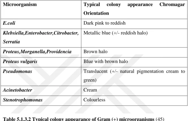
21 5.1.3 CHROMagar Orientation (Becton Dickenson, USA):
For the discovery and segregation of Gram positive and Gram negative pathogenic microorganisms CHROMagar Orientation was assessed.
BBL™ CHROMagar™ Orientation medium(Becton Dickenson,USA) can also be defined as a nonchoosy distinguished standard for the segregation, distinguishing and naming and listing of urinary tract pathogens separately. It is a superior to commonly used differential media for the isolation, differentiation and counting of UTI pathogens (42).
22 Table 5.1.3.1 Typical colony appearance of Gram (-) microorganisms (45)
Table 5.1.3.2 Typical colony appearance of Gram (+) microorganisms (45)
Microorganism Typical colony appearance Chromagar
Orientation
E.coli Dark pink to reddish
Klebsiella,Enterobacter,Citrobacter, Serratia
Metallic blue (+/- reddish halo)
Proteus,Morganella,Providencia Brown halo
Proteus vulgaris Blue with brown halo
Pseudomonas Translucent (+/- natural pigmentation cream to green)
Acinetobacter Cream
Stenotrophomonas Colourless
Microorganism Typical colony appearance Gram (+)
Enterococcus Turquoise blue
S.aureus Golden, opaque, small
S.epidermidis Cream, pinpoint colonies
S.saprophyticus Pink, opaque, small
23 5.1.4 BBL CHROMagar Candida Medium (Becton Dickenson, USA):
This media is a choosy and distinguishable medium for the separation of fungi. With the addition of chromogenic substrates in the medium, the colonies of C.albicans,
C.tropicalis and C.krusei generate dissimilar colors, thus permitting the straight
discovery of these yeast species on the separation plate. 1-6 Colonies of C.albicans emerge light to medium green, C.tropicalis colonies come into view blue-greenish to metallic-blue, and C.krusei colonies appear light rose with a whitish border (42).
24 Table 5.1.4 .1Typical colony appearance of candida species for microorganisms (47)
Microorganism Typical colony appearance
C.albicans Green
C.tropicalis Metallic blue
C.krusei Pink, fuzzy
C.kefyr, C.glabrata Mauve-brown
Other species White to mauve
5.2Biochemical Tests
5.2.1 Catalase test (Osel, Turkey)
To determine organisms that produce catalase enzyme, Catalase test was applied. This enzyme detoxifies hydrogen peroxide by breaking it down into water and oxygen gas. The bubbles appearing from production of oxygen gas obviously indicate a catalase positive result. The sample on the right below shows catalase positive. The Staphylococcus spp. and the Micrococcus spp. are catalase positive. The Streptococcus spp. and Enterococcus spp. are catalase negative (48).
25 5.2.2 Bile Esculin Agar (BEA) (Becton Dickenson, USA):
BEA is a choosy and differential medium which is presumptively utilized to determine Enterococci and group D Streptococci founded on the capability of an organism to hydrolyze esculin. Bile esculin agar includes oxygall (bile salts, first selective ingredients) to restrain from the growth of other Gram-positive organisms excluding Enterococci and group D Streptococci. Bile esculin disk is exercised for the quick finding of esculin hydrolysis in existence of bile for differentiating group D Streptococci from non-group D Streptococci (50)
26 5.2.3 Coagulase test (Oxoid Limited, USA)
Coagulase test is one of the utilized biochemical tests. It is extremely fundamental test in the microbiology. The coagulase test recognizes whether an organism gives the exoenzyme coagulase, which brings about the fibrin of blood plasma to clot. Coagulase responds with prothrombin in the blood. It causes blood to clot by alerting fibrinogen to fibrin. The coagulase test was exploited to distinguish the essentially pathogenic species Staphylococcus aureus from the regularly non-pathogenic species Staphylococcus epidermis. The S.aureus potentially pathogenic in human beings and animals, but S. epidermis is commonly not pathogenic (48).
5.2.4 Cytochrome Oxidase test (Oxoid Limited, USA)
The study also utilized the oxidase test. It is the final enzyme in the respiratory electron transport chain of mitochondria that spots the existence of a cytochrome oxidase system that will catalyze the transport of electrons between electron donors in the bacteria and a redox dye- tetramethyl-p-phenylene-diamine. The dye was reduced to deep purple color. This test was employed to help the determination of Pseudomonas,
Neisseria, Alcaligens, Aeromonas, Campylobacter, Vibrio, Brucella and Pasteurella, all
of which produce the enzyme cytochrome oxidase. Acinetobacter and
Enterobacteriaceae family do not produce this enzyme (48).
Its major function is to convert molecular oxygen to water and aid in establishing mitochondrial membrane potential. Cytochrome c oxidase locates to the inner membrane which segregates the mitochondrial matrix from the intermembrane space. This colorimetric assay is founded on surveillance of the reduction in absorbance at 550 nm of Ferro cytochrome c brought about by its oxidation to ferricytochrome c by cytochrome c oxidase (48).
27 5.2.5 MIO (Motility, Indole, Ornithine) Medium (Salubris Inc.,Turkey)
We utilized MIO (Motility, indole production, and ornithine decarboxylation) which are three distinguishing tests that are supplied in the one culture tube. It is recommended to use in testing motility, indole production, and ornithine-decarboxylase activity of enteric bacilli. We employed this test to differentiate the motility of the diverse Enterobacteria that bring about the lower respiratory infections. MIO medium is a semisolid medium utilized in the differentiation of the Enterobacteriaceae group by motility, ornithine decarboxylase activity and indole production (52).
Gelatin and casein peptones offer nitrogen, vitamins, minerals and amino acids vital for growth. They also give tryptophan required for the creation of indole. Yeast extract is a source of vitamins, especially of the B-group; Dextrose is the fermentable carbohydrate providing carbon and energy. L-ornithine is added to test the existence of the enzyme ornithine decarboxylase. If the organisms have such enzyme, it will be activated in an acid environment created by the initial fermentation of dextrose. Once the amino acid is decarboxylated, diamine putescine is produced. The result is an alkalinization of the medium, which turns it a dark blue. Organisms without the enzyme will stay acidic because of the fermentation, resulting in a yellow color in the medium. Bromocresol purple is a pH indicator to specify decarboxylase activity; the low concentration of bacteriological agar is for motility (52).
The bacteria are inoculated by stabbing the MIO medium and incubated in an aerobic atmosphere for 18 - 24 hours at 35 ± 2°C. If the indole reaction is negative, it is incubator for additional 24 hours. The motility and ornithine decarboxylase reactions were read before adding the Kovac‟s Reagent for the indole test. The motility was pointed out by cloudiness in the media or growth extending away from the line of inoculation. Ornithine decarboxylation is specified by a purple color in the medium. A negative ornithine reaction produces a yellow color at the bottom of the tube (52).
28 For the indole test, 3 to 4 drops of Kovac‟s Reagent was supplemented and the tube shaken carefully. The appearance of a red or pink color in the reagent layer is a positive indication of indole. Kovac‟s Reagent finds out the microorganism competent of cleaving the tryptophan. When these microorganisms are found in the medium, they liberate indole that responds with 4-dimethylaminobenzaldehyde to shape a dark red dye (52).
5.2.6 Citrate utilization test (Salubris Inc., Turkey)
Simmon‟s citrate media is composed of citrate only. If the bacteria grows in this media this means that bacteria can utilize the citrate to obtain from it all its requirements (Carbon + Nitrogen). It presents in bottles or tubes as slope media (48).
The purpose of slope is to obtain large area of surface for the bacteria, it contains indicator (Bromothymol blue). The color of media is green. If the bacteria utilizes the citrate, it produces alkaline sub of the pH of media will change. The media thus changes from green to blue (48).
Photo: 5.2.6.1 Citrate Test (53).
29 5.2.7Urea test (Salubris Inc., Turkey)
It contains urea and the indicator is phenol red. If the bacteria secrete the urease enzyme that break down the urea in the medium, the pH of media will change to alkaline and color of media will change from yellow or orange to pink (Neutral or acid to alkaline). This media is also presented in bottles or tubes (48).
30 5.2.8 Triple Sugar Iron Agar (TSI) (Salubris Inc., Turkey)
TSI Contain: Glucose + lactose + Sucrose + Iron Indicator: - They contain two types of indicator: Phenol red: Fermentation
Ferric ammonium citrate: H2S Production
In this medium the bacteria begin to take Glucose then lactose then sucrose then protein. Mostly the Enterobacteriaceae break down the glucose.
Break down of sugar: Acidic (Low pH) Break down of protein: Alkaline (High pH)
The color of medium after preparation is red (Alkaline) when the bacteria utilize the sugar it produce acid, the color of media will change from Red (alkaline) to yellow (acidic) 1. A/A 2. K/A 3. K/No change A: Acidic (Yellow) K: Alkaline (Red)
A/A: - The color of whole blood is yellow It‟s mean glucose fermenter & lactose fermenter. K/A: - The color of slope is red (No change) The color of butt is yellow (48).
31 A) Non-lactose fermenter:
This is the case that non-lactose fermenter represents 90% of the possibilities; this means that the bacteria fermented the glucose and did not ferment the lactose and then transferred directly to the protein and produced K (48).
B) Quick-lactose fermenter (L.F) Such as Klebsiella spp.
This kind of bacteria ferments the glucose and lactose and transfers directly to the protein (48).
C) Late lactose fermenter: Such as Shigella sonnei.
The lactose is regarded to be difficulty to ferment for the reason of these bacteria. K/No change
The color of whole media is red Not break down for any sugar
The bacteria break down protein directly In this media we read:
1. Fermentation: - A/A or K/A or K/no change 2. Gas Production: - Crack or bubble
Results: - Gas positive or Gas negative 3. H2S Production:-
-We can detect the H2S production by the indicator (Ferric ammonium) -By the presence of black color in the media.
- H2S production produce in acidic media K/A - Results: - H2S Positive or H2S Negative (48).
32 5.2.9 Mueller Hinton Agar Medium (Becton Dickenson, USA):
This media includes beef infusion, casamino acids, and starch. The levels of tetracycline and magnesium, thymidine, thymine, sulfonamide inhibitors, and calcium ions, are controlled so in order not to get in the way susceptibility testing and to yield good growth. The Kirby-Bauer antimicrobial disk diffusion procedure was used with Mueller Hinton Agar plates (48).
5.2.10 Antimicrobial Susceptibility Testing (Kirby Bauer Method)
Antimicrobial susceptibility testing (AST) is pointed out for pathogens contributing to an infectious process that warrants antimicrobial therapy. Otherwise susceptibility to antimicrobials could not be discovered consistently based on knowledge of their identity. Such tests are majorly employed when the etiologic agents are members of species competent of demonstrating resistance to generally prearranged antibiotics. Some organisms have predictable susceptibility to antimicrobial agents (ie, Streptococcus pyogenes to penicillin), and empirical therapy for these organisms is usually utilized. Thus, AST for such pathogens is seldom needed or conducted. Many laboratory approaches are available to characterize the in vitro susceptibility of bacteria to antimicrobial agents (48).
Phenotypic methods for discovering precise antimicrobial resistance mechanisms are mainly being used to complement AST (ie, inducible clindamycin resistance among several Gram-positive bacteria ) and to supply clinicians with preliminary direction for antibiotic selection pending results generated from standardized AST (ie, β-lactamase tests) (48).
33 The Kirby-Bauer (K-B) test employs small filter disks impregnated with a recognized concentration of antibiotic. The disks are put on a Mueller-Hinton agar plate that is inoculated with the test microorganism. Upon incubation, antibiotic diffuses from the disk into the surrounding agar. If susceptible to the antibiotic, the test organism will be incapable to develop in the spot straight away surrounding the disk, exhibiting an area of inhibition (see figure below). The size of this area is reliant on a number of aspects, including the sensitivity of the microbe to the antibiotic, the rate of diffusion of the antibiotic through the agar, and the depth of the agar. Microorganisms that are resistant to an antibiotic will not display an area of inhibition (growing right up to the disk itself) or show a comparatively small zone (48).
Photo: 5.2.10.1 Antimicrobial Susceptibility Testing (56). Results
Results may be read after 18-24 hours of incubation. Subsequent incubation, the zone sizes are evaluated to the nearest millimeter (mm) utilizing a ruler or caliper; including the diameter of the disk in the measurement. We used Clinical Laboratory Standards Institute (CLSI) criteria for AST evaluation, CLSI 2016 (57).
34 5.3 Used Instruments (VITEK) (BioMerieux, France)
The VITEK 2 is an automated microbial identification system which gives greatly precise and reproducible findings as displayed in several independent researches, with its colorimetric reagent cards, and related hardware and software advances.
They also offer an alternative of automatic pipetting and dilution for antimicrobial susceptibility testing (58).
5.4 Specimen Collection and Handling:
1. Sputum, tracheal aspirate, broncheoalveolar lavage (BAL) fluid:
a. Optimal timing: Specimens were preferably acquired before commencement of antimicrobial therapy although they might be gained at any time throughout the clinical course.
b. Specimen types: The study included the following satisfactory lower respiratory tract specimens such as sputum, tracheal aspirate, BAL fluid. Specimens with possibility for upper airway contamination (i.e., BAL fluid, pleural fluid, lung biopsy) are preferred. c. Specimen collection:
i. BAL fluid, tracheal aspirate: The study used sterile containers in which specimens were collected. Each of these specimen containers was labeled with the patient‟s name, ID number, the specimen type, and the date when theses specimens were collected. ii. Sputum: Patients were precisely informed regarding the distinction between sputum and oral emissions, after that they were instructed to wash the mouth with water and then expectorate deep cough sputum exactly into a sterile screw-cap collection cup or sterile dry container.
35 Specimen rejection criteria:
The following samples were rejected based on the following criteria: Without barcode specimens
No source on List Leaked specimens Non sterile cup
Inappropriate storage conditions (More than 1 hour and >4+O C) Same day more than one specimen
5.5 Specimen Inoculation
Specimen processes were worked out in biological safety cabinet, as aerosols can result in laboratory acquired respiratory infections.
1. All specimen processed were rapidly as possible to maintain viability of pathogen and avoid putting the patient at risk for repeated procedure.
2. We selected the purulent or most blood- tinged portion of the specimen. 3. Prepared Gram stain for details on preparation and reading of smears. 4. Used sterile swap, stick, loop or pipette.
5. An optochin disk was added to media.
Streak Plate:
Sputum and tracheal aspiration specimens were inoculated on the Medias with streaking method with 10µl sterile loop.
1. Specimen was spread over a portion of the culture media surface wisely.
2. The loop dragged from the inoculated section and spread it out into a second section. 3. The loop dragged again from the section 2 and then spread out into the third section. The same was done for the third and the fourth section. Sections 1 and 4 were ensured that were not overlapped. The inoculation loop used was disposed into a suitable container.
36 4. The lid was replaced and the streaked agar plate incubated at the appropriate temperature in an inverted position to avoid condensation.
5. Over the agar surface the inoculum was streaked in such a way that it “thins out” the bacteria.
6. Streaked plates were incubated at 37°C for 24 hours and examined the colonies grown in the plate carefully.
Photo: 5.5.1Streaking Technique (59).
5.5.1 BAL Inoculation: Dilution method
BAL: Utilizing a sterile loop each agar plate is inoculated with the deposit of the centrifuge sample. Centrifuged BAL is re-suspended in the fluid and three serial dilutions are made (1/10, 1/1000 and 1/100,000) 0.1mL of each dilutions is plated out. Optionally a calibrated loop is employed. For BAL fluids tests, quantitative calibrated loops intended for the conveyance of 0.010 and 0.001 µL are made use of. The colonies are calculated on the plates following incubation, and the number of CFU per milliliter is identified by multiplying the number of colonies by the dilution factor and 100. Diagnostic threshold is 104cfu/mL for BAL. Specimens from patients who have got antibiotics may also provide false-negative findings. Finding: Number of colony x104 CFU/mL.
37 5.5.2 TA and sputum inoculation:
TA and sputum samples were inoculated on the following media: Chrome agar (Becton Dickenson, USA), Blood agar (Becton Dickenson, USA), Chocolate agar (Becton Dickenson, USA), Chrome candida agar (Becton Dickenson, USA).
According to specimens, the study used colony counting but according to chocolate agar it was added optochin disk and incubated aerobically and made on Gram stain. 24 h later we observed the colony counting, if the amount of colony counting is less than 1000 CFU/mL, it is considered not significant but if the amount of colony is 104 or more than it is considered significant.
The study also used subculture and biochemical tests then put incubation with 24 h after this time it was read again and used antibiogram test to obtain specific type of bacteria. Incubation Plates were incubated at 35 to 37OC in 5% CO2 for a minimum of 48 h to 72 h as it is preferred.
5.6 Investigation of Gram Stain and Culture Results
The study employed a Gram stain (Salubris Inc., Turkey) technique which is a method that distinguishes Gram positive from Gram negative bacteria where the former showed purple color and the latter exhibited pink color.
Gram positive bacteria (thick layer of peptidoglycan-90% of cell wall) stains purple Gram negative bacteria (thin layer of peptidoglycan-10% of cell wall and high lipid
content) stains red/pink
Gram staining technique: Crystal violet stain, lugol‟s iodine (mordant), acetone-alcohol decolorizer (more rapid, not over decolorize smear), fuchsin (counter stain)
38 Report of Gram Reaction
1. Number of bacteria present (scanty – few - moderate- many) 2. Gram reaction of bacteria (Gram positive or Gram negative)
3. Morphology of bacteria (Cocci- diplococcic- rod – coccobacilli-bacilli) Investigation of Bronchoalveolar Lavage
Streaked plates were incubated at 37°C for 24 hours and the colonies examined for grown in the plate carefully the next day. If the amount of colony count is less than 1000 CFU/mL, it is considered not significant but if the amount of colony is 104 or more than it is considered significant. Then we decided to do subculture and biochemical tests applied from Isenberg guide.
Investigation of Tracheal Aspirate
Streaked plates were incubated at 37°C for 24 hours before we examined the colonies grown on the plate carefully the next day.
Subsequent to incubation, the colonies were calculated on the plates and the number of CFU per milliliter is identified by multiplying the number of colonies. Predominantly if we get 104 CFU/mL we decided infection for determination of sub culture, biochemical tests, AB, and Vitek to obtain the exact of bacteria.
Investigation of Sputum
Streaked plates were incubated at 37°C for 24 hours and examined for the colonies grown in the plate carefully the next day. After incubation the dominantly growth colonies were identified.
Interpretation and Reporting of the Results
Employing the published CLSI guiding principles, the susceptibility or resistance of the organism to each drug tested is determined.
One the recording sheet for each drug, it is displayed whether the area size is susceptible (S), intermediate (I), or resistant (R) according to the explanation chart.
The findings of the Kirby-Bauer disk diffusion susceptibility test are stated only as susceptible, intermediate, or resistant.