Can chondral healing be improved following microfracture? The effect of
adipocyte tissue derived stem cell therapy
Hasan H. Ceylan
a, Kerem Bilsel
b, Nur Buyukpinarbasili
c, Hamid Ceylan
d, Mehmet Erdil
e,⁎
,
Ibrahim Tuncay
b, Cengiz Sen
fa
LNB State Hospital, Department of Orthopaedics and Traumatology, Istanbul, Turkey
b
Bezmialem Vakif University Medical School, Department of Orthopaedics and Traumatology, Istanbul, Turkey
c
Bezmialem Vakif University Medical School, Department of Medical Pathology, Istanbul, Turkey
d
Ataturk University Science Faculty, Department of Molecular Biology and Genetics, Erzurum, Turkey
eIstanbul Medipol University, Department of Orthopaedics and Traumatology, Istanbul, Turkey f
Istanbul University Istanbul Medical School, Department of Orthopaedics and Traumatology, Istanbul, Turkey
a b s t r a c t
a r t i c l e i n f o
Article history: Received 17 January 2015
Received in revised form 13 September 2015 Accepted 26 November 2015
Background: We aimed to investigate the effect of adipose tissue-derived mesenchymal stem cells (ADSCs) on chondral healing using the microfracture (MF) technique.
Methods: Thirty male rabbits were randomly divided into three groups. Standard cylindrical osteochondral defects (OCDs) were created in the weight-bearing areas of the medial condyles of all the right knees; the defects were four millimeters in diameter and two millimeters in depth. The control group (group A) was restricted to spontaneous healing. For group B, we performed MF with a 1.5-mm drill. For group C, we applied MF using the same method and then applied 3 × 10⁶ADSCs to the defect area. At eight weeks post-operation, the subjects
were sacrificed, and the distal femoral joint surfaces were evaluated histopathologically for chondral healing. The samples were scored according to the International Cartilage Repair Society (ICRS) scale.
Results: The results for group C were significantly better than those for group A in terms of the surface properties (p = 0.003). The matrix evaluation was better for group A than for group C (p = 0.01). The cell distribution, cell viability and subchondral bone parameters were similar between the groups (p = 0.198, p = 0.387 and p = 0.699). The cartilage mineralization parameter was better for group C than for group A (p = 0.001). The signs of healing were better for group C than for group B, but the differences were not significant (p = 0.185). Conclusions: Improvements with additional ADSC treatments were not statistically significant in cases in which ADSC treatment was compared with isolated MF treatment.
Clinical Relevance: Additional ADSCs treatment may have positive effect on chondral healing but it doesn’t seem significant.
© 2015 Elsevier B.V. All rights reserved.
Keywords:
Adipocyte tissue derived stem cell Osteochondral defect
Microfracture Chondral healing Cartilage repair
1. Introduction
Selection of the ideal treatment for articular cartilage pathologies is a challenging issue for orthopedic surgeons[1–4]. The microfracture (MF) technique is frequently used and is based on pluripotent stem cells (SCs) formingfibrocartilage wound tissue after subchondral bone stimulation[1–4]. This technique has been successfully performed, particularly in cartilage lesions that are small, well contained, and Outerbridge 3 to 4[4].
Although defects can be treated with MF, the newly formed tissue is poor in hyaline cartilage; it is structurally and biomechanically deficient
compared with articular cartilage[2,5]. Treatment success is related to the degree of histological and functional similarities of the repaired tis-sue to the original tistis-sue. Additional studies are being conducted to im-prove the quality of newly formed cartilage tissue forfilling defects
[6–11].
The ideal source of SCs is controversial. With the discovery that adult human adipose tissue is a potential source of SCs, a new source of SCs has become available[9,12–15]. Adipose tissue-derived mesenchymal stem cells (ADSCs) appear to be advantageous for the following reasons: they are easily obtained with the least amount of morbidity to the donor site, their reproducibility in a culture environment is simple, they do not lose their multipotency during earlier culture and passaging, and their chondrogenic potential is maintained[8,15–17]. By contrast, obtaining SCs from bone marrow is invasive, difficult and painful. The number of fi-broblast colony forming units (CFUs) is three times greater in adipose tis-sue than in bone marrow, and adipose tistis-sue is more expendable than bone marrow in the culture environment[18].
⁎ Corresponding author at: Medipol University Medical School, Department of Orthopaedics and Traumatology, 34200 Istanbul, Turkey. Tel.: +90 530 696 6045; fax: +90 212 476 4259.
E-mail address:[email protected](M. Erdil).
http://dx.doi.org/10.1016/j.knee.2015.11.008 0968-0160/© 2015 Elsevier B.V. All rights reserved.
Contents lists available atScienceDirect
Recently, the use of ADSC therapy for chondral pathologies has in-creased among knee specialists. Most surgeons who use ADSC therapy believe that this procedure affects the healing process and contributes to cartilage regeneration. In this experimental animal study, we aimed to evaluate the contribution of ADSC application to cartilage repair in the MF procedure.
2. Materials and methods
In this study, which was approved by the Institutional Animal Care and Use Committee prior to performing the study, according to the power analysis we used 30 identically bred seven-month-old white male New Zealand rabbits that weighed an average of 3420 ± 230 g (min. 3120 to max. 3580) each and were bred under standard laboratory
conditions. Cell culture procedures to acquire the ADSCs were conducted in a cell laboratory using the technique described by Zuk et al.[15].
In addition to the experimental animals, one male rabbit with identical characteristics was used as the source of the adipose tissue. For sedation and analgesia, 15 mg/kg of xylazine (Rompun, Bayer, Le-verkusen, Germany) was administered intramuscularly (IM). For anesthesia, 45 mg/kg of ketamine hydrochloride (Ketalar, Zentiva, Kirklareli, Turkey) was administered intramuscular (IM) 10 min later. A two-centimeter bikini incision was made to the inguinal region, from which a 2 × 2-cm subcutaneous adipose tissue sample was obtained.
2.1. Cell culture procedure
Approximately 100 mg of adipose tissue that was acquired from the subject was delivered to the laboratory under sterile conditions and washed with a physiological saline solution (PSS) three times. After the PSS was discarded, the adipose tissue samples were incubated at 37 °C for two hours in three milliliter 0.075% phosphate buffered saline (PBS) solution with type 2 collagenase. After three hours of centrifuga-tion in an 800-g falcon tube, separacentrifuga-tion of the tissue from the stem cells occurred. The acquired cells were cultured in DMEM-LG in a T-150flask with one percent antibiotic and 10% fetal bovine serum (FBS). After the cells in the primary culture covered≥70% of the flask bottom, the cells were passaged. The cells that were adherent to theflask bottom were lifted with trypsin and washed with PBS. Using identical medium and environmental conditions, the cells were re-cultured on a wider surface area. Throughout the passage, the cells were observed once every three days under a microscope; the medium was changed, and the cells in the flask were expected to reach 70% confluence. After the primary culture stage was completed and≥70% confluence was reached, the cells were lifted with trypsin; after washing with PBS, the cells were placed in identical medium (DMEM-LG containing one percent penicillin and 10% FBS) for further incubation. The cells collected with trypsinization after thefirst passage were used for the quality verification stage.
The desired outcomes from the quality verification stage were as fol-lows: 1) survival at the end of production ofN80% of the number before production with 2 × 10⁴of at least 2 × 10⁷cells; 2) current cytometric
analysis performed on a sample taken at the end of the second passage showing that the mesenchymal SCs are CD73, CD105 and CD90 positive and CD34, CD45 and HLA-DR negative (purity); 3) maintenance of the sterility of the culture area throughout the study and the absence of my-coplasma; 4) apyrogenicity; 5)fixation of the collagen synthesis of the activity analysis atfive micrograms per milliliter per 1 × 10⁷cells per
day; 6) maintenance of RTA (relative telomere enzyme activity) at b1.2; and 7) viability N70% before application (stability). The MSCs were produced and resuspended in 0.1 cm3PBS (contains one percent
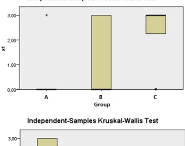
Fig. 1. To obtain the standard defects, we used a four-millimeter-diameter drill that allowed us to create a controlled two-millimeter-depth defect in all of the objectives. Graphic 1. The graphic shows the surface morphology (top), matrix composition (middle)
FBS) solution containing 3 × 10⁶cells in 30-gauge injectors that were
transported to the operating room under cold chain conditions. 2.2. Experimental groups
Upon power analysis we used 8 ± 2 animals for each group. The animals were randomly selected and assigned to three groups of ten subjects each. Only osteochondral defects (OCDs) were applied to the knees of the rabbits in the control group (group A), and spontaneous healing was investigated. OCDs were created and MF was performed in the subjects in the second group (group B). In the third group (group C), after creating OCDs and performing MF, ADSCs were applied to the defect.
2.3. Surgical procedure
The surgical procedures were performed by one surgeon, using the same technique for each knee. A standard anesthesia regimen, surgical
approach, technique to create the cartilage defect, and wound covering were applied in all of the rabbits.
For sedation and analgesia, 15 mg/kg of xylazine (Rompun, Bayer, Leverkusen, Germany) was administered IM to the rabbits. For anesthesia, 45 mg/kg of ketamine hydrochloride (Ketalar, Zentiva, Kirklareli, Turkey) was administered IM 10 min later. The distal areas of the inguinal region were shaved, and after the region was cleaned with a 10% polyvinylpyr-rolidone iodine solution (Batticon, Adeka, Turkey), it was covered in a sterile manner. Immediately following the operations, 0.5 mg/kg of amox-icillin trihydrate (Clamoxyl, Pfizer, USA) was applied subcutaneously to the immediate vicinity of the incision.
A standard longitudinal parapatellar incision was made in the right knee joint. Standard cylindrical four-millimeter-diameter and two-mil-limeter-deep OCDs were created in the medial condyle weight-bearing region. After the medial condyle was exposed, we always placed the sleeve at the center of the condyle, perpendicular to the chondral sur-face and with the same axis to the femoral shaft so that all of the defects were at the same location. These standard defects were made using a sleeve-controlled drill (Fig. 1). The drill bit was four millimeters in diameter and two millimeters longer than the sleeve. When the sleeve was positioned against the chondral surface, we were able to create a two-millimeter deep defect. Only OCDs were applied to the knees of the rabbits in the control group (group A), and spontaneous healing was examined. OCDs were created and MF was performed in the rabbits in the second group (group B); in the third group (group C), after creat-ing OCDs and performcreat-ing MF, ADSCs were applied to the defect. 2.3.1. Standard surgical procedures common to all three groups
The animals were brought into the operating room. For sedation and analgesia, 15 mg/kg of xylazine (Rompun, Bayer, Leverkusen, Germany) was administered IM to the rabbits. For anesthesia, 45 mg/kg of ketamine hydrochloride (Ketalar, Zentiva, Kirklareli, Turkey) was administered IM 10 min later. The knee was shaved carefully to avoid harming the dermis and marked with a pen to indicate the precise incision.
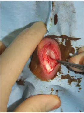
The entire leg was stained with povidone iodine three times and allowed to dry. With a perforated sterilized surgical cover, all body parts were covered except the knee where the operation was to be per-formed. A longitudinal incision was performed in the medial parapatellar area. The subcutaneous layer and articular capsule were passed with a longitudinal incision. The joint was observed, and the pa-tella was dislocated laterally (Fig. 2). While the knee wasflexed, it was placed on the femur medial condyle cartilage perpendicular to the guide articular surface. By carefully passing a four-millimeter-diameter drill through the guide, a two-millimeter-deep cartilage OCD was formed (Figs. 1 and 3). To avoid thermal damage, the surgical drill motor was run at the lowest cycle, and during drilling, the defect area was washed
Fig. 3. A photo of a medial condyle in which we created a four-millimeter-diameter and two-millimeter-deep OCD. The middle illustration shows a distal femur after an OCD was created with a drill. The right image shows a sagittal view of the same defect.
Fig. 2. After the parapatellar skin incision and capsulotomy, the patella was luxated later-ally, and the medial condyle was directly visualized.
with isotonic NaCl. The size of the formed defect and its parallel position to the femur axis were checked. The cartilage debris in the defect was removed with a mini-curette. The defect area and the interior of the joint were power washed with isotonic NaCl. The joint capsule and the dermis were closed. The wound sides were wiped with povidone iodine, and 0.5 mg/kg of prophylactic amoxicillin trihydrate (Clamoxyl, Pfizer, USA) was injected subcutaneously. To avoid potential damage to the sutures and the wound by the rabbit, the sutured and covered wound was wrapped with a bandage (Tensoplast, BSN Medical, England). One-third of the animals were not treated to allow for spontaneous healing (group A).
2.3.2. The surgical procedure performed in the MF group (group B) In addition to the procedures described above, a single MF was performed with a 1.5-mm drill from exactly the center of the defect, ensuring that it was in the same plane as the femoral medulla (Fig. 4). Leakage of blood and adipose tissue from the medulla was observed, and the joint was irrigated and sutured.
2.3.3. The surgical procedure performed in the ADSC treatment group (group C)
The surgical procedure performed on group B was repeated on this group. Additionally, after the joint defect area was washed, the 0.1-cm3
semi-gel ADSC solution, which was brought to the operating room follow-ing the MF in a 30-gauge injector under cold chain conditions, was care-fully administered into the center of the defect without overflow from the knee joint (Fig. 5). The knee joint capsule was tightly sutured. 2.4. Post-operative care
Standard post-operative analgesia, wound care, cage surveillance, feeding and sacrifice were performed similarly for all of the animals. Post-operative care of the rabbits was conducted in cages in the animal laboratory. The animals were fed ad libitum under normal living condi-tions, and they drank tap water. Post-operative examination for pain and infection was performed. None of the subjects required additional analgesic or antibiotic treatment. The surgical area was examined for adhesiveness, discharge, rubor, and edema, and in two animals, the su-tures in the wound area were found open on the second day. The dermal wounds were immediately cleaned and re-sutured. No infection was observed in the animals. One animal in the control group refused food from the third post-operative day and died on thefifth day.
2.5. Sacrifice
The animals that remained alive at eight weeks post-surgery were administered 15 mg/kg of xylazine IM (Rompun, Bayer, Leverkusen,
Germany) for sedation and analgesia. For anesthesia, 45 mg/kg of keta-mine hydrochloride (Ketalar, Zentiva, Kirklareli, Turkey) was adminis-tered IM 10 min later. The animals in which anesthesia was started were given 1.5 cm3of xylazine (Rompun, Bayer, Leverkusen, Germany)
intracardially and then were sacrificed. The rabbit cadavers were placed in medical waste boxes and sent for disposal.
2.6. Histopathological examination
The acquired samples of the distal femur joint were divided into five-millimeter slices in the coronal plane using physical methods. The slices werefixed with a 10% neutral formaldehyde dipping technique and transported to the pathology laboratory. The samples were kept in acid for one week for decalcification. A tissue block that included the slice that centered on the cartilage defect was acquired from each subject. The paraffin blocks were divided into five-micrometer slices with a microtome (Leika RM2245, Wetzlar, Germany). For deparaffinization, the slices were maintained in an incubator at 60 °C. Post-incubation, the slices were exposed to xylene and were incubated in alcohol in a series of concentrations decreasing from 96% to 70% for rehydration. The slices retained in the alcohol series were washed with distilled water. After staining with hematoxylin (33230, Riedel-de Haen, Germany) for two minutes, the slices were washed inflowing water forfive minutes to remove the extra stain from the tissue and then stained with eosin (1345, Merck, Darmstadt, Germany) for 30 s. After being washed underflowing water for five minutes in an identical man-ner, the slices that passed through the alcohol series at concentrations of
Fig. 5. The implantation of AMSCs in the defect area.
Fig. 4. The microfracture procedure for the medial condyle. After a standard-sized OCD was created, a 1.5-millimeter diameter drill was used to create a microfracture. The right image shows medullary bleeding after the procedure.
70%, 80% and 96% were air-dried and incubated in xylene for 30 min each for transparency; then, they were covered with Entellan (UN 1866, Merck, Darmstadt, Germany).
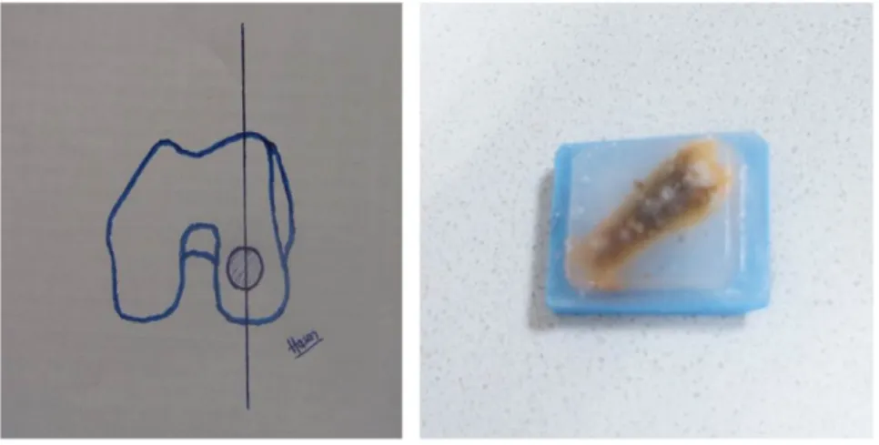
A sagittal slice passing through the defect center was examined
(Fig. 6) for each medial condyle sample. The samples were graded by
a pathologist blinded to the experimental groups, in accordance with the parameters of the International Cartilage Repair Society (ICRS) scale[19]. The slices were separately graded according to the ICRS, and the ICRS points were determined for each subject.
The samples, which were acquired by slicing along the sagittal plane from the exact center of the area in which the cartilage defect had been formed, were stained with hematoxylin & eosin and evaluated by a pathologist blinded to the experimental groups. The samples were graded for cartilage tissue regeneration in accordance with the ICRS scale.
3. Results
All of the cartilage defects tended to close from the periphery to the center, and the defects in treatment groups B and C werefilled almost completely. In the ADSC group, the healing andfilling of the defect with repair tissue were slightly smoother than in the MF group. However, there was no objective scoring difference in the macroscopic compar-ison of the groups, and thus, the examination entailed only general observations (Fig. 7).
3.1. Histopathological comparison of the treatment results
The histopathological analysis of the samples in group A (the control group) showed that the surface of the repair tissue was irregular in all the samples, with the exception of one sample. Whereas the samples in this group displayed a matrix composition of hyaline
weight in three subjects, healing was achieved through mixed cartilage formation in four subjects and throughfibrocartilage tissue in two subjects. The cell distribution was gener-ally in the form of a column–cluster mixture, and no sample was composed completely of colonized cells. Cell viability was found in all the subjects. In the subchondral bone, many of the subjects showed reformation. Whereas in one subject there was normal cartilage mineralization, the other subjects exhibited abnormal mineralization (Fig. 8).
The results of the examination of the MF group (group B) were as follows: in the histopathological analysis of the samples in the group that received classical treatment, the surface of the repair tissue was regular in four subjects and irregular in the other subjects. In the examination of the matrix composition of the samples of this group, three subjects showed repair by mixed cartilage formation and six subjects showed repair byfibrocartilage tissue. In one subject, repair by fibrous tissue was observed. The cell distribution was generally in the form of a column–cluster mixture, and no sample was composed completely of colonized cells in this group. Except for one subject, the subjects showed cell viability. Whereas the subchondral bone showed a tendency for reformation infive of the subjects, four subjects showed granulation tissue. Whereas six subjects showed normal cartilage mineralization, abnormal mineralization was observed in four subjects (Fig. 8).
The histopathological analysis of the samples in the ADSC group (group C) revealed that repair tissue was regular in eight subjects and irregular in two subjects. In the exam-ination of the matrix composition in the samples of this group, six subjects showed repair by mixed cartilage formation, and four subjects showed repair byfibrocartilage tissue. There was no evidence of repair byfibrous tissue in any of the subjects. The cell distribu-tion was in the form of a column–cluster mixture in six subjects, and no repair tissue was composed completely of colonized cells in this group. Cell viability was observed in all the subjects. Whereas the subchondral bone was entirely normally shaped in one subject, it showed a tendency for reformation infive subjects, and it showed a granulation tissue pattern in four subjects. Except for one subject, the subjects displayed normal cartilage mineralization (Fig. 8).
3.2. Statistical comparison of the treatment results
After the histopathological evaluation of the samples according to the ICRS scale, statistical analyses of the results were performed using Statistical Package for the Social Sciences (SPSS). The statistical significance value was set at p b 0.05. For descriptive statis-tics, the mean and standard deviation were used (Table 1). Because the ICRS grading tech-nique is an approximate scale and the number of samples was small, the results of the histopathological evaluation of the groups were analyzed using the Kruskal–Wallis non-parametric test (Table 2,Graphic 1). The variables that displayed a significant difference were compared using the post hoc-Dunn test (Table 3).
Surface: When the groups were compared in terms of the surface of the repair tissue, group C had a better mean than groups A and B (Table 1). The comparison using the post hoc-Dunn test revealed that group C showed significantly better healing than group A, which underwent spontaneous healing (p = 0.003). No significant differences were observed in the surface morphology between group A and group B or between group B and group C (p = 0.21 and p = 0.077).
Matrix: When the groups were compared in terms of the matrix of the newly formed cartilage, it was observed that in group A, which underwent spontaneous healing, the matrix was significantly better than in group C (p = 0.01). No significant differences were observed in the matrix evaluation between group A and group B or between group B and group C (p = 0.21 and p = 0.17).
In terms of cell distribution, cell viability, and subchondral bone, there were no significant differences between the groups (Kruskal–Wallis test; p = 0.198, p = 0.387, p = 0.699, respectively).
Cartilage mineralization: The comparison of the existence of close-to-normal cartilage mineralization among the groups revealed that group C showed a significantly better Fig. 7. One of the distal femur samples from the ADSC treatment group. The defect area
wasfilled with newly repaired tissue.
Fig. 6. The distal femur sagittal samples obtained for the histopathological evaluation. Five-millimeter slices that were perpendicular to the joint surface and parallel to the femoral diaph-ysis were obtained using a microtome. The right image shows the slice, which was embedded in paraffin before slicing.
result than group A (p = 0.001). Similarly, group B had a significantly better result than that of group A (p = 0.036). Group C had a better mean score; however, the mean score of group C was not significantly better than that of group B (p = 0.185).
4. Discussion
In this study, we investigated the contribution of SC recruitment, applied as ADSCs, in addition to MF to defect healing for the treatment of cartilage defects. Cartilage defect healing in the subjects in our groups was evaluated using the parameters in the ICRS scale. Comparing the results of the group that received ADSCs post-MF to those of the group with MF only, the former group had higher mean results for the surface, matrix, cell viability, subchondral bone and cartilage mineralization
(Table 1). However, our study showed no statistical difference
(Table 3). Comparing the results of the MF group to those of the control group, the MF group had higher mean results for the surface, cell distri-bution and cartilage mineralization (Table 1). However, only the carti-lage mineralization parameter was significantly better in MF group (Table 3).
The functions of chondrocytes are susceptible to hormonal effects
[20]. Whereas growth hormone, thyroxin and testosterone accelerate glycosaminoglycan synthesis, cortisone, hydrocortisone and estradiol decelerate it. The hypophyseal growth hormone somatotropin stimulates the growth of cartilage cells indirectly by stimulating
somatomedin C synthesis in the liver[20]. In this experimental study, we used male rabbits to ensure that the results would not be affected by possible hormone changes and increases in body weight from pregnancy.
Different approaches regarding the number of applied SCs have been used in studies of cartilage defect repair with SCs, with the number of SCs varying from one to 5 × 10⁶[7,11,14]. In our study, we applied
3 × 10⁶SCs around the defect.
It has been shown in the rabbit knee that cartilage defects with a diameter of four millimeters or greater do not close spontaneously, whereas cartilage defects with a diameter of less than four millimeters do heal spontaneously[13,21]. In the OCD group in our study, although we observed that the defect showed a tendency to heal from the periph-ery to the center, there was no full closure. To verify the effectiveness of our treatment, we created OCDs at a size (four millimeters) at which the medial femoral condyle in the rabbits could not spontaneously close on the joint surface.
The mean cartilage thickness at the anteromedial femoral condyle has been reported as 0.3 ± 0.07 mm in the literature[22]. Although there is no standard depth when creating such defects, there are published studies with defects ranging from one to four millimeters in depth[23]. In one study, defects with a two-millimeter depth were cre-ated in the knees of rabbits[24]. In another study, general defects with a three-millimeter depth were created in cartilage on the knees of rabbits
Table 2
The Kruskal–Wallis test showed significant differences only in the surface, matrix and cartilage mineralization parameters between the three groups. All of the groups were similar in terms of cell distribution, cell viability and subchondral bone.
Kruskal–Wallis Surface Matrix Cell distribution Cell viability Subchondral bone Cartilage mineralization
Chi square 8.915 6.720 3.242 1.900 0.716 11.647
P 0.012 0.035 0.198 0.387 0.699 0.003
Bold values indicate significance at pb0.05. Table 1
Mean, SD and median values for each group according to the ICRS scale.
Group Surface Matrix Cell
distribution Cell viability Subchondral bone Cartilage mineralization A N 9 9 9 9 9 9 Mean 0.33 2.11 1.22 3.00 1.56 0.33 SD 1.00 0.78 0.83 0.00 0.73 1.00 Median 0.00 2.00 1.00 3.00 2.00 0.00 B N 10 10 10 10 10 10 Mean 1.20 1.20 1.80 2.80 1.40 1.80 SD 1.55 0.63 0.42 0.63 0.70 1.55 Median 0.00 1.00 2.00 3.00 1.50 3.00 C N 10 10 10 10 10 10 Mean 2.40 1.60 1.60 3.00 1.70 2.70 SD 1.26 0.52 0.52 0.00 0.67 0.95 Median 3.00 2.00 2.00 3.00 2.00 3.00
Fig. 8. Post-mortem histopathological evaluation of each of the three groups at the eighth week post-operation. The microscopic images show the chondral healing differences between the OCD (left), OCD + MF (middle) and OCD + MF + AMSC (right image) groups. Smooth surface, mixed hyaline andfibrocartilage healing areas were observed in OCD + MF + AMSC (right image). H&E 200.
[25]. In additional studies, four-millimeter-diameter and three-millime-ter-deep full-thickness cartilage defects and 4.5-mm-diameter and three-millimeter-deep defects were created[11,13]. In yet another animal study, a four-millimeter-deep cartilage defect was created in a rabbit knee[23]. In our study, we created OCDs that were four millime-ters in diameter and two millimemillime-ters deep in accordance with the previous literature. We operated on the medial condyle of the rabbit knee, which is the most frequently used model in cartilage defect man-agement studies[1,11,25]. It is possible that such a wide defect would not be applicable to standard clinical scenarios compared with the human knee, but we primarily aimed to determine the differences in chondral repair between groups.
The local environment is crucial for the differentiation of SCs[26,27]. Additionally, progenitors in the synovialfluid play a significant role in SC differentiation[28]. Because local growth factors (the autocrine effect) or factors originating from the synovia (the paracrine effect) are active in cartilage metabolism[26–28]. In addition, systemic hor-mones affect chondrocyte metabolism through diffusion via the synovi-alfluid[29]. The existence of potent SCs in synovialfluid, which are one hundred times more efficient in chondrogenic potential than those in ADSCs and Bone Marrow derived mesenchymal Stem Cells (BMSCs), has been shown through in vitro cultures[10,30,31]. We did not apply a barrier cover, such as a collagen membrane, periosteum, or perichondri-um, over the defect. Following the application of these barriers, stability of the barrier is not guaranteed, and a coverage loss ratio of up to 70% may occur[1].
In thefirst stage, SC cultures may be unable to adapt to the tissue to which they are applied, and synthetic or biological scaffolds are used. In these scaffolds, many features are important, such as mechanical prop-erties that are suitable for the tissue to be applied, size, degradation time, vascularity tolerance, and a suitable surface for cell adsorption. These polymers are difficult to acquire, they are expensive, and they lead to bioadaptability problems[32]. Application of SCs directly into the joint is a simple osteoarthritis treatment. Previous studies have shown that SCs can live without scaffold and that the cells injected into the joint can maintain their viability[6]. Many animal studies have reported the application of SCs directly into joints[14,33,34]. In a well-known study, Lee et al. directly injected SCs into joints[33]. In our study, it was appropriate to apply SCs directly to the defect without using a scaffold.
Cartilage tissue is adapted to a pressurized environment, and it re-quires recurring movement cycles to obtain nourishment and remove metabolites [8,29,35]. The application of hydrostatic pressure to chondrocytes results in a significant increase in the total collagen content and shaping[8,36]. At the end of eight weeks of pressure appli-cation, the GAG content in a pressurized group increased significantly
[36]. Significant proliferation of chondrocytes was shown in cultures grown under shear force rather than in a static culture environment, and an increase in TGF-beta synthesis under shear forces was reported
[37]. Based on data from previous studies, we did not immobilize our animals and we permitted free movement in our study.
There were limitations in our study. Although additional time may be needed for chondral healing, we terminated the experiment in the eighth week based on the general practice in the literature (between six and 12 weeks). Additional time could have been allotted to observe long-term changes. Another uncertain aspect in the literature is the appropriate number of cells to be used because other researchers have reported their results using different numbers. The low number of
cells may be one reason that we could not obtain significant cartilage healing. We do not know the precise correlation between the outcome of the applied treatment and the number of cells. A membrane or scaffold like material may help the persistence of these cells around the defect. We did not use such a barrier because of previously reported high failure rates. We harvested all of the ADSCs from a single, separate animal to prevent additional post-operative complications in the exper-imental animals. We do not know whether ADSCs obtained from the ac-tual injured animal (i.e., if each rabbit provided its own stem cells) would yield better results. In addition, the animals were not genetically identical and there might be potential for immunogenicity of the stem cells. Due to technical inadequacy, we were only able to utilize hema-toxylin & eosin staining, which enables observation of the general cellu-lar architecture. If we could have stained the samples with Safranin-O to determine the proteoglycan content of the cartilage tissue and the ma-trix, we could have revealed the quality of the newly formed cartilage in more detail. Maybe surface repair chondrocytes come into the joint from the node of Ranvier, also we could not demonstrate this due to technical inadequecies again. Another unexpected result of our study was that, group A had significantly more matrix than group C and a trend to have more matrix than group B. We could not explain the rea-son of this with our knowledge and current literature.
In our study, in terms of histopathological improvement, the MF + ADSC group did not show statistically significant superiority to the MF group. We believe that the application of ADSCs after MF did not contribute significantly to chondral healing. In light of these results, further research is needed to investigate the clinical necessity of addi-tional ADSC treatment in patients with MF. The specific indications should be defined for this treatment, which is relatively expensive, and the treatment should be practiced in a manner that has been shown scientifically to be effective.
Conflict of interest statement
All authors of our study disclose nofinancial and personal relation-ships with other people or organizations like employment, consultancies, stock ownership, honoraria, paid expert testimony, patent applications/ registrations or other fundings that could potentially and inappropriately influence (bias) our work and conclusions.
Acknowledgments
This study was supported by the Istanbul Bezmialem Vakif University Scientific Research Projects Department. Grant number was 9.2012/11. We thank Omer Uysal for the statistical analysis; Fatma Eyuboglu for the cell culture procedure assistance; Safak Sayar, Oznur Inan and Onder Huseyin Bas for the animal laboratory assistance.
References
[1]Gunes T, Sen C, Erdem M, Koseoglu RD, Filiz NO. Combination of microfracture and periosteal transplantation techniques for the treatment of full-thickness cartilage defects. Acta Orthop Traumatol Turc 2006;40:315–23.
[2]Hunziker EB. Articular cartilage repair: basic science and clinical progress. A review of the current status and prospects. Osteoarthritis Cartilage 2002;10:432–63. [3]Rodrigo J, Steadman JR, Silliman JE, Fulstone HA. Improvement of full thickness
chondral defect healing in the human knee after debridment and microfracture using continous passive motion. Am J Knee Surg 1994;7:109–16.
[4]Williams III RJ, Harnly HW. Microfracture: indications, technique, and results. Instr Course Lect 2007;56:419–28.
Table 3
The Dunn test shows the significant changes between groups.
Dunn Surface Matrix Cell distribution Cell viability Subchondral bone Cartilage mineralization
A vs. B 0.21 0.21 NS NS NS 0.036
A vs. C 0.003 0.01 NS NS NS 0.001
[5]Jakob RP, Franz T, Gautier E, Mainil-Varlet P. Autologous osteochondral grafting in the knee: indication, results, and reflections. Clin Orthop Relat Res 2002;401: 170–84.
[6]Barry FP. Mesenchymal stem cell therapy in joint disease. In: Bock G, Goode J, edi-tors. Tissue engineering of cartilage and bone. Chichester: John Wiley & Sons; 2002. p. 886–966.
[7]Jung M, Kaszap B, Redöhl A, Steck E, Breusch S, Richter W, et al. Enhanced early tissue regeneration after matrix-assisted autologous mesenchymal stem cell transplantation in full thickness chondral defects in a minipig model. Cell Transplant 2009;18:923–32. [8]Kessler MW, Grande DA. Tissue engineering and cartilage. Organogenesis 2008;4:
28–32.
[9]Nathan S, Das De S, Thambyah A, Fen C, Goh J, Lee EH. Cell-based therapy in the repair of osteochondral defects: a novel use for adipose tissue. Tissue Eng 2003;9: 733–44.
[10]Sakaguchi Y, Sekiya I, Yagishita K, Muneta T. Comparison of human stem cells derived from various mesenchymal tissues: superiority of synovium as a cell source. Arthritis Rheum 2005;52:2521–9.
[11]Yan H, Yu C. Repair of full-thickness cartilage defects with cells of different origin in a rabbit model. Arthroscopy 2007;23:178–87.
[12]Afizah H, Yang Z, Hui JH, Ouyang HW, Lee EH. A comparison between the chondrogenic potential of human bone marrow stem cells (BMSCs) and adipose-derived stem cells (ADSCs) taken from the same donors. Tissue Eng 2007;13: 659–66.
[13]Li Q, Tang J, Wang R, Bei C, Xin L, Zeng Y, et al. Comparing the chondrogenic potential in vivo of autogeneic mesenchymal stem cells derived from different tissues. Artif Cells Blood Substit Immobil Biotechnol 2011;39:31–8.
[14]Toghraie FS, Chenari N, Gholipour MA, Faghih Z, Torabinejad S, Dehghani S, et al. Treatment of osteoarthritis with infrapatellar fat pad derived mesenchymal stem cells in Rabbit. Knee 2011;18:71–5.
[15]Zuk PA, Zhu M, Mizuno H, Huang J, Futrell JW, Katz AJ, et al. Multilineage cells from human adipose tissue: implications for cell-based therapies. Tissue Eng 2001;7: 211–28.
[16]Jakobsen RB, Shahdadfar A, Reinholt FP, Brinchmann JE. Chondrogenesis in a hyaluronic acid scaffold: comparison between chondrocytes and mesenchymal stem cell from bone marrow and adipose tissue. Knee Surg Sports Traumatol Arthrosc 2010;18:1407–16.
[17]Wolter TP, Von Heimburg D, Stoffels I, Groeger A, Pallua N. Cryopreservation of ma-ture human adipocytes: in vitro measurement of viability. Ann Plast Surg 2005;55: 408–13.
[18]Mitchell JB, McIntosh K, Zvonic S, Garrett S, Floyd ZE, Kloster A, et al. Immunophenotype of human adipose-derived cells: temporal changes in stromal-associated and stem cell-associated markers. Stem Cells 2006;24:376–85.
[19]Mainil-Varlet P, Aigner T, Brittberg M, Bullough P, Hollander A, Hunziker E, et al. His-tological assessment of cartilage repair: a report by the Histology Endpoint
Committee of the International Cartilage Repair Society (ICRS). J Bone Joint Surg Am 2003;85(Suppl. 2):45–57.
[20]Junqueira LC, Carneiro J. Basic Histology: Text & Atlas. New York: McGraw-Hill/ Lange; 2003.
[21]Carranza-Bencano A, Garcia-Paino L, Armas Padron JR, Cayuela Dominguez A. Neochondrogenesis in repair of fullthickness articular cartilage defects using free autogenous periosteal grafts in the rabbit. A follow-up in six months. Osteoarthritis Cartilage 2000;8:351–8.
[22]Rasanen T, Messner K. Regional variations of indentation stiffness and thickness of normal rabbit knee articular cartilage. J Biomed Mater Res 1996;31:519–24. [23]Sun Y, Feng Y, Zhang CQ, Chen SB, Cheng XG. The regenerative effect of platelet-rich
plasma on healing in large osteochondral defects. Int Orthop 2010;34:589–97. [24]Calandruccio RA, Gilmer WS. Proliferation, regeneration and repair of articular
carti-lage of immature animals. J Bone Joint Surg Am 1962;44:431–55.
[25]Chu CR, Szczodry M, Bruno S. Animal models for cartilage regeneration and repair. Tissue Eng Part B Rev 2010;16:105–15.
[26]Bajada S, Mazakova I, Richardson JB, Ashammakhi N. Updates on stem cells and their applications in regenerative medicine. J Tissue Eng Regen Med 2008;2:169–83. [27]Watt FM, Hogan BL. Out of Eden: stem cells and their niches. Science 2000;287:
1427–30.
[28]Duynstee ML, Verwoerd-Verhoef HL, Verwoerd CD, Van Osch GJ. The dual role of perichondrium in cartilage wound healing. Plast Reconstr Surg 2002;110:1073–9. [29]Ulrich-Vinther M, Maloney MD, Schwarz EM, Rosier R, O'Keefe RJ. Articular cartilage
biology. J Am Acad Orthop Surg 2003;11:421–30.
[30]Hipp J, Atala A. Sources of stem cells for regenerative medicine. Stem Cell Rev 2008; 4:3–11.
[31]Yokoyama A, Sekiya I, Miyazaki K, Ichinose S, Hata Y, Muneta T. In vitro cartilage for-mation of composites of synovium-derived mesenchymal stem cells with collagen gel. Cell Tissue Res 2005;322:289–98.
[32]Yang S, Leong KF, Du Z, Chua CK. The design of scaffolds for use in tissue engineering. Part I. Traditional factors. Tissue Eng 2001;7:679–89.
[33]Lee KB, Hui JH, Song IC, Ardany L, Lee EH. Injectable mesenchymal stem cell therapy for large cartilage defects— a porcine model. Stem Cells 2007;25:2964–71. [34]Murphy JM, Fink DJ, Hunziker EB, Barry FP. Stem cell therapy in a caprine model of
osteoarthritis. Arthritis Rheum 2003;48:3464–74.
[35]Salter RB, Simmonds DF, Malcolm BW, Rumble EJ, MacMichael D, Clements ND. The biological effect of continuous passive motion on the healing of full-thickness de-fects in articular cartilage. An experimental investigation in the rabbit. J Bone Joint Surg Am 1980;62:1232–51.
[36]Hu JC, Athanasiou KA. The effects of intermittent hydrostatic pressure on self-assembled articular cartilage constructs. Tissue Eng 2006;12:1337–44.
[37]Malaviya P, Nerem RM. Fluid-induced shear stress stimulates chondrocyte prolifer-ation partially mediated via TGF-β1. Tissue Eng 2002;8:581–90.