HIC-net: A deep convolutional neural network model for classification of histopathological breast images
Tam metin
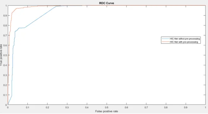
Şekil

Benzer Belgeler
Kahire'deki İngiliz Yüksek Komiseri Mısır hanedamm 'Taht kabul edilmediği takdirde, Kahire'de bir otelde kalmakta olan Ağa H an'm Mısır Kralı yapılacağı' tehdidiyle,
ALLAHIN EMRİ İLE PEVGAMBEßlN KAVLİ ]|_E ŞİRİN j-IANIM KI ZIMIZ) OĞLUMUZ FER HADA ISTI YO DUZ... BUYUCUN
Bir kişi dahi ortaya çıkıp Mustafa Kemal'in seninle yakınlaş masından söz etmedi" diye üs teleyince "Kitabın ses getirmesi için gerekti" şeklindeki yanıtı
Anıtlar Yüksek Kurulu'nun restorasyon çalışmasına onay vermesi halinde mart ayı başında hizmete açılacak kulede, çay 350 - 500 bin lirayı aşmayacak.. Turizm
Adana-Mersin Demiryolu yapılırken. Valinin konuş masından sonra dualarla tö ren başladı. Ne var ki uzak tan istasyona soluyarak giren lokomotifi gören halk çil
Arap memleketleri bile b> matemi Müslüman dünyasına maı ederek teessürümüze iştirâk eder lerken bizim bu gafletimizin ma.. zur görülecek tarafı kalır
Basın Müzesi'nin kuruluş çalışmalarına katılan ve arşivindeki pek çok belge ve yayın koleksiyonunu müzeye devreden tarihçi yazar Orhan Koloğlu (altta); bu
Diğer taraftan kırsal alanda daha çok kadının tarımdan arta kalan zamanlarında çoğunlukla tarımsal hammaddeyi değerlendirerek geleneksel bir üretim tekniği
