Introduction
A hydatidiform mole (HM) is a gestational trophoblas-tic tumor (GTN) originating from the placental site with a potential for local invasion and distant spread. The HM is classified as complete (CHM) and partial (PHM) sub-types according to the histopathological criteria [1]. Sialic acid (SA) is a generic term for derivatives of neu-raminic acid [2]. SA plays an important role in ensuring the proper and healthy functioning of biological systems. In humans, the alteration of SA levels is known to be as-sociated with various disorders and conditions such as cardiovascular diseases, inflammatory diseases, en-docrine diseases, and neurologic diseases. Free SA is very rarely observed in organisms [3]. Sialic acid levels are increased in certain types of cancer [4]. Increases in the levels of total serum SA (TSA) and lipid-bound SA (LSA) have been observed in various pathologies such as advanced ovarian cancer [5], cervical cancer [6], breast cancer [7] and endometrial cancer [8]. To the best of the present authors’ knowledge, SA activities have not been reported previously in patients with HM in the Eng-lish literature. In this preliminary study, they aimed to in-vestigate whether HM disease with malignant potential is significantly associated with increased SA levels.
Materials and Methods
Between April 2009 and November 2009, a total of 114 women who were admitted to Gaziantep University, Faculty of Medicine, Obstetrics and Gynecology Outpatient Clinic were prospectively analyzed. Patients were divided into three groups including HM (Group 1, n=34), control group including non-pregnant healthy patients (Group 2, n=42), and another control group including healthy pregnant patients with a single viable fetus within the first trimester of pregnancy (Group 3, n=38). Informed consents were obtained from the patients. The study protocol was approved by the Ethics Committee for Clinical Research of Gaziantep Univer-sity, Faculty of Medicine.
The diagnosis of HM (complete or partial HM) was based on histopathological examination of the molar tissue samples, which were taken by suction curettage under mask anesthesia. Blood samples (six ml of whole blood) were drawn from the cubital vein into regular tubes. The samples were centrifuged at 1,600 r.p.m. for five minutes and stored at -80 °C until used.
Serum SA operating procedure
Serum-free SA levels were measured using the Sialic Acid Quantitation Kit, a commercial enzymatic, photometric method in accordance with manufacturer instructions.
Test Principle:
First, N-Acetylneuraminic acid (NANA) aldolase catalyzes the reversible reaction (Reaction 1):
1) NANA £ N-Acetylmannosamine + pyruvic acid The pyruvic acid can be reduced to lactic acid, β-NADH, and lactic dehydrogenase (Reaction 2)
2) Pyruvic acid + β-NADH £ Lactic acid + β-NAD Under the proper conditions, the first forward reaction pre-dominates, and when coupled with β-NADH, the reaction is
com-Revised manuscript accepted for publication February 17, 2015
Evaluation of sialic acid levels in patients
with hydatidiform mole: a preliminary study
H.Ç. Özcan1, E. Öztürk2, S. Sucu3, M.G. Uğur1, I. Kutlar1, Ö. Balat1, B. Erbağci41 Gaziantep University, School of Medicine, Department of Obstetrics and Gynecology, Gaziantep 2 Medipol University, School of Medicine, Department of Obstetrics and Gynecology, Istanbul 3 Gaziantep Cengiz Gokcek Maternity Hospital, Department of Obstetrics and Gynecology, Gaziantep
4 Gaziantep University, School of Medicine, Department of Biochemistry, Gaziantep (Turkey)
Summary
Purpose of investigation: This study aims to investigate whether hydatidiform mole (HM) disease with malignant potential is sig-nificantly associated with increased sialic acid (SA) levels. Materials and Methods: A total of 114 women were enrolled in this study. Patients were divided into three groups including HM (Group 1, n=34), control group including non-pregnant healthy patients (Group 2, n=42), and another control group including healthy pregnant patients within 12 weeks of gestation (Group 3, n=38). Serum-free SA levels were measured. Results: There was a statistically significant difference in serum-free SA levels among the groups (p ≤ 0.001). Patients with HM had significantly higher levels compared to the control groups. Conclusion: The present study results showed that there was a significant correlation between HM and serum SA level.
Key Words: Hydatidiform mole; Sialic acid; Pregnancy.
CEOG
Clinical and ExperimentalObstetrics & Gynecology
7847050 Canada Inc. www.irog.net
Clin. Exp. Obstet. Gynecol. - ISSN: 0390-6663 XLIII, n. 3, 2016
H.Ç. Özcan, E. Öztürk, S. Sucu, M.G. Uğur, I. Kutlar, Ö. Balat, B. Erbağci 415
pleted. β-NADH oxidation at 340 nm and NANA samples can be accurately measured spectrophotometrically.
Statistical analysis
Statistical analysis was performed using SPSS v17.0 software. Categorical variables were expressed in number and percentage, while continuous variables were expressed in mean and standard deviation (SD) (in minimum and maximum, if necessary). A one-way analysis of variance (ANOVA) was used for the comparison of continuous variables when the assumptions were met, whereas the Kruskal-Wallis test was carried out when the assumptions were not met. The t-test or Mann-Whitney U test performed used to com-pare differences between two independent groups with Bonferroni correction. The Spearman’s correlation coefficient was used to as-sess the strength of the correlation among these continuous vari-ables producing abnormal distribution. A p value of < 0.05 was considered statistically significant.
Results
There was no difference in mean age between the sub-jects (p = 0.256).
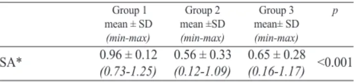
Serum SA levels were significantly higher in the HM group (Group 1) than compared to the controls (Group 2 and Group 3; p = 0.001) (Table 1). In the HM group, 23 subjects had CHM, while 11 had PHM. There was no difference in SA levels between the CHM and PHM subgroups (Table 2). Discussion
A HM has a potential for local invasion (15%) and distant metastasis (4%) [9]. Normal trophoblasts exhibit a broad range of synthetic activities, including the synthesis of steroid hor-mones and glycoproteins. The most effective approach for identifying GTN is assessing the level of hCG. The serum and urine hCG levels are closely parallel to the number of viable tumor cells. As the level of hCG is associated with the activ-ity of viable tumor cells, monitoring increases or decreases in hCG levels is more important than employing radiological or
other diagnostic methods. The monitoring of the disease or the planning of treatment does not require histological diagnosis; as such, decisions regarding the monitoring/treatment ap-proach are based on hCG values [10]. SA can be found freely or bound to proteins and lipids (gangliosides) at the terminal positions of oligosaccharides [11]. SAs are generally located in the inner and outer surfaces of lysosomal membranes as well as at the terminal positions of the main and side chains of oligosaccharides. SA, therefore, is the primary molecule, which is encountered by biochemical compounds, which in-teracts with cells and other cells. This feature of SA and its being negatively charged at physiological pH are directly as-sociated with the functions of SA in the organism [12]. Poly-sialic acid (PolySia) is an anionic large homopolymer composed of α-2,8-linked SA residues that are normally found on cell surfaces. PolySia, which has been subject to consider-able study in the nervous system, is involved in the modulation of cell development (by increasing cell migration), and in the regulation cell differentiation. PolySia is also observed in the immune cells of children and adults, and serves as an indica-tor for numerous cancer pathologies [13]. Hromatka et al. [13] identified PolySia in the trophoblast of human placenta. Cy-totrophoblasts and syncytiotrophoblasts express PolySia dur-ing the initial trimester of pregnancy, although PolySia expression gradually decreases as the pregnancy progresses. In a model of chorionic villous growth, it has been observed that PolySia causes the migration of cytotrophoblasts. Further-more, in an in vitro model, it was determined that removing PolySia from the cytotrophoblasts’ environment had the effect of decreasing their ability to penetrate and invade basement membranes. In addition, biopsies from patients with gesta-tional trophoblastic pathologies (such as malignant choriocar-cinomas and benign molar pregnancies) exhibited overexpression of PolySia. These findings suggest that Poly-Sia assumes an active and functional role in the normal de-velopment of the human placenta, and that PolySia is also involved in the invasion of trophoblast tumors. In this context, the aim of the present study was to demonstrate clinically the strong relationship between SA and trophoblast tumors.
The SA level was increased in parallel to increased tumor burden and degree of metastatic diseases [4]. Some studies demonstrated that SA levels might be increased in cancer patients without clinical symptoms [14]. In addition, many studies showed that measurement of SA level might be used in cancer patients in the assessment of progression and re-gression of the disease, when combined with other bio-markers, particularly. The function of SA as a tumor biomarker is associated with abnormal glycolization of can-cer cell membranes through the activation of new glycosyl-transferases, a specific characteristic of tumor cells. The role of SA in the distant metastasis is associated with the increase in the capacity of endothelial binding [15]. There are also some studies in the literature conducted on this subject.
Shimizu et al. [16] observed that serum SA levels were increased in gynecologic tumors including myomas, benign Table 1. — SA levels among the groups.
Group 1 Group 2 Group 3 p
mean ± SD mean ±SD mean± SD
(min-max) (min-max) (min-max)
SA* 0.96 ± 0.12 0.56 ± 0.33 0.65 ± 0.28 <0.001 (0.73-1.25) (0.12-1.09) (0.16-1.17)
*SA Unit: µmol/L
Table 2. — SA levels of complete and partial mole preg-nancies.
Group 1 - C Group 1 - P Group 2 Group 3 p
mean ± SS mean ± SS mean ± SS mean ± SS
(min-max) (min-max) (min-max) (min-max)
SA* 0.98 ± 0.11 0.91 ± 0.13 0.56 ± 0.33 0.65 ± 0.28 0.301 (0.86-1.24) (0.73-1.25) (0.12-1.09) (0.16-1.17)
Evaluation of sialic acid levels in patients with hydatidiform mole: a preliminary study
416
ovarian tumors, cervical cancer, endometrial cancer, and ovarian cancer using an enzymatic method. Cancer patients with poor prognosis exhibited considerably higher SA lev-els than cancer patients with good prognosis, regardless of the type of treatment being employed. In addition, higher SA levels were indicative of the cancer’s clinical course. For patients receiving combination therapies and who re-quire monitoring for extended periods of time, achieving effective follow-up by using tumor markers alone may prove extremely difficult. However, SA will serve as a use-ful marker even for the follow-up of such patients, since it is a non-specific marker for cancers of different histologies. Similarly, Yue et al. [17] reported that serum LSA was more cost-efficient and easy-to-use than Ca125 and might be used in patients with ovarian cancer. In another study in-vestigating a biochemical index for diagnosis and treatment of cervical cancer, Patel et al. [18] measured serum TSA, LSA, and lactate dehydrogenase using high-specific spec-trophotometric methods. Compared to the control group, the level of all markers were significantly higher (p ≤ 0.001) in the cervical cancer group. No significant changes were observed in the markers for early (1-2) and advanced (3-4) stages. Among patients who were non-responsive to radiotherapy, the TSA and LSA values were significantly higher compared to patients responsive to radiotherapy (p < 0.05 and p < 0.01, respectively). The authors reported that TSA was the most sensitive biomarker (90.74%) and might be helpful to identify patients with cervical cancer and follow their treatment responses, when combined with other biomarkers. On the other hand, Vivas et al. [5] con-cluded that TSA and LSA levels were not a contributing factor to early diagnosis of cervical cancer or clinical stag-ing of the tumor. In the present study, the authors demon-strated significant differences in serum SA levels among the HM group, healthy pregnant controls, and non-pregnant controls. Serum SA levels were significantly higher in the HM group, compared to control groups. However, they ob-served no significant differences in SA levels between the CHM and PHM subgroups.
Conclusion
In conclusion, SA level measurement alone appears to have a limited value in the preliminary diagnosis of a ma-lignant disease. On the other hand, SA level may be help-ful in the assessment of the progression and regression of a disease during therapy, when combined with other mark-ers such as HCG, particularly. To become clinically useful, however, assay methods need to be refined. To confirm these views, the present authors have planned a second prospective study that will demonstrate the importance of SA levels in potentially invasive and metastatic molar pa-tients, and which will demonstrate the response to admin-istered chemotherapeutic medications based on variations in SA levels.
Acknowledgements
This study was granted by the project research unit of Gaziantep University (project number TF. 09. 21)
References
[1] Berkowitz R.S., Goldstein D.P.: “Current management of gestational trophoblastic diseases”. Gynecol. Oncol., 2009, 112, 654. [2] Varki A.: “Diversity in the sialic acids”. Glycobiology 1992, 2, 25. [3] Reuter G., Gabius H.J.: ‘Sialic acids structure-analysis-
metabolism-occurrence-recognition”. Biol. Chem. Hoppe Seyler, 1996, 377, 325. [4] Sillanaukee P., Ponnio M., Jaaskelainen I.P.: “Occurrence of sialic
acids in healthy humans and different disorders”. Eur. J. Clin.
In-vest., 1999, 29, 413.
[5] Schutter E.M., Visser J.J., van Kamp G.J., Mensdorff-Pouilly S., van Dijk W., Hilgers J., Kenemans P.: “The utility of lipid-associ-ated sialic acid (LASA or LSA) as a serum marker for malignancy. A review of the literature”. Tumour Biol., 1992, 13, 121.
[6] Vivas I., Spagnuolo L., Palacios P.: “Total and lipid-bound serum sialic acid as markers for carcinoma of the uterine cervix”. Gynecol.
Oncol., 1992, 46, 157.
[7] Romppanen J., Eskelinen M., Tikanoja S., Mononen I.: “Total and lipid-bound serum sialic acid in benign and malignant breast dis-ease”. Anticancer Res., 1997, 17, 1249.
[8] Paszkowska A., Berbec H., Semczuk A., Cybulsk M.: “Sialic acid concentration in serum and tissue of endometrial cancer patients”.
Eur. J. Obstet. Gynecol. Reprod. Biol., 1998, 76, 211.
[9] Berkowitz R.S., Goldstein D.P.: “Chorionic tumors”. N. Engl. J.
Med., 1996, 335, 1740.
[10] Tyrey L.: “Human chorionic gonadotropin: properties and assay methods”. Semin. Oncol., 1995, 22, 121.
[11] Taniuchi K., Chifu K., Hayashi N., Nakamachi Y., Yamaguchi N., Miyamoto Y., et al.: “A new enzymatic method for the determination of sialic acid in serum and its application for a marker of acute phase reactants”. Kobe J. Med. Sci., 1981, 27, 91.
[12] Schauer R., Kelm S., Reuter G., Roggentin P., Shaw L.: “Biochem-istry and role of sialic acids”. In: Rosenberg A. (ed). Biology of the
sialic acids. New York: Plenum, 1995, 7.
[13] Hromatka B.S., Drake P.M., Kapidzic M., Stolp H., Goldfien G.A., Shih IeM., Fisher S.J.: “Polysialic acid enhances the migration and invasion of human cytotrophoblasts”. Glycobiology, 2013, 23, 593. doi: 10.1093/glycob/cws162. Epub 2012 Dec 3.
[14] Gatchev O., Rastam L., Lindberg G., Gullberg B., Eklund G.A., Törnberg S.: “Tumors of the central nervous system and serum sialic acid concentration in men and women”. Br. J. Cancer, 1993, 68, 425. [15] Narayanan S.: “Sialic acid as a tumor marker”. Ann. Clin. Lab. Sci.,
1994, 24, 376.
[16] Shimizu Y., Hasumi K., Masubuchi K., Okudaira Y.: “Management of patients with gynecologic cancer by serum sialic acid determina-tion”. Gynecol. Oncol., 1989, 33, 231.
[17] Yue K., Bian M., Zhu D., Liu W., Siu S.: “Serum lipid-associated sialic acid (LSA) in diagnosing and monitoring ovarian cancer”.
Zhongguo Yi Xue Ke Xue Yuan Xue Bao, 1995, 17, 128.
[18] Patel P.S., Rawal G.N., Balar D.B.: “Importance of serum sialic acid and lactate dehydrogenase in diagnosis and treatment monitoring of cervical cancer patients”. Gynecol. Oncol., 1993, 50, 294.
Address reprint requests to: H. ÇAĞLAYAN ÖZCAN, M.D.
Gaziantep University, Faculty of Medicine Department of Obstetrics and Gynecology En route to Kilis
Sahinbey, Gaziantep 27010 (Turkey) e-mail: [email protected]