Oxidative stress markers are increased in non-alcoholic fatty liver
disease patients with high serum alanine aminotransferase levels
Podwyższenie wartości markerów stresu oksydacyjnego u pacjentów
z niealkoholową stłuszczeniową chorobą wątroby z wysokim stężeniem
aminotransferazy alaninowej w surowicy
Suzan Muratoglu Severcan
1, Ayse Bilgihan
2, Gulbanu Erkan
3, Ugur Ercin
2, Cinar Severcan
41Department of Medical Biochemistry, Institute of Health Sciences, Gazi University, Ankara, Turkey Head of the Department: Prof. Dr. Neslihan Bukan
2Department of Medical Biochemistry, Medical Faculty, Ufuk University, Ankara, Turkey Head of the Department: Prof. Dr. Ayse Bilgihan
3Division of Gastroenterology, Department of Internal Medicine, Istanbul Medipol University, Istanbul, Turkey Head of the Division: Prof. Dr. Esin Korkut
4Department of Biochemistry, Faculty of Pharmacy, Zonguldak Bulent Ecevit University, Zonguldak, Turkey Head of the Department: Assist. Prof. Ayse Ceylan Hamamcioglu
Medical Studies/Studia Medyczne 2019; 35 (4): 283–287 DOI: https://doi.org/10.5114/ms.2019.91245
Key words: inflammation, oxidative stress, alanine aminotransferase, non-alcoholic fatty liver disease.
Słowa kluczowe: stan zapalny, stres oksydacyjny, aminotransferaza alaninowa, niealkoholowa stłuszczeniowa choroba wątroby.
Abstract
Introduction: Non-alcoholic fatty liver disease (NAFLD) is a pathological disease that causes chronic damage in the liver without alcohol consumption.
Aim of the research: To determine oxidative stress and inflammation statuses (NAFLD) with increased alanine aminotrans-ferase (ALT).
Material and methods: Oxidative stress (malondialdehyde (MDA), advanced oxidation protein products (AOPP), inflamma-tion (serum tumour necrosis factor-α (TNF-α)), interleukin-6 (IL-6), and hyaluronic acid (HA) were measured by the ELISA method or manually. A total of 133 non-obese and non-diabetic individuals were included. Patients diagnosed with NAFLD with normal ALT levels were included in group 1 (n = 53), and those with high ALT levels were included in group 2 (n = 35). The control group included individuals without any known systemic disease (n = 45).
Results: We found that the serum MDA levels were significantly elevated in group 2 in comparison to the control group (p < 0.05). However, there were no significant changes in the serum AOPP levels in any group (p > 0.05). The serum TNF-α and IL-6 levels of group 1 and group 2 were higher than those in the control group (p < 0.05). The serum HA levels in group 2 increased significantly in comparison to the other groups (p < 0.05).
Conclusions: HA and MDA were enhanced with high ALT levels and with elevated TNF-α and IL-6 levels without high ALT levels in NAFLD.
Streszczenie
Wprowadzenie: Niealkoholowa stłuszczeniowa choroba wątroby (NAFLD) jest schorzeniem wywołującym przewlekłe uszko dzenie wątroby, którego przyczyną są inne czynniki niż nadmierne spożycie alkoholu.
Cel pracy: Oznaczenie stresu oksydacyjnego i występowania stanu zapalnego u pacjentów z NAFLD z podwyższoną aktywnością aminotransferazy alaninowej (ALT).
Materiał i metody: Markery stresu oksydacyjnego (aldehyd dimalonowy (MDA) i zaawansowane produkty utleniania białek (AOPP)) oraz stanu zapalnego (czynnik martwicy nowotworów α (TNF-α)), interleukina-6 (IL-6) i kwas hialuronowy (HA)) w surowicy oznaczano testem ELISA lub metodą manualną. Do badania włączono ogółem 133 osoby bez nadwagi i cukrzycy. Pacjentów z rozpoznaniem NAFLD, u których stężenie ALT było prawidłowe, włączono do grupy 1. (n = 53), natomiast chorych z NAFLD i wysokim stężeniem ALT – do grupy 2. (n = 35). Grupa kontrolna obejmowała osoby, u których nie występowały żadne znane choroby ogólnoustrojowe (n = 45).
Wyniki: Stwierdzono, że stężenia MDA w surowicy w grupie 2. są istotnie podwyższone w porównaniu z grupą kontrolną (p < 0,05). Nie odnotowano natomiast istotnych zmian w stężeniach AOPP w surowicy w żadnej z grup (p > 0,05). Stężenia
TNF-α i IL-6 w surowicy w grupie 1. i 2. były wyższe niż w grupie kontrolnej (p < 0,05). Stężenie HA w surowicy w grupie 2. było istotnie podwyższone w stosunku do pozostałych grup (p < 0,05).
Wnioski: W badaniu wykazano podwyższone stężenia HA i MDA u pacjentów z NAFLD z wysokim stężeniem ALT oraz podwyższone stężenia TNF-α i IL-6 u osób z NAFLD z prawidłowym stężeniem ALT.
Introduction
Non-alcoholic fatty liver disease (NAFLD) causes chronic damage in the liver [1]. NAFLD is a pathologi-cal condition that is seen in persons who do not drink alcohol but are still susceptible to fat accumulating around the liver. The pathogenesis of NAFLD is mul-tifactorial, which explains the “two-hit” traditional hypothesis. The first hit consists of aggregation of fatty acids/triglycerides (TGs) in the liver and impair-ment of insulin resistance (IR) response. The second hit is increased oxidative stress and inflammatory response. Eventually, two hits result in liver injury [2, 3]. An increased serum alanine aminotransferase (ALT) level is an indicator of NAFLD if auto-immune diseases, viruses, and medicines are excluded. How-ever, in some patients, despite the normal ALT levels, these diseases may be seen in histopathology [4].
Lipid peroxidation begins by removing hydrogen atoms contained in the α-methylene group of poly-unsaturated fatty acid chains from the membrane structure, as a result of the free radical effect. Lipid peroxidation is terminated with lipid hydroperoxide that is converted to aldehydes and other carbonyl compounds. The level of one of these compounds, malondialdehyde (MDA), is measured by the thio-barbituric acid test, and this method is often used to detect lipid peroxide levels [5]. A membrane pro-tein that has an enzymatic function and the recep-tor are sensitive to free radical modification. This causes protein oxidation with cell and membrane defects. Determination of protein carbonyl is widely used to recognise the damage to the protein struc-ture. Advanced oxidation protein products (AOPP) are a marker of protein oxidation and defined as cross-linked protein products that contain dityro-sine. They are known as valuable markers of oxida-tive protein modifications [6]. The oxidaoxida-tive stress increases with advanced levels of NAFLD, and it is observed that it has a role in the disease’s steatosis and advancement into steatohepatitis [7]. Hyal-uronic acid (HA) is a natural polysaccharide, which is made out of the disaccharide units of glucuronic acid and N-acetylglucosamine. It is stated that HA may be beneficial in distinguishing people who have non-alcoholic fatty liver disease and those who have non-alcoholic steatohepatitis, and it is acknowledged that HA is an important parameter for showing liver fibrosis in chronic hepatitis [8].
TNF-α (26 kDa) is a transmembrane protein (TNF-α) in adipocytes. TNF-α regulates many inflam-matory and autoimmune processes. For persons suffer-ing from obesity, the elevated level of TNF-α depends
on hyperinsulinaemia, which induces insulin resis-tance in peripheral tissues [9]. Interleukin-6 (IL-6) is a cytokine synthesised by mononuclear phagocytes, vascular endothelial cells, fibroblasts, epithelial cells, and activated T cells [10]. IL-6 is significantly correlated with increasing visceral fat and insulin resistance, and it independently predicts increasing hepatic inflamma-tion. Interleukin-6 and TNF-α are pro-inflammatory cytokines. It is considered that TNF-α constitutes the second hit, which is determinant in the transformation of fatty liver to progressive disease forms [11].
Aim of the research
This study classified NAFLD patients based on their increased ALT levels. The aim of this study was to determine the oxidative stress and inflammation statuses in NAFLD by examining MDA, AOPP, TNF-α, IL-6, and HA levels. These parameters play some roles in the pathogenesis and progression of this disease.
Material and methods
This study included a total of 133 non-obese and non-diabetic individuals who visited clinics with a dyspeptic symptom or for routine check-up proce-dures. The groups were categorised based on their serum ALT levels and ultrasonography findings. The groups were formed as follows: patients diagnosed with non-alcoholic fatty liver disease with normal ALT levels as were defined as group 1 (n = 53); patients diagnosed with non-alcoholic fatty liver disease with high ALT levels as group 2 (n = 35); and the control group included individuals without any known sys-temic disease (n = 45). The inclusion criteria were as follows: presence of hepatosteatosis in USG for groups 1 and 2; the presence of negative markers such as vi-ral, autoimmune, and metabolic liver diseases; and no ethanol intake. The exclusion criteria included dia-betes mellitus, drugs that can lead to type 2 diadia-betes (steroids), and drugs that can cause hepatosteatosis (e.g. steroids, oral contraceptives, methotrexate, tet-racycline, amiodarone). The serum level of ALT was measured by using an autoanalyser in routine bio-chemistry laboratory.
Lipid peroxidation determines the reaction that occurs between MDA and thiobarbituric acid under acidic conditions, and the pink-coloured product is measured spectrophotometrically at 532 nm. MDA con-centration was recorded in units of “nmol MDA/ml”. The AOPP level was determined by spectrophotomet-ric methods. Procedures were performed for chlori-nated oxidants and crosslinking products of serum
protein, which are oxidised by potassium iodide. Next, the absorbance of the released chemical potassium io-dide was immediately measured against the PBS solu-tion at 340 nm. Chloramine-T solusolu-tions had levels of concentration from 6.25 μmol/l to 100 μmol/l. These figures were used in drawing a standard curve. AOPP concentrations were found in chloramine-T equiva-lents, measured in μmol/l units. The coefficient of variation HA (Cusabio, catalogue no: CSB-E04805h), TNF-α (Diasource, catalogue no: KAP 1751), and IL-6 (Diasource, catalogue no: KAP1261) serum levels of all patients were studied by using the ELISA method.
Statistical analysis
SPSS (Statistical Package for the Social Sciences) version 18.0 was used to analyse the data, which were obtained and tabulated. Shapiro-Wilk test was used to test the parametric test conditions. The data showed non-parametric test conditions according to the Shapiro-Wilk test. Consequently, the Mann-Whit-ney U test, which is a non-parametric test, was chosen to compare two independent groups. In all statisti-cal analyses, the significance level was accepted as
p ≤ 0.05. Correlation analyses were performed using Spearman’s correlation test.
Results
Table 1 shows the mean and standard deviation values of the demographic variables, and Table 2 shows the mean and standard deviation values for the biochemical variables of all groups.
The mean ages of the control, group 1, and group 2 were 34.75, 56.28, and 45.06, the mean body mass in-dex (BMI) values of these groups were 22.57, 23.36, and 24.25 kg/m2, and the mean ALT levels of these groups
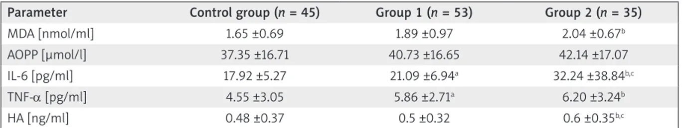
were 17.71, 22.96, and 72.91, respectively (Table 1). The serum MDA levels increased significantly in group 2 (high level of ALT) in comparison to the con-trol group (p = 0.012), while there was no significant difference between group 1 (low level of ALT) and group 2 (p > 0.05). There were no significant chang-es in the AOPP levels amongst the groups (p > 0.05). The serum TNF-α levels in groups 1 and 2 were sig-nificantly higher than those in the control group (p = 0.01 and p = 0.011, respectively). However, there
was no significant difference between groups 1 and 2 (p > 0.05). A significant change in the serum IL-6 lev-els was also found amongst all groups (p = 0.001). The serum IL-6 levels increased significantly in groups 1 and 2 in comparison to the control group (p = 0.01 and
p = 0.001, respectively), and the values in group 2 were significantly higher than those in group 1 (p = 0.001). A significant rise of HA levels was measured in group 2 in comparison to the control group and group 1 (p = 0.002 and p = 0.038, respectively) (Table 2).
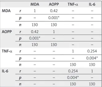
On other hand, according to the Spearman’s cor-relation test results, the MDA and AOPP levels in-creased simultaneously. There was a strong correla-tion between the MDA and AOPP levels (r2 = 0.422, p < 0.01). There was also a strong correlation between the TNF-α and IL-6 levels (r2 = 0.422, p < 0.01) (Table 3).
Discussion
Major metabolites (glucose, fatty acids, and cho-lesterol) play a key role in the well-being of the liver. Obesity as a result of poor dietary habits leads to in-creased insulin resistance of the peripheral tissues, which causes an accumulation of fat in the liver [12– 14]. As a result of this, NAFLD may spread [14–17]. NAFLD is a disorder that is frequently found within blood abnormalities. Younossi et al. [18] determined high aminotransferase enzyme levels in the US gener-al population, measured at 1% to 9%, and the majority of patients who were diagnosed with hypertransami-nasaemia had levels at 3% to 5% and reported symp-toms of NAFLD. NAFLD is generally associated with insulin resistance, which leads to type 2 diabetes and obesity. Ghamar-Chehreh et al. [19] reported that, in NAFLD, increased levels of ALT were not correlated Table 1. List of mean levels of demographic characters of the all groups
Parameter Control group
(n = 45) Group 1 (n = 53) Group 2 (n = 35)
Age 34.75 56.28 45.06
BMI 22.57 23.36 24.25
ALT [IU/l] 17.71 22.96 72.91 N – number of patients, BMI – body mass index, ALT – alanine ami-notransferase.
Table 2. List of mean ± standard deviation levels and the coefficient of variations biochemical values of all groups. Parameter Control group (n = 45) Group 1 (n = 53) Group 2 (n = 35)
MDA [nmol/ml] 1.65 ±0.69 1.89 ±0.97 2.04 ±0.67b
AOPP [µmol/l] 37.35 ±16.71 40.73 ±16.65 42.14 ±17.07
IL-6 [pg/ml] 17.92 ±5.27 21.09 ±6.94a 32.24 ±38.84b,c
TNF-α [pg/ml] 4.55 ±3.05 5.86 ±2.71a 6.20 ±3.24b
HA [ng/ml] 0.48 ±0.37 0.5 ±0.32 0.6 ±0.35b,c
n – number of patients, asignificance p ≤ 0.05; difference between control group and group 1, bsignificance p ≤ 0.05; difference between
with dyslipidaemia but showed a correlation with insulin resistance. In livers with progressed damage, reduced hyaluronate receptors of sinusoidal endo-thelial cells induce high HA levels. Serum HA levels are a predictor of the progression of hepatic fibrosis. Accordingly, determining serum HA is a good mark-er to help diagnose hepatic fibrosis [20]. Suziki et al. [21] and Pawitpok et al. [22] reported that serum HA and ALT levels increase together, and that the disease changes from steatosis to fibrosis.
According to our results, the serum HA levels in this study significantly increased (p < 0.05) in group 2 in comparison to the control group and group 1. It was concluded that elevated ALT levels may induce HA without diagnosis of fibrosis in NAFLD. Logu-ercio et al. [23] stated that inflammation parameters such as TNF-α and IL-6 increased significantly, along with the insulin resistance in NAFLD patients who had abnormally high serum ALT levels. In parallel to this, Jang et al. also showed increased ALT levels with inflammation markers [24]. Nevertheless, Wong
et al. [25] did not mention any change reagrding an
increase in ALT levels with TNF-α and IL-6. In this study, the TNF-α and IL-6 levels increased signifi-cantly in groups 1 and 2 in comparison to the control group (p < 0.05). The IL-6 levels of group 2 were also measured, and it was noticed that they significantly increased in comparison to group 1 (p < 0.05). These results suggested that NAFLD may trigger a high level of inflammation, whereas increases in the ALT levels did not stimulate inflammatory parameters. In other words, these findings showed that TNF-α and IL-6 levels increased together (p < 0.01).
The levels of lipid peroxidation products are im-portant in determining steatosis. Araya et al. [26] re-ported that the serum MDA levels increased signifi-cantly in comparison to the control group of steatosis
and non-alcoholic steatohepatitis (NASH) patients, but there was no significant correlation in the serum ALT levels between these groups. Kumar et al. [27] reported that the serum levels of MDA were signifi-cantly higher among NAFLD patients with raised ALT levels in comparison to the control groups. Mousavi
et al. [28] found similar results in a rat model. There
was a contradiction between these studies in terms of the ALT levels. According the results in this study, while there were significantly increased serum MDA levels in group 2 in comparison to the control group, we did not find any significant difference with group 1. This result showed that elevated ALT levels in NAFLD may increase lipid peroxidation. Protein damage oc-curs as a result of oxidative stress, as well as lipid per-oxidation, in a fatty liver. There are a few studies in the literature on the AOPP levels in fatty liver disease. Nevertheless, different protein carboxylation and oxidation products were reported to increase [26, 29]. Araya et al. [26] observed that protein carboxylation increased significantly in the control group of NAFLD and NASH patients, but they could not measure any significant correlation between these groups. Videla
et al. [29] showed that protein oxidation increased
sig-nificantly in NAFLD patients in comparison to NASH and the control group. In our study, it was not possible to observe the significance of the differences of the AOPP levels of groups 1 and 2 in comparison to the control group (p > 0.05). However, there were strong positive correlations between MDA and AOPP (p < 0.01). Both MDA and AOPP levels increased together in NAFLD.
Conclusions
This study showed that HA and MDA were en-hanced with high ALT levels and on elevated TNF-α and IL-6 levels without high ALT levels in NAFLD.
HA and MDA levels may be used as valuable mark-ers for the diagnosis of NAFLD. Further studies are needed to clarify high ALT levels and biochemical pa-rameters in the pathogenesis of NAFLD.
Acknowledgments
This project was approved by Gazi Univer-sity Board of Local Ethics under the code G.Ü. ET. 070220122 and was supported financially by Gazi University Scientific Research Projects Unit under the project code 01/2012-14.
Conflict of interest
The authors declare no conflict of interest. References
1. Fiorucci S, Biagioli M, Distrutti E. Future trends in the tre-atment of non-alcoholic steatohepatitis. Pharmacol Res 2018; 134: 289-298.
Table 3. Correlation analyses among the parameters
MDA AOPP TNF-α IL-6
MDA r 1 0.42 – – p – 0.001* – – n 130 130 – – AOPP r 0.42 1 – – p 0.001* – – – n 130 130 – – TNF-α r – – 1 0.254 p – – – 0.004* n – – 130 130 IL-6 r – – 0.254 1 p – – 0.004* – n – – 130 130
2. Del Ben M, Polimeni L, Baratta F, Pastori D, Angelico F. The role of nutraceuticals for the treatment of non-alcoholic fatty liver disease. Br J Clin Pharmacol 2017; 83: 88-95. 3. Engin A. Non-alcoholic fatty liver disease. Adv Exp Med
Biol 2017; 960: 443-467.
4. Tolman KG, Fonseca V, Ton MH, Dalplaz A. Narrative review: hepatobiliary disease in type 2 diabetes mellitus. Ann Intern Med 2004; 141: 946-956.
5. Bellanti F, Villani R, Facciorusso A, Vendemiale G, Servid-dio G. Lipid oxidation products in the pathogenesis of non-alcoholic steatohepatitis. Free Radic Biol Med 2017; 111: 173-185.
6. Cristani M, Speciale A, Saija A, Gangemi S, Minciullo PL, Cimino F. Circulating advanced oxidation protein pro-ducts as oxidative stress biomarkers and progression me-diators in pathological conditions related to inflamma-tion and immune dysregulainflamma-tion. Curr Med Chem 2016; 23: 3862-3882.
7. Spahis S, Delvin E, Borys JM, Levy E. Oxidative stress as a critical factor in nonalcoholic fatty liver disease patho-genesis. Antioxid Redox Signal 2017; 26: 519-541. 8. Gudowska M, Cylwik B, Chrostek L. The role of serum
hyaluronic acid determination in the diagnosis of liver fibrosis. Acta Biochim Pol 2017; 64: 451-457.
9. Dludla PV, Nkambule BB, Jack B, Mkandla Z, Mutize T, Silvestri S, Orlando P, Tiano L, Louw J, Mazibuko-Mbe- je SE. Inflammation and oxidative stress in an obese state and the protective effects of gallic acid. Nutrients 2018; 11: pii: E23.
10. Mauer J, Chaurasia B, Goldau J, Vogt MC, Ruud J, Nguy-en KD, Theurich S, HausNguy-en AC, Schmitz J, Brönneke HS, Estevez E, Allen TL, Mesaros A, Partridge L, Febbraio MA, Chawla A, Wunderlich FT, Brüning JC. Signaling by IL-6 promotes alternative activation of macrophages to limit endotoxemia and obesity-associated resistance to insulin. Nat Immunol 2014; 15: 423-430.
11. Farrell GC, Larter CZ. Nonalcoholic fatty liver disease: from steatosis to cirrhosis. Hepatology 2006; 43 (2 Suppl 1): 99-112.
12. Suliga E, Kozieł D, Cieśla E, Rębak D, Głuszek S. Dietary pat-terns in relation to metabolic syndrome among adults in poland: a cross-sectional study. Nutrients 2017; 9: pii: E1366. 13. Suliga E, Kozieł D, Cieśla E, Rębak D, Głuszek S. Coffee consumption and the occurrence and intensity of me-tabolic syndrome: a cross-sectional study. Int J Food Sci Nutr 2017; 68: 507-513.
14. Ashtari S, Pourhoseingholi MA, Zali MR. Non-alcohol fat-ty liver disease in Asia: prevention and planning. World J Hepatol 2015; 7: 1788-1796.
15. Kontogianni MD, Tileli N, Margariti A, Georgoulis M, Deutsch M, Tiniakos D, Fragopoulou E, Zafiropoulou R, Manios Y, Papatheodoridis G. Adherence to the Mediter-ranean diet is associated with the severity of non-alcoho-lic fatty liver disease. Clin Nutr 2014; 33: 678-683. 16. Shahroukh HD, Davoodi M, Rahmani O, Roushanaei A,
Seydeyan SM, Adel SMH. Association of nonalcoholic fat-ty liver with carotid intima-media thickness in patients with dyslipidaemia. Med Stud 2018; 34: 226-231. 17. Koo SH. Nonalcoholic fatty liver disease: molecular
me-chanisms for the hepatic steatosis. Clin Mol Hepatol 2013; 19: 210-215.
18. Younossi ZM, Koenig AB, Abdelatif D, Fazel Y, Henry L, Wymer M. Global epidemiology of nonalcoholic fatty
li-ver disease-meta-analytic assessment of prevalence, inci-dence, and outcomes. Hepatology 2016; 64: 73-84. 19. Ghamar-Chehreh ME, Amini M, Khedmat H, Moayed
Ala-vian S, Daraei F, Mohtashami R, Hadi R, Beyram BA, Taheri S. Elevated alanine aminotransferase activity is not associa-ted with dyslipidemias, but relaassocia-ted to insulin resistance and higher disease grades in non-diabetic non-alcoholic fatty li-ver disease. Asian Pac J Trop Biomed 2012; 2: 702-706. 20. Orasan OH, Ciulei G, Cozma A, Sava M, Dumitrascu DL.
Hyaluronic acid as a biomarker of fibrosis in chronic liver diseases of different etiologies. Clujul Med 2016; 89: 24-31. 21. Suzuki A, Angulo P, Lymp J, Li D, Satomura S, Lindor K.
Hyaluronic acid, an accurate serum marker for severe hepatic fibrosis in patients with non-alcoholic fatty liver disease. Liver Int 2005; 25: 779-786.
22. Pawitpok C, Kongtawelert P, Ua-arayaporn S, Punyarit P, Piy-anirun W, Chutaputti A. Efficacy of serum hyaluronic acid level for predicting liver fibrosis in patients with nonalcoho-lic fatty liver disease. Thai J Gastroenterol 2006; 7: 83-87. 23. Loguercio C, De Simone T, D’Auria MV, de Sio I, Federi-
co A, Tuccillo C, Abbatecola AM, Del Vecchio Blanco C. Non-alcoholic fatty liver disease: a multicentre clinical study by the Italian Association for the Study of the Liver. Dig Liver Dis 2004; 36: 398-405.
24. Jiang W, Wu N, Wang X, Chi Y, Zhang Y, Qiu X, Hu Y, Li J, Liu Y. Dysbiosis gut microbiota associated with in-flammation and impaired mucosal immune function in intestine of humans with non-alcoholic fatty liver dise-ase. Sci Rep 2015; 5: 8096.
25. Wong VW, Wong GL, Choi PC, Chan AW, Li MK, Chan HY. Disease progression of non-alcoholic fatty liver disease: a prospective study with paired liver biopsies at 3 years. Gut 2010; 59: 969-974.
26. Araya J, Rodrigo R, Videla LA, Thielemann L, Orellana M, Pettinelli P, Poniachik J. Increase in long-chain polyunsa-turated fatty acid n-6/n-3 ratio in relation to hepatic ste-atosis in patients with non-alcoholic fatty liver disease. Clin Sci 2004; 106: 635-643.
27. Kumar R, Prakash S, Chhabra S, Singla V, Madan K, Gupta SD. Association of pro-inflammatory cytokines, adipokines and oxidative stress with insulin resistance and non-alcoholic fatty liver disease. Indian J Med Res 2012; 136: 229-236.
28. Mousavi SN, Faghihi A, Motaghinejad M, Shiasi M, Iman-parast F, Amiri HL, Shidfar F. Zinc and selenium co-sup-plementation reduces some lipid peroxidation and an-giogenesis markers in a rat model of NAFLD-Fed high fat diet. Biol Trace Elem Res 2018; 181: 288-295.
29. Videla LA, Rodrigo R, Orellana M, Fernandez V, Tapia G, Quiñones L, Varela N, Contreras J, Lazarte R, Csendes A, Rojas J, Maluenda F, Burdiles P, Diaz JC, Smok G, Thiele-mann L, Poniachik J. Oxidative stress-related parameters in the liver of non-alcoholic fatty liver disease patients. Clin Sci 2004; 106: 261-268.
Address for correspondence: Suzan Muratoglu Severcan Department of Medical Biochemistry Institute of Health Sciences
Gazi University
06500 Besevler, Ankara, Turkey Phone: +90 55399837774