Factors affecting mortality among victims of electrical burns
Çağrı Tiryaki, M.D.,1 Mustafa Celalettin Haksal, M.D.,2 Murat Burç Yazıcıoğlu, M.D.,1 Ali Çiftçi, M.D.,1 Osman Esen, M.D.,3 Hamdi Taner Turgut, M.D.,1 Abdullah Yıldırım, M.D.,4 Murat Güven, M.D.5 1Department of General Surgery, Kocaeli Derince Training and Research Hospital, Kocaeli-Turkey2Department of General Surgery, Medipol University Faculty of Medicine, İstanbul-Turkey
3Department of Anesthesiology and Reanimation, Kocaeli Derince Training and Research Hospital, Kocaeli-Turkey 4Department of Burn Treatment Center, Kocaeli Derince Training and Research Hospital, Kocaeli-Turkey
5Department of Plastic and Reconstructive Surgery, Kocaeli Derince Training and Research Hospital, Kocaeli-Turkey
ABSTRACT
BACKGROUND: The aim of this study was to determine the factors affecting mortality rate among patients with an electrical burn. METHODS: A total of 115 patients admitted to the emergency department and hospitalized in the Burn Treatment Center or In-tensive Care Unit (ICU) due to the electrical burn, were included in the study.
RESULTS: A total of 115 patients (4 female and 111 male) with a mean age of 32.88±12.87 years were included in the study. The mean hospitalization period was 25.03±20.50 days, and the mean total body surface area burned (% TBSA) was 22.83±15.54%. Among those patients, 9 (8.5%) expired, and the remaining 106 were discharged after treatment. In a logistic regression analysis, TBSA >20% (p=0.02, OR: 11.7, CI: 1.38–99.16); ICU requirement (p=0.005, OR: 1.28, CI: 1.08–1.58); erythrocyte transfusion requirement (p=0.02, OR: 12.48, CI: 1.44–107.83); fresh frozen plasma (FFP) requirement (p=0.03, OR: 10.23, CI: 1.18–88.17); albumin requirement (p=0.02, OR: 12.60, CI: 1.44–109.85); admission serum albumin level <3.5 mg/dl (p=0.04, OR: 7.25, CI: 0.82–63.64); and admission hemoglobin level <12 mg/dl (p=0.01, OR: 8.29, CI: 1.57–43.61) were determined as risk factors for mortality in patients with electrical burns. CONCLUSION: In clinical practice, defining a mortality risk analyzer using these factors may be helpful in the management of pa-tients with electrical burns. Additional, more comprehensive studies are required to define the risk factors for mortality and long-term morbidities in patients with electrical burns.
Keywords: Albumin; burn extent; calcium; electrical burns; hemoglobin; mortality.
Electrical burns are most commonly reported among young men causing long-term hospitalization and high cost rates.
[5] They are still important causes of work-power loss with
high rates of morbidity and mortality especially in developing countries.[6] As high as 21.7% of the mortality rate has also
been reported in electrical burn patients.[7] Recently Ghavami
et al. investigated 682 electrical burn patients and reported the mortality rate as 2.5%, while those requiring amputation was 23.7%.[8]
The aim of this study was to determine the factors affecting mortality rates among the victims of an electrical burn. In this way, we aimed to draw attention to these points for the prompt and improved management of patients.
MATERIALS AND METHODS
This retrospective study was conducted in the Derince Train-ing and Research Hospital, between January 2012 and De-cember 2015. The study was approved by the local ethics committee. A total of 115 patients, admitted to the
emer-Address for correspondence: Murat Burç Yazıcıoğlu, M.D. Derince Eğitim ve Araştırma Hastanesi, Genel Cerrahi Kliniği, Kocaeli, Turkey
Tel: +90 262 - 319 50 79 E-mail: [email protected]
Ulus Travma Acil Cerrahi Derg 2017;23(3):223–229
doi: 10.5505/tjtes.2016.29166 Copyright 2017
TJTES
INTRODUCTION
Electrical burns are one of the causes of important health burdens throughout the world with incidences varying be-tween 4–18% of all burns.[1–3] In the pathogenesis, with the
passage of electric through the human body, the biochemi-cal compositions of the body are altered together with many electro-traumas. Necrosis on the skin and deeper tissues may cause the dysfunction of the affected limbs and life-threat-ening organ complications including renal failure or sepsis.[4]
gency department and hospitalized in the General Surgery Department or Intensive Care Unit (ICU) due to the electri-cal burns, was included in the study.
The total body surface area was calculated by the rule of nines, and the maximally affected burn area was also record-ed. Burns are graded as: Grade 1: the superficial thickness of the skin is involved; Grade 2: the full thickness of skin is destroyed; Grade 3: the skin, subcutaneous tissues, fat, and muscles are destroyed; Grade 4: the skin, subcutaneous tis-sues, and bones are destroyed.
After an initial assessment, intravenous fluid resuscitation was started with the monitoring of the urine output. Erythrocyte or fresh frozen plasma (FFP) was transfused when necessary. Surgical interventions including debridement, escharatomies, fasciotomies, or a flap coverage were also managed where required. Demographic data, the hospitalization period, the surgical interventions, and the laboratory data including a complete blood count, renal, and liver function tests, C-reactive protein, blood coagulation parameters, and serum electrolyte levels were recorded. The corrected calcium level was calculated based on the formula
Corrected calcium = serum calcium + 0.8 * (4 - serum albumin). Patients with incomplete records were not included in the study.
The Statistical Analysis
Statistical analyses of the results were performed using the SPSS software (version 21, IBM SPSS Statistics). The results were presented as a mean±SD for continuous variables and as a number and proportions (%) for categorical variables. The student’s t-test or the chi square test was used for the analyses. The risk level was assessed by a logistic regression analysis. A p value of less than 0.05 was considered statisti-cally significant.
RESULTS
A total of 115 patients (4 female and 111 male) with a mean age of 32.88±12.87 (16–82) years were included in the study. The accident took place at the home in 19 cases and at work in 81 cases, as well as a traffic accident in 1 case. In the re-maining 14 cases, the accident happened in other places. The patients were admitted to the hospital by ambulance (n=98), by their own cars (n=14) or by air ambulance (n=3).
Among those patients, 65 were directly hospitalized in the intensive care unit. A tracheotomy was required in one pa-tient. An inhalation injury accompanying an electric burn was present in one patient. The mean hospitalization period was 25.03±20.50 (1–163) days, and the mean burn extent was 22.83±15.54% (1.0–66.0%). Among those patients, 9 (8.5%)
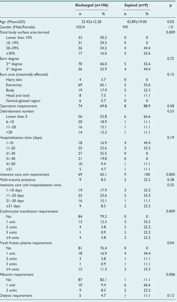
expired, and the remaining 106 were discharged after treat-ment. The general characteristics of the expired and dis-charged patients are summarized in Table 1.
The mean age of the expired group was older (42.89±19.00 vs 32.42±12.28 years, p=0.03); the mean total body surface area burned (% TBSA) was larger (40.75±18.79 vs 20.46±14.62, p=0.001); the ICU requirement was more common (100% vs 65.1%, p=0.05), and the mean of the required erythro-cyte suspensions (4.14±5.52 vs 1.19±2.53, p=0.01), the fresh frozen plasma (6.00±10.03 vs 2.47±5.38, p=0.04) and the al-bumin (1.62±1.84 vs 0.52 vs 0.76, p=0.003) were also signifi-cantly higher in the expired group.
The laboratory data of the study participants at admission are summarized in Table 2. The mean of serum creatinine (1.14±0.59 vs 0.81±0.21, p=0.03), phosphorus (4.85±3.10 vs 3.22±1.11, p=0.005) and aspartate amino transferase (AST) (466.71±549.61 vs 213.55±254.55, p=0.03) levels were signif-icantly higher; while serum calcium (6.90±1.37 vs 8.32±1.05 p=0.01), albumin (2.68±0.85 vs 3.46±0.73, p=0.01), corrected calcium (8.74±0.88 vs, p=0.04) and hemoglobin (9.89±4.90 vs 13.05±2.43, p=0.002) levels were significantly lower in the expired group.
A logistic regression analysis was performed for these param-eters, with their laboratory limits defined as normal, to de-termine the risk factors in mortality (Table 3). In the logistic regression analysis, TBSA >20% (p=0.02, OR: 11.7, CI: 1.38– 99.16); ICU requirement (p=0.005, OR: 1.28, CI: 1.08–1.58); erythrocyte transfusion requirement (p=0.02, OR: 12.48, CI: 1.44–107.83); FFP requirement (p=0.03, OR: 10.23, CI: 1.18– 88.17); albumin requirement (p=0.02, OR: 12.60, CI: 1.44– 109.85); admission serum albumin level <3.5 mg/dl (p=0.04, OR: 7.25, CI: 0.82–63.64); admission hemoglobin level <12 mg/dl (p=0.01, OR: 8.29, CI: 1.57–43.61) were determined as risk factors for mortality in patients with electrical burns.
DISCUSSION
Electrical burns are devastating conditions. In this study we have evaluated the risk factors for mortality in victims of elec-trical burns and TBSA >20%; ICU requirement, erythrocyte FFP or albumin transfusion requirements, admission albumin level <3.5 mg/dl and admission hemoglobin level <12 mg/dl were determined as the risk factors for mortality. In patients with electrical burns, since morbidity, and mortality rates are high, defining risk factors is important for the prompt and precise management and especially to reveal preventive mea-sures in high-risk patients. To the best of our knowledge, this is one of the studies evaluating the largest demographic and laboratory features in the prediction of mortality in patients with electrical burns.
Epidemiological data obtained in our study was concomitant with the literature. In a study of Iqbal et al.,[9] among 13,295
Table 1. The general characteristics of the study participants
Discharged (n=106) Expired (n=9) p n % n %
Age (Mean±SD) 32.42±12.28 42.89±19.00 0.03
Gender (Male/Female) 102/4 9/0 1.0
Total body surface area burned 0.009
Lower than 10% 32 30.2 0 0 10–19% 31 29.3 0 0 20–29% 26 24.5 4 44.4 ≥30% 17 16.0 5 55.6 Burn degree 0.72 2nddegree 70 66.0 5 55.6 3rddegree 36 33.9 4 44.4
Burn area (maximally affected) 0.15
Hairy skin 4 3.7 0 0
Extremity 69 65.1 5 55.6
Body 19 17.9 3 33.3
Head and neck 8 7.5 1 11.1
Genital-gluteal region 6 5.7 0 0 Operation requirement 74 69.8 8 88.9 0.58 Debridement number 0.53 Lower than 5 56 52.8 6 66.6 6–10 20 18.9 1 11.1 11–20 16 15.1 1 11.1 >20 14 13.2 1 11.1
Hospitalization time (days) 0.19
1-10 18 16.9 4 44.4 11-20 25 23.6 3 33.3 21-30 27 25.5 0 0 31-40 21 19.8 0 0 41-50 10 9.4 1 11.1 ≥51 5 4.7 1 11.1
Intensive care unit requirement 69 65.1 9 100 0.005 Multi-trauma presence 9 8.5 2 22.2 0.38 Intensive care unit hospitalization time 0.55
1–10 days 19 17.9 3 33.3
11–20 days 25 23.6 3 33.3
21–30 days 16 15.1 1 11.1
≥31 days 9 8.5 2 22.2
Erythrocyte transfusion requirement 0.009
No 84 79.2 0 0
1 unit 13 12.3 3 33.3
2 units 4 3.8 2 22.2
3 units 1 0.9 2 22.2
≥4 units 4 3.8 2 22.2
Fresh frozen plasma requirement 0.04
No 81 76.4 0 0 1 unit 18 16.9 4 44.4 2 units 3 2.8 1 11.1 3 units 1 0.9 1 11.1 ≥4 units 12 11.3 3 33.3 Albumin requirement 0.006 No 87 82.1 1 11.1 1 unit 10 9.4 6 66.6 2 units 9 8.5 2 22.2 Dialysis requirement 5 4.7 1 11.1 0.12
patients admitted with burns, about 10% were with elec-trical burns. The patients with elecelec-trical burns were mainly (98.6%) males as in our study. The mortality rate was 14% in all of the hospitalized burn patients while in the electrical burn sub-group, the mortality rate was reported to be lower than 1%. Kym et al.[10] retrospectively analyzed the clinical
re-cords of 625 patients with a mean age of 33.4±18.2 years ad-mitted with an electrical injury. They determined the male/ female ratio as 13.5 and the burn extent as 14.0%±13.8%. Mohammadi et al.[11] reported the mean age as 30.5 years
and the mean burn extent as 27.5% with a 95.3% male pre-dominance among electrical burn patients and defined that 59.3% of them occurred at the work site. Similarly Ghavami et al.[7] assessed 682 electrical burn patients (~10.8% of all
burn patients); with a mean age of 29.4 years and 97.8% of a male predominance. They also defined that most of the electrical burns took place at work. The young age and high percentage of the male gender may be explained by the work
conditions that result in the higher exposure to electrical devices.
The mortality rate has been defined as varying between 0–21.7% in electrical burns in previous studies.[11–13] We have
reported an 8.5% mortality rate which may be regarded as not low. We believe that since our hospital is a tertiary center, more complicated cases are being admitted, and in this study, we only have investigated the hospitalized patients which are considered as the main reasons of this not low rate.
The risk factors of mortality in patients with electrical burns have been studied in several previous studies. Agbenorku et al.[14] investigated 13 electrical burn patients (11 male, 2
fe-male; with a mean age of 37.8 years) and reported an older age and TBSA >20% as risk factors for mortality. The overall mortality rate reported was 23.1% in that study. Saracoglu et al.[15] investigated the factors affecting the mortality rate of
Table 2. Laboratory data of the study participants at their admission to the hospital
Discharged (n=106) Expired (n=9) p Mean±SD Mean±SD Creatinine (mg/dL) 0.81±0.21 1.14±0.59 0.03 Urea (mg/dL) 29.29±11.89 34.55±23.96 0.28 Glucose (mg/dL) 132.25±56.29 167.85±47.31 0.11 Calcium (mg/dL) 8.32±1.05 6.90±1.37 0.01 Corrected calcium (mg/dL) 8.74±0.88 7.95±1.30 0.04 Chloride (mEq/L) 104.82±6.27 104.85±4.14 0.99 Total protein (mg/dL) 6.01±1.09 4.81±1.55 0.01 Albumin (mg/dL) 3.46±0.73 2.68±0.85 0.01 Phosphorus (mg/dL) 3.22±1.11 4.85±3.10 0.005 Uric acid (mg/dL) 4.49±1.82 3.69±1.70 0.11
Aspartate amino transferase (IU/l) 213.55±254.55 466.71±549.61 0.03 Alanine aminotransferase (IU/l) 103.50±174.10 136.71±119.37 0.62
Total bilirubin (mg/dL) 0.83±0.49 0.90±0.54 0.71 Potassium (mEq/L) 3.83±0.68 4.04±0.87 0.44 Sodium (mEq/L) 136.88±4.98 136.57±5.13 0.87 C-reactive protein 75.88±62.40 72.10±70.73 0.86 Hemoglobin (g/dL) 13.05±2.43 9.89±4.90 0.002 Lymphocyte count 15.18±7.93 9.72±6.54 0.09
Mean platelet volume 8.44±1.90 8.40±0.75 0.92
Neutrophil (%) 74.73±9.75 66.85±32.41 0.12
Red cell distribution width 14.39±1.88 14.76±2.24 0.61
Platelet count 228.43±82.68 259.50±136.63 0.35
White blood cell count count 16.96±6.73 18.15±6.98 0.64 International normalized ratio 1.11±0.32 1.22±0.14 0.34 Activated partial thromboplastin time (sec) 29.95±21.08 29.98±5.24 0.99 SD: Standard deviation.
patients presenting with electrical burn wounds in a retro-spective study and reported the mortality rate as high as 26% in 101 patients. Similar with our study, they also reported that 72% of the burns took place at work. In this study, all of the expired patients were men, and the mean age, creatine kinase, and creatine kinase-MB levels, TBSA, the hospitaliza-tion period in the intensive care unit, and the intubahospitaliza-tion rate were significantly higher in the expired group.
The mortality associated risk factors have been studied more extensively in all types of burn patients. Quite recently, Huang et al.[16] developed a mathematical model of
predict-ing mortality based on the admission characteristics includpredict-ing gender, age, the total burn area, the full thickness burn area, the inhalation injury, shock, and the period before admission in 6220 burn cases. Ceniceros et al.[17] investigated the
fac-tors associated with mortality in burn patients and reported that age, the burn extent, and the SOFA score at the onset of bacteremia and recurrent bacteremia were significantly as-sociated with the mortality. We did not investigate the bac-teremia status of the participants. In our study, although the mean age was statistically significantly different between the expired and discharged patients, we did not determine age as a risk factor for mortality, but the TBSA >20% in the logistic regression analysis.
Nevertheless, the association of laboratory data at admission with mortality has only been studied in a limited number of previous studies. Aguayo-Becerra et al.[18] reported that burn
patients with albumin levels <2 g/dL at admission had a mor-tality risk of >80%, with 84% sensitivity and 83% specificity. Maldonado et al.[19] investigated the role of some biochemical
variables including albumin and calcium levels in the predic-tion of mortality in 143 patients with major burns hospital-ized in the ICU but reported that in a multi-variate analysis, only age, the total body surface area burned, the pH value,
and the magnesium levels were independently associated with mortality. Hamilton et al. reported that there was not any sig-nificant difference regarding 30-day mortality rates between patients with a hemoglobin level of higher or lower than 10 mg/dl among critically ill burn patients.[20] We have defined the
admission albumin level <3.5 mg/dl and admission hemoglobin level <12 mg/dl as the risk factors for mortality in electrical burns. There are some mechanisms of the hypo-albuminemia development in burn patients. All types of burns result in hy-per-catabolic responses that decrease serum albumin levels, and also with an increase in TBSA%, an extracellular fluid loss induces vascular permeability and a plasma albumin loss from the wound exudations. When developed, hypoalbuminemia is associated with edema, disturbances in wound healing, and an increased susceptibility to sepsis that may play a role in mor-tality.[21] On the other hand, anemia directly reduces the
oxy-gen delivery and worsens the multi-organ failure, especially an acute kidney injury if present which may be associated with the increased mortality.[22]
The effects of transfusion requirements in defining mortal-ity risk factors in burns also were not studied considerably previously. In concomitant with our results, Caleman et al.[23]
defined the albumin transfusion requirement as a risk factor for mortality in burn patients. Lu et al.[24] did not define the
erythrocyte or plasma transfusion as a predictor of mortality in patients with a burn injury. Nevertheless, we have deter-mined that erythrocyte, FFP, or albumin transfusion require-ments were all associated with the increased risk of mortality in electrical burn patients. Similar to our results, Palmieri et al.[25] also defined an association of increased mortality with a
blood product use in severe burns in childhood.
There are some limitations of this study that should be men-tioned. High voltage burns have been defined as a risk fac-tor for mortality in previous studies, but we did not classify
Table 3. The logistic regression analysis
p Odd’s ratio (95% confidence interval)
Age >33 years of age 0.18 2.74
Total body surface area burned >20% 0.02 11.7 (1.38–99.16) Intensive care unit requirement 0.005 1.28 (1.08–1.58) Erythrocyte transfusion requirement 0.02 12.48 (1.44–107.83) Fresh frozen plasma requirement 0.03 10.23 (1.18–88.17)
Albumin requirement 0.02 12.60 (1.44–109.85)
Admission creatinine >1.1 0.10 4.72
Admission albumin <3.5 mg/dL 0.04 7.25 (0.82–63.64)
Admission phosphorus> 4.5 mg/dL 0.20 3.25
Admission calcium <8.5 mg/dL 0.07 7.24
Admission aspartate amino transferase >40 0.60 1.57 Admission hemoglobin <12 mg/dL 0.01 8.29 (1.57–43.61)
the burns at a high or low voltage. Secondly, the long-term complications could not be defined since the follow-up re-cords were not examined. Lastly, the bacteremia status of the participants was not investigated which may also have some effects on mortality.
Conclusion
In this study, we have evaluated the risk factors for mortality in victims of electrical burns and reported that TBSA >20%; ICU requirement, erythrocyte, FFP or albumin transfusion re-quirements, admission albumin level <3.5 mg/dl and admission hemoglobin level <12 mg/dl are the risk factors for mortal-ity. In clinical practice, defining a mortality risk analyzer us-ing these factors may be helpful in the management of these patients. Additional, larger studies are required to define the risk factors for mortality and long-term morbidities in pa-tients with electrical burns.
Conflict of interest: None declared.
REFERENCES
1. Burn Incidence and Treatment in the United States: 2015. Available from: http://www.ameriburn.org/resources_factsheet.php.
2. Aldemir M, Kara IH, Girgin S, Güloglu C. Factors affecting mortality and epidemiological data in patients hospitalised with burns in Diyarba-kir, Turkey. S Afr J Surg 2005;43:159–62.
3. Sun CF, Lv XX, Li YJ, Li WZ, Jiang L, Li J, Feng J, et al. Epidemiological studies of electrical injuries in Shaanxi province of China: a retrospective report of 383 cases. Burns 2012;38:568–72. [CrossRef ]
4. Buja Z, Arifi H, Hoxha E. Electrical Burn Injuries. An Eight-year Review. Ann Burns Fire Disasters 2010;23:4–7.
5. Patil SB, Khare NA, Jaiswal S, Jain A, Chitranshi A, Math M. Chang-ing patterns in electrical burn injuries in a developChang-ing country: should prevention programs focus on the rural population? J Burn Care Res 2010;31:931–4. [CrossRef ]
6. Sahin I, Ozturk S, Alhan D, Açikel C, Isik S. Cost analysis of acute burn patients treated in a burn centre: the Gulhane experience. Ann Burns Fire Disasters 2011;24:9–13.
7. Ghavami Y, Mobayen MR, Vaghardoost R. Electrical burn injury: a five-year survey of 682 patients. Trauma Mon 2014;19:18748. [CrossRef ]
8. Acosta AS, Azarcon-Lim J, Ramirez AT. Survey of electrical burns in Phil-ippine General Hospital. Ann N Y Acad Sci 1999;888:12–8. [CrossRef ]
9. Iqbal T, Saaiq M, Ali Z. Epidemiology and outcome of burns: early expe-rience at the country’s first national burns centre. Burns 2013;39:358–62. 10. Kym D, Seo DK, Hur GY, Lee JW. Epidemiology of electrical injury:
Differences between low- and high-voltage electrical injuries during a 7-year study period in South Korea. Scand J Surg 2015;104:108–14. 11. Mohammadi AA, Amini M, Mehrabani D, Kiani Z, Seddigh A. A survey
on 30 months electrical burns in Shiraz University of Medical Sciences Burn Hospital. Burns 2008;34:111–3. [CrossRef ]
12. Rai J, Jeschke MG, Barrow RE, Herndon DN. Electrical injuries: a 30-year review. J Trauma 1999;46:933–6. [CrossRef ]
13. Luz DP, Millan LS, Alessi MS, Uguetto WF, Paggiaro A, Gomez DS, et al. Electrical burns: a retrospective analysis across a 5-year period. Burns 2009;35:1015–9. [CrossRef ]
14. Agbenorku P, Agbenorku E, Akpaloo J, Obeng G, Agbley D. Electrical burns: The trend and risk factors in the Ghanaian population. Ann Burns Fire Disasters 2014;27:176–83.
15. Saracoglu A, Kuzucuoglu T, Yakupoglu S, Kilavuz O, Tuncay E, Ersoy B, et al. Prognostic factors in electrical burns: a review of 101 patients. Burns 2014;40:702–7. [CrossRef ]
16. Huang Y, Zhang L, Lian G, Zhan R, Xu R, Huang Y, et al. A novel math-ematical model to predict prognosis of burnt patients based on logistic regression and support vector machine. Burns 2016;42:291–9. [CrossRef ]
17. Ceniceros A, Pértega S, Galeiras R, Mourelo M, López E, Broullón J, et al. Predicting mortality in burn patients with bacteraemia. Infection 2016;44:215–22. [CrossRef ]
18. Aguayo-Becerra OA, Torres-Garibay C, Macías-Amezcua MD, Fuentes-Orozco C, Chávez-Tostado Mde G, Andalón-Dueñas E, et al. Serum albumin level as a risk factor for mortality in burn patients. Clinics (Sao Paulo) 2013;68:940–5. [CrossRef ]
19. Maldonado AA, Sillero A, Küntscher M. Prediction of mortality in pa-tients with major burns: clinical and biochemical factors. Ann Plast Surg 2011;67:226–31. [CrossRef ]
20. Hamilton JA, Mora AG, Chung KK, Bebarta VS. Impact of Anemia in Critically Ill Burned Casualties Evacuated From Combat Theater via US Military Critical Care Air Transport Teams. Shock 2015;44 Suppl 1:50–4. [CrossRef ]
21. Lehnhardt M, Jafari HJ, Druecke D, Steinstraesser L, Steinau HU, Klatte W, et al. A qualitative and quantitative analysis of protein loss in human burn wounds. Burns 2005;31:159–67. [CrossRef ]
22. Han SS, Baek SH, Ahn SY, Chin HJ, Na KY, Chae DW, et al. Anemia Is a Risk Factor for Acute Kidney Injury and Long-Term Mortality in Criti-cally Ill Patients. Tohoku J Exp Med 2015;237:287–95. [CrossRef ]
23. Caleman G, Morais JF, Puga ME, Riera R, Atallah AN. Use of albumin as a risk factor for hospital mortality among burn patients in Brazil: non-concurrent cohort study. Sao Paulo Med J 2010;128:289–95. [CrossRef ]
24. Lu RP, Lin FC, Ortiz-Pujols SM, Adams SD, Whinna HC, Cairns BA, et al. Blood utilization in patients with burn injury and association with clinical outcomes (CME). Transfusion 2013;53:2212–21.
25. Palmieri TL, Sen S, Falwell K, Greenhalgh DG. Blood product transfu-sion: does location make a difference? J Burn Care Res 2011;32:61–5.
OLGU SUNUMU
Elektrik yanıklarında mortaliteye etki eden faktörler
Dr. Çağrı Tiryaki,1 Dr. Mustafa Celalettin Haksal,2 Dr. Murat Burç Yazıcıoğlu,1 Dr. Ali Çiftçi,1
Dr. Osman Esen,3 Dr. Hamdi Taner Turgut,1 Dr. Abdullah Yıldırım,4 Dr. Murat Güven5
1Kocaeli Derince Eğitim ve Araştırma Hastanesi, Genel Cerrahi Kliniği, Kocaeli 2Medipol Üniversitesi Tıp Fakültesi, Genel Cerrahi Anabilim Dalı, İstanbul
3Kocaeli Derince Eğitim ve Araştırma Hastanesi, Anestezi ve Reanimasyon Kliniği, Kocaeli 4Kocaeli Derince Eğitim ve Araştırma Hastanesi, Yanık Tedavi Merkezi, Kocaeli
5Kocaeli Derince Eğitim ve Araştırma Hastanesi, Plastik ve Rekonstrüktif Cerrahi Kliniği, Kocaeli
AMAÇ: Bu çalışmanın amacı elektrik yanığı olan hastalarda mortalite oranlarına etki eden faktörleri belirlemektir.
GEREÇ VE YÖNTEM: Elektrik yanığı nedeniyle Derince Eğitim ve Araştırma Hastanesi Acil Servisi’ne kabul edilip yanık tedavi merkezi veya yoğun bakıma (YB) yatırılan 115 hasta çalışmaya dahil edildi.
BULGULAR: Ortalama yaşı 32.88±12.87 yıl olan toplam 115 hastanın (4 kadın, 111 erkek) çalışmaya alındı. Ortalama hastanede yatış süresi 25.03±20.50 gün ve yanan ortalama toplam vücut yüzey alanı (%TVYA) ise %22.83±15.54 idi. Bu hastaların 106’sı tedavi sonrası taburcu edilirken, dokuzu (%8.5) hayatını kaybetti. Lojistik regresyon analizinde, TVYA >%20 (p=0.02, odds ratio (OR): 11.7, confidence interval (CI): 1.38–99.16), YB gerekenler (p=0.005, OR: 1.28, CI: 1.08–1.58); eritrosit transferi gerekenler (p=0.02, OR: 12.48, CI: 1.44–107.83); Taze donmuş plazma (TDP) gereksinimi olanlar (p=0.03, OR 10.23, CI: 1.18–88.17); albümin gereksinimi olanlar (p=0.02, OR: 12.60, CI: 1.44–109.85); kabulde serum albümin seviyesi <3.5 mg/dL (p=0.04, OR: 7.25, CI: 0.82–63.64); kabul hemoglobin seviyesi <12 mg/dL (p=0.01, OR: 8.29, CI: 1.57–43.61) hastalarda mortaliteyi belirleyen risk faktörleri olarak belirledik.
TARTIŞMA: Klinik uygulamada, elektrik yanığı olan hastalarda bu faktörlerin analiz edilmesi mortalite oranını belirlemede yararlı olabilir. Elektrik yanığı olan hastalarda mortalite risk faktörlerini ve uzun dönem morbiditeleri belirlemek için daha geniş çalışmalara gereksinimi vardır.
Anahtar sözcükler: Albümin; elektrik yanığı; hemoglobin; kalsiyum; mortalite; yanık derecesi.
Ulus Travma Acil Cerrahi Derg 2017;23(3):223–229 doi: 10.5505/tjtes.2016.29166