Letters to the Editor
Depressive Disorder After
Pontine Ischemic Stroke:
Clinicoradiologic Correlates
To the Editor: It is widely known that the occurrence of poststroke mood disorders, espe-cially depression, is one of the most frequent complications of stroke.1 Studies evaluating the association of focal lesions with depressive disor-der have shown that lesions involving the frontal lobe, caudate, or putamen were more likely to lead to depression than comparable isolated lesions of the brainstem.2,3To the best of our knowledge, there are no studies in the literature showing the metabolic correlates of poststroke depression that is secondary to an isolated brainstem infarction. Case Report
A 61-year-old man experienced depressive symp-toms after he sustained a left-sided pontine stroke 1 month earlier. The patient described having severe physical fatigue, moderate in-somnia, and suicidal thoughts in the last few weeks. He felt sad and lost practically all interest in doing things, which led him to retreat from daily work activities. The patient was co-operative, alert, and fully oriented during the psychiatric examination. He displayed a de-pressed mood state. His speech was slowed, and he spoke in a moderate depressive voice. The patient scored 24 points on the original Beck Depression Inventory, which indicated moderate depression. The neurological examination result
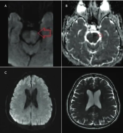
was normal except for right-sided mild motor weakness (41/5). A diffusion-weighted MRI scan of the brain performed on the admission day showed an acute infarct on the left pontine area and a corresponding apparent diffusion coefficient (Figure 1, A and B). By contrast, reduced glucose uptake in [18 F]fluorodeox-yglucose and positron emission tomography scans also involved the left frontal and temporoparietal cortices (Figure 2), whereas there were no abnormalities in the left frontal and temporopari-etal cortical regions on MRI (Figure 1C).
Discussion
The brainstem has traditionally been associated with vital func-tions, reflexive emotional reacfunc-tions, and motor performance.
Although the neuronal activity within the brainstem with motor areas of the cerebral cortex was found to correlate highly with parameters of movement, the role of the brainstem in the de-velopment of depression is still unclear. In the case of our patient, functional abnormalities did not entirely parallel morphological changes and were also found in the frontal, temporal, and parietal regions, which appeared to be rather unaffected on MRI. This indicates that the reduced glucose uptake observed in the re-spective cortical regions may reflect secondary deficits as a result of diminished functions of emotional circuits involving the brainstem. This was suggested by previous studies showing that with a depressive mood state, increases in limbic-paralimbic blood flow and decreases in neocortical regions were identified in de-pression.4Ourfindings suggest that the functional integrity of
FIGURE 1. Diffusion-Weighted MRI Scana
a(A) The diffusion-weighted MRI scan displays hyperintensity on the left pontine area,
which corresponds with an area of decreased apparent diffusion coefficient located in the (B) left pons and theoretically represents the area of cytotoxic edema and acute infarct (red arrows). (C) On diffusion-weighted MRI andfluid-attenuated inversion re-covery sequences, respectively, there were no abnormalities in the left frontal and temporoparietal cortical regions that might explain the apparent hypometabolism revealed by [18F]fluorodeoxyglucose and positron emission tomography imaging.
J Neuropsychiatry Clin Neurosci 28:1, Winter 2016 neuro.psychiatryonline.org e1
pathways linking the cortex and brainstem may be integral to the normal regulation of mood; these results also indicate a relationship between the brainstem noradrenergic system and depression. This type of quantitative analysis can provide that information, unlike a subjective radiologic evaluation limited with MRI. A greater understanding of the func-tional activity of the underlying regions affected by post-stroke depression may help to not only provide insight into the specific networks involved, but it may also support thefinding that disruption of the neurochemistry
of noradrenergic transmission plays an im-portant role in the pathophysiology of major depression.5
REFERENCES
1. Paolucci S, Antonucci G, Grasso MG, et al: Post-stroke depression, antidepressant treatment and rehabilitation results. A case-control study. Cere-brovasc Dis 2001; 12:264–271
2. Robinson RG, Spalletta G: Poststroke depression: a review. Can J Psychiatry 2010; 55:341–349 3. Nys GM, van Zandvoort MJ, van der Worp HB,
et al: Early depressive symptoms after stroke: neuropsychological correlates and lesion charac-teristics. J Neurol Sci 2005; 228:27–33
4. Mayberg HS, Liotti M, Brannan SK, et al: Reciprocal limbic-cortical function and negative mood: con-verging PET findings in depression and normal sadness. Am J Psychiatry 1999; 156:675–682 5. Klimek V, Stockmeier C, Overholser J, et al:
Re-duced levels of norepinephrine transporters in the locus coeruleus in major depression. J Neurosci 1997; 17:8451–8458
Burak Yulug, M.D. Ahmet Mithat Tavlı, M.D. Tansel Cakir, M.D. Lütfü Hanoglu, M.D.
From the Depts. of Neurology (BY, AMT, LH) and Nuclear Medicine (TC), University of Istanbul-Medipol, Istanbul, Turkey.
Send correspondence to Burak Yulug, M.D.; e-mail: burakyulug@gmx.de Received Jan. 14, 2015; revisions received Feb. 20 and March 16, 2015; accepted March 17, 2015.
J Neuropsychiatry Clin Neurosci 2016; 28:e1–e2; doi: 10.1176/appi. neuropsych.15010011
FIGURE 2. [18F]fluorodeoxyglucose and Positron Emission Tomography Imagesa
a(A) NeuroQ software (version 3.5; Syntermed, Inc., Atlanta, GA) was used to process the
(B) raw [18F]fluorodeoxyglucose and positron emission tomography data. The average pixel values in standardized regions of interest were automatically calculated. Area/whole brain ratios were compared with the normal values in the database. Significant hypo-metabolism is seen on the left frontal parietal and temporal cortical areas.
e2 neuro.psychiatryonline.org J Neuropsychiatry Clin Neurosci 28:1, Winter 2016
