Romanian Neurosurgery | Volume XXX | Number 2 | 2016 | April-June
Article
Statistical analysis of associated vertebra and
costal anomalies in spina bifida patients
Ibrahim Alatas, Huseyin Canaz, Ayten Saracoglu, Haluk Kafali, Gokhan Canaz, Mehmet Tokmak
Istanbul, Turkey
258 | Alatas et al - Associated vertebra and costal anomalies in spina bifida patients
Statistical analysis of associated vertebra and costal
anomalies in spina bifida patients
Ibrahim Alatas
1, Huseyin Canaz
1, Ayten Saracoglu
2, Haluk Kafali
2,
Gokhan Canaz
3, Mehmet Tokmak
41Spina Bifida Research Center, Sisli Florence Nightingale Hospital, Istanbul Bilim University, Istanbul 2Department of Anesthesiology, Sisli Florence Nightingale Hospital, Istanbul Bilim University, Istanbul 3Department of Neurosurgery, Haseki Training and Research Hospital, Istanbul
4Department of Neurosurgery Medipol University, Istanbul
Abstract: Objective: Spina bifida is one of the most severe birth defects and can happen as a result of disrupted primary neurulation. Congenital vertebra and costa anomalies are more frequently seen with spina bifida, and associated anomalies significantly affect the prognosis of affected children. In this study, we aimed to determine the incidence of scoliosis, costal anomalies, and vertebral deformations seen at the time of diagnosis and to statistically evaluate their concomitancies. Methods: Gender and mean ages of the patients were determined. The spina bifida patients were examined for deformation anomalies, butterfly vertebra, hemivertebra, wedge vertebra, costal anomalies and scoliosis. The relationships between these anomalies were evaluated. Results: 94 patients with a mean age of 11,5 months examined. The incidence of scoliosis was 21.8% among female infants and 17.9% among males. Rates of scoliosis with vertebra anomalies (hemivertebra, wedge vertebra) and costal anomalies did not differ significantly (P > 0.05). Wedge vertebra were the most frequent vertebra anomaly type with 38.2% ratio. Costal anomalies were detected in 25.5% of females and 20.5% of male infants. Hemivertebra and wedge vertebra were seen significantly more frequently in this group. Gender distribution did not differ between with and without any vertebra types. Conclusion: Congenital vertebra and costa anomalies are more frequently seen with spina bifida. We believe that these anomalies and relationship with spina bifida may demonstrate differences among different ethnic groups or locations. More detailed multi-centered studies performed on this issue will aid in the determination of etiologies, genetics, and treatment principles of these congenital anomalies.
Romanian Neurosurgery (2016) XXX 2: 258-266 | 259
Introduction
Spina bifida can happen as a result of the disruption of any stage of primary neurulation, which terminates at the 4th week of intrauterine life, or secondary neurulation, which terminates at the 11th week. In patients with spina bifida, higher incidence of vertebral formation, segmentation anomalies, scoliosis, kyphosis, and costal anomalies have been detected.
Associated anomalies significantly affect the prognosis of children with myelomeningocele. Congenital vertebral anomalies can affect any vertebral segment or involve one or more than one segment (31). Vertebral anomalies lead to skeletal deformities and consequently complicate the clinical picture (19). Vertebral anomalies complicate primary surgery and affect pathogenesis and monitorization of tethered spinal cord syndrome during long-term follow-up. Costal anomalies are usually associated with vertebral anomalies in patients with spina bifida. Congenital costal anomalies complicate vertebral surgery, lead to pulmonary problems at an early stage, and increase mortality rates. In patients with spina bifida, scoliosis can be congenital or develop secondary to paresthesis. In this patient group, scoliosis has a progressive course and induces severe restriction of the range of motion. In this case, re-planning of rehabilitation and treatment processes can be necessary.
In this study, we aimed to determine the incidence of scoliosis, costal anomalies, and vertebral deformations seen at the time of diagnosis in children with open or closed
spinal dysraphism, independently of the type of spinal dysraphism. The concomitancies were statistically evaluated.
Material and method
The spina bifida patients included in the study were delivered in Bakırkoy, Yenimahalle, Women’s & Children’s Hospital and Kanuni Sultan Suleyman Training and Research Hospital and diagnosed firstly on an ambulatory basis. Gender and mean ages of the patients were determined. The patients were examined for spinal deformations, scoliosis, and costal anomalies associated with spina bifida. The relationships between associated anomalies were evaluated.
The mean, standard deviation, rate, and frequencies were used as descriptive statistics of the data. Distribution of variables was controlled with the Kolmogorov-Smirnov test. The Mann-Whitney U test was used in the quantitative analysis of data. Qualitative analysis of data was performed using a chi-square test. When the criteria for this test were not met, Fisher’s exact test was used. Statistical analysis was performed using SPSS 21.0.
Results
A total of 94 patients with a mean age of 11.5 months (55 female and 39 male) were included in the study. Vertebra were examined for deformation anomalies, which revealed butterfly vertebra (n=8; 8.6%), hemivertebra (n = 13; 13.8%), and wedge vertebra (n = 31; 33%) (Table 1). Costal anomalies (n = 22; 23.4%) and scoliosis (n = 19; 20.2%) were also found.
260 | Alatas et al - Associated vertebra and costal anomalies in spina bifida patients
The incidence of scoliosis was 21.8% among female infants and 17.9% among males. The mean ages of those with and without scoliosis were 9.7 and 11.9 months, respectively. The distributions of age and gender of those with and without scoliosis were not significant (P ˃ 0.05). Rates of scoliosis in patients with and without butterfly vertebra, hemivertebra, wedge vertebra, and costal anomalies did not differ significantly (P ˃ 0.05) (Table 2).
Costal anomalies (rib anomalies) were detected in 25.5% of females and 20.5% of male infants. The mean ages of the patients with and without costal anomalies were 7.68 and 12.64 months, respectively, without any significant difference between the two groups (P ˃ 0.05). The rate of butterfly vertebra was significantly higher in patients with costal anomalies when compared to those without (P < 0.05). Hemivertebra were seen significantly more frequently in infants with costal anomalies relative to those without (P < 0.05). Wedge vertebra were significantly more frequently observed in the group with costal anomalies when compared to those without (P < 0.05) (Table 3).
Hemivertebra were detected in 14.5% of females and 12.8% of male infants. The mean
ages of the patients with and without hemivertebra were 7.85 and 12.06 months, respectively. Gender distribution in patients with and without hemivertebra did not demonstrate significant differences (P ˃ 0.05).
Butterfly vertebra were detected in 7.3% of females and 10.3% of male infants. Mean ages of the patients with and without butterfly vertebra were 5.88 and 12.00 months, respectively. Gender distribution among patients with and without butterfly vertebra did not demonstrate significant differences (P ˃ 0.05).
Wedge vertebra were detected in 38.2% of females and 38.2% of male infants. Mean ages of the patients with and without wedge vertebra were 11.13 and 11.65 months, respectively. Gender distribution among patients with and without wedge vertebra did not demonstrate significant differences (P ˃ 0.05) (Table 4).
Table I
Numbers of patients according to diagnosis
Table II Analysis of Scoliosis Scoliosis (-) Scoliosis (+) p med. ± sd./n-% med.± sd./n-% Age 11,9 ± 17,2 9,7 ± 16,4 0,371 Butterfly vertebrae 6 %75 2 %25 0,661 n % Butterfly vertebrae 8 %8,6 Hemivertebrae 13 %13,8 Wedge vertebrae 31 %33 Rib Anomalies 22 %23,4 Scoliosis 19 %20,2
Romanian Neurosurgery (2016) XXX 2: 258-266 | 261
Hemivertebrae 9 %69,24 4 %30,76 0,307
Wedge vertebrae 22 %73,3 8 %27,7 0,135
Rib anomalies 15 %68,2 7 %31,8 0,121
Table III
Analysis of Rib Anomalies (Costal Anomalies)
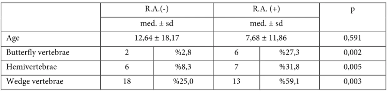
R.A.(-) R.A. (+) p med. ± sd med. ± sd Age 12,64 ± 18,17 7,68 ± 11,86 0,591 Butterfly vertebrae 2 %2,8 6 %27,3 0,002 Hemivertebrae 6 %8,3 7 %31,8 0,005 Wedge vertebrae 18 %25,0 13 %59,1 0,003 Table IV
Distribution of Scoliosis, Rib Anomalies and Vertebrae Formation Anomalies According to Gender
Female (n=55) Male (n=39) n % n % p Butterfly vertebrae 4 %7,3 4 %10,3 0,61 Hemivertebrae 8 %14,5 5 %12,8 0,811 Wedge vertebrae 21 %38,2 10 %25,6 0,203 Rib anomalies 14 %25,5 8 %20,5 0,577 Scoliosis 12 %21,8 7 %17,9 0,645
Discussion
For comprehension of the congenital anomalies of vertebra and their associated anomalies, normal embryological development of the vertebral axis should be known. A complex association exists between neural elements of the spinal cord and its supportive mesenchymal elements (19). Development of vertebra and the spinal cord begins from the third week of embryonic life and is completed at 20 years of age (24).
Problematic development for any reason causes incomplete closure of any region of the neural tube, which generally takes place within critically important days after fertilization (i.e., the 23rd. and 28th days) and leads to the formation of neural tube defects. Neural tube defects are heterogeneous and complex congenital anomalies of the central nervous system. Neural tube defects constitute a group of cerebral and spinal cord anomalies caused by incomplete closure of cerebral and spinal cord structures within the first weeks of
262 | Alatas et al - Associated vertebra and costal anomalies in spina bifida patients
embryonic life. Normally, closure of the neural tube is simultaneously realized irregularly in five different regions of the spinal cord, both towards the cephalad and in the caudal direction. The cephalad and caudal openings of the neural tube are closed at 25 and 27 days after fertilization, respectively. Dysfunctional primary neurulation causes formation of open neural tube defects (spina bifida aperta), while disruption of the secondary neurulation leads to the development of closed neural tube defects (spina bifida occulta).
Exposure to teratogenic agents can lead to specific anomalies during certain phases of embryonic life. These include impairments in the development of notochords, unsegmented mesoderm, and differentiation of sclerotomes (24). During progression of the neurulation process, the notochord aids in the formation of mesenchymal elements of the spinal cord (5). Mesodermal layers on both sides of the notochord mainly differentiate into paraxial, middle, and lateral regions (10, 20). The notochord also induces differentiation of the mesoderm into somites through a longitudinal segmentation process (22, 23, 26). Somites are paired structures localized on both sides of the embryonic midline that are constructed from mesoderm-derived epithelial blocks. They take their final shape following segmentation of the presomitic mesoderm (2, 4, 9, 25, 27). Vertebrae, ribs, intervertebral discs, related skeletal muscles, and connective tissue originate from these somites. Segmental alignment of the vertebrae stems directly from the segmental structure of the somites (15).
Deformations develop as a result of a deficiency of structural elements of the
vertebrae. Anterior, anterolateral, posterior, posterolateral, or lateral parts of the vertebral ring can be affected. Malformations can be partial or complete. Partial malformation manifests as wedge-shaped vertebrae, while its complete forms can appear as hemivertebra, butterfly vertebrae, or vertebral aplasia (19).
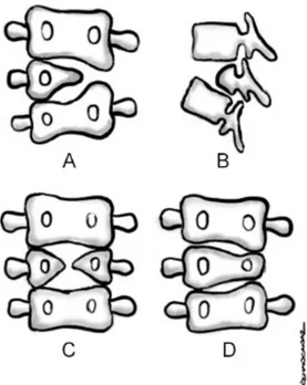
Hemivertebra are one of the most frequently seen vertebral anomalies. Since one side of the vertebra is not formed, it is characterized by an incomplete vertebral body, a single pedicle, and hemilamina. A hemivertebra is not an extra vertebra but a partially developed vertebral remnant (Figure 1A, 1B) (17).
Butterfly vertebra result from an inability of bilateral ossification foci. With a central cleft, they resemble a cleaved bilateral hemivertebra. Normally, the notochord is localized in the central part of a disc, and persistence of the notochord during the formation of vertebrae leads to the development of butterfly vertebra (Figure 1C) (19).
Wedge vertebra form as a result of dysplasic development of the vertebral body. However, on the affected side, the presence of the pedicle is maintained. It stems from one-sided partial developmental impairment of one of the chondrification foci (Figure 1D) (19).
Total aplasia of the vertebral body can rarely be seen and leads to the development of kyphosis. The embriological pathology leading to this anomaly is not yet clearly defined. However, in this condition, late or early ossification phases of the central part of the vertebral body could conceivably be
Romanian Neurosurgery (2016) XXX 2: 258-266 | 263
dysfunctional. Clear-cut data are not available about the incidence of concomitancy between congenital vertebral anomalies and myelomeningocele. However, there is a correlation between multifocal complex anomalies and the risk of neural tube defect formation (13).
Figure 1 - Illustrating defects of formation. A. Lateral hemivertebrae. B. Dorsal hemivertebrae. C. Butterfly
vertebrae. D. Wedge vertebrae
In the literature, vertabral malformations have been more frequently reported in patients with myelomeningocele, but there is no literature data concerning the incidence in all patients with spina bifida. In our study, all study populations with the diagnosis of open (spina bifida aperta) or closed (spina bifida occulta) neural tube defects were reviewed. The most frequently encountered vertebral
malformation was wedge vertebra (33%), followed by hemivertebra (13.8%) and butterfly vertebra (8.5%). Gender distribution among patients with vertebral malformation did not demonstrate any statistically significant difference.
Non-coincidental correlations between costovertebral malformations and neural tube defects have been reported (6, 7). Costal anomalies associated with myelomeningocele have been described as costal deficiency, costal fusion, and irregular or bicephalic ribs (3, 30). In our series, costal anomalies were detected in 22 patients (23.4%). Gender distribution did not demonstrate a statistically significant difference among patients with costal anomalies, but the rates of hemivertebra, butterfly vertebra, and wedge vertebra were significantly higher. As discussed, embriological development of the ribs and vertebra stems from the same origin, and an interaction persists between the two structures. As a result of this phenomenon, the incidence of costal anomalies increases in conditions that affect embriological development and lead to the formation of vertebral anomalies. Vertebral anomalies progress to scoliosis, kyphosis, lordosis, and mixed skeletal anomalies and lead to clinical symptoms and signs (19).
Scoliosis is a three-dimensional deviation of the spine on a frontal plane of more than 10 degrees (Cobb angle > 10o). This deformity can develop secondary to idiopathic factors, congenital vertebral malformations, tumors, or neuromuscular diseases. Adolescent idiopathic scoliosis is the most frequently seen form. However, neuromuscular scoliosis
264 | Alatas et al - Associated vertebra and costal anomalies in spina bifida patients
causes more severe spinal deformities and demonstrates a progressive course. In combination with the effects of the underlying disease, it leads to more severe restriction of mobility (1).
The incidence of scoliosis, kyphosis, and lordosis is higher in children with myelomeningocele (8). Among these, the most frequently seen is scoliosis, while kyphosis is the rarest (11). Most of these deformities occur during pediatric ages and secondary to paralysis, and nearly 15% of them are congenital (28). Spinal curvature in myelomeningocele emerges at an earlier age relative to many developmental anomalies. It is seen at 2 and 3 years of age and can worsen at 7 years of age (8, 12, 28).
Helpful definitions for the incidence and prevalence of scoliosis in children with myelomeningocele have been developed by Trivedi et al. (29)(28). In their survey, a patient population with a Cobb angle of more than 20 degrees was determined as cases with scoliosis. Most of the spinal curvatures develop during early stages of life, while nearly 40% of them occur after age 9. A small proportion is seen after age 15 (29).
Scoliosis in patients with myelomeningocele can develop secondary to congenital, idiopathic, or spinal dysraphism. It can also be directly or indirectly related to subsequent paralysis. In patients with spinal dysraphism involving thoracic vertebrae, the incidence of scoliosis rises to 90% (12, 28, 29). 85% of these curvatures are greater than 45 degrees. The incidence of scoliosis in patients decreases with paralysis involving lower vertebral levels. The incidence of scoliosis
stemming from L4 level drops down to 60%, and only 40% require surgical intervention. When the level of deficit involves levels below L4, the incidence of scoliosis regresses to 10% (14, 29).
Acquired scoliosis in patients with myelomeningocele has a greater tendency to regress. Muller et al. reported that acquired scoliosis in these patients worsens at a mean annual rate of 5 degrees (21). The angle of curvature and the patient’s age are risk factors for disease progression.
Various etiological factors for scoliosis have been described in patients with myelomeningocele. C-shaped scoliosis can generally stem from muscle weakness due to high-level paraplegia. Paralysis affecting asymmetrical levels or spastic hemiplegia due to hydrocephalus can cause this type of scoliosis. These types of scoliosis accompany kyphosis rather than lordosis. These curvature patterns typically first appear at a younger age and frequently during infancy and lead a progressive course. If present in these cases with scoliosis a surgical procedure aiming at severe spasticity can be required (16).
Another reason for scoliosis in this population is uncompensated hydrocephalus
secondary to hydromyelia or
hydrosyringomyelia. In these patients, S-shaped scoliosis is typically observed at the thoracic and thoracolumbar levels. Shunt dysfunction or hydomyelia can present with scoliosis at any age even during early childhood. Typical symptoms of hydromyelia may not be detected in the affected patient. In patients with scoliosis with a Cobb angle of less than 50 degrees, shunt replacement has been
Romanian Neurosurgery (2016) XXX 2: 258-266 | 265
demonstrated to have a regressive effect on congenital deformations from scoliosis. Segmentation defects of vertebrae are also among the etiological factors in children with scoliosis. These malformations can accompany hydromyelia, tethered cord, or muscle paralysis. Therefore, the attending physician should consider each one of the components of scoliosis when arranging a treatment program (18).
We detected scoliosis in 19 (20.2%) out of 75 patients. A statistically significant difference was not seen in the age and gender distribution of patients with and without scoliosis. The rates of scoliosis did not demonstrate differences among patients with and without costal anomaly, butterfly vertebrae, hemivertebrae, or wedge vertebra. The lower incidence of scoliosis in our study compared with the literature findings was evaluated, which revealed to be related to the inclusion of both patients with myelomeningocele and those with closed spinal dysraphism. Our patient population was also younger than 12 years of age. When the progressive pattern of neuromuscular scoliosis in patients with spina bifida is considered, we can admit that assessments in future years might detect increased incidence of scoliosis in this patient group.
Since our study had a retrospective design, and some of the patients did not attend the follow-up visits after a while, we could not include follow-up results of all patients in this study. Our study did not aim to determine the incidence of overall rates of scoliosis in patients with open and closed neural tube defects. On the contrary, we statistically
evaluated patients for the presence of scoliosis at the time of diagnosis of neural tube defects. Because of a lack of demographic and follow-up data of some patients, we could not differentiate between congenital and neuromuscular scoliosis in all patients, and our data relevant to these issues were not included in this study.
Conclusion
Vertebral deformities associated with or developed secondary to spinal dysraphism make the clinical picture of this disease more challenging or even complicate treatment and follow-up planning. The incidence of neural tube defects with their partially elucidated embriological and genetic mechanism demonstrate regional differences. We think that neural tube defects and associated anomalies of the vertebrae and other organ can demonstrate differences with respect to their locations and ethnic groups. More detailed multi-centered studies performed on this issue will aid in the determination of etiologies, genetics, and treatment principles of these congenital anomalies.
Correspondence
Dr. Gokhan Canaz
Haseki Reseach and Training Hospital Department of Neurosurgery
34087 Fatih/İstanbul Türkiye +90 212 529 44 00
E-mail: [email protected]
References
1.Allam AM, Schwabe AL. Neuromuscular scoliosis. PM & R: the journal of injury, function, and rehabilitation 2013; 5: 957-963.
266 | Alatas et al - Associated vertebra and costal anomalies in spina bifida patients
2.Aulehla A, Pourquie O. Oscillating signaling pathways during embryonic development. Current opinion in cell biology 2008; 20: 632-637.
3.Cetinkaya M, Ozkan H, Koksal N, et al. Spondylocostal dysostosis associated with diaphragmatic hernia and neural tube defects. Clinical dysmorphology 2008; 17: 151-154.
4.Dequeant ML, Pourquie O. Segmental patterning of the vertebrate embryonic axis. Nature reviews Genetics 2008; 9: 370-382.
5.Egelhoff J, Prenger EC. The Spine, in Ball WS (ed), Pediatric neuroradiology, Philadelphia, Lippincott-Raven, 1997.
6.Etus V, Ceylan S, Ceylan S. Association of spondylocostal dysostosis and type I split cord malformation. Neurological sciences: official journal of the Italian Neurological Society and of the Italian Society of Clinical Neurophysiology 2003; 24: 134-137.
7.Giacoia GP, Say B. Spondylocostal dysplasia and neural tube defects. Journal of medical genetics 1991; 28: 51-53. 8.Herring JA, Tachdjian MO. Tachdjian's pediatric orthopaedics, Philadelphia, Saunders, 2002.
9.Iimura T, Denans N, Pourquie O. Establishment of Hox vertebral identities in the embryonic spine precursors. Current topics in developmental biology 2009; 88: 201-234.
10.Jinkins JR. Atlas of neuroradiologic embryology, anatomy, and variants, Philadelphia, Lippincott Williams & Wilkins, 2000.
11.Karol LA. Orthopedic management in
myelomeningocele. Neurosurgery clinics of North America 1995; 6: 259-268.
12.Lindseth RE. Myelomeningocele, in Lovell WW, Winter RB, Morrissy RT, Weinstein SL (eds), Lovell and Winter's pediatric orthopaedics, Philadelphia, Lippincott Williams & Wilkins, 2001, pp601-632.
13.Loder RT. Congenital scoliosis and kyphosis, in Dewald RL (ed), Spinal deformities: the comprehensive text, New York, Thieme, 2003, pp684-693.
14.Mackel JL, Lindseth RE. Scoliosis in myelodysplasia. The Journal of Bone & Joint Surgery 1975; 57: 1031. 15.Makino Y, Kaneko K, Yamaguchi A, et al. Developmental biology and etiology of axial skeleton: Lessons from a mouse model of spondylocostal dysostosis and spondylothoracic dysostosis. Journal of Oral Biosciences 2013; 55: 175-179.
16.McLaughlin TP, Banta JV, Gahm NH, et al. Intraspinal rhizotomy and distal cordectomy in patients with myelomeningocele. The Journal of bone and joint surgery American volume 1986; 68: 88-94.
17.McMaster MJ, David CV. Hemivertebra as a cause of scoliosis. A study of 104 patients. The Journal of bone and joint surgery British volume 1986; 68: 588-595.
18.Autho: Management of Vertebral Problems and Deformities, in Memet Özek M, Cinalli G, Maixner WJ (eds): The Spina Bifida Management and Outcome. Milano: Springer-Verlag Italia, 2008, pp305-318. 19.MemetÖzek M, Belirgen M. Vertebral Anomalies and Spinal Malformations in Myelomeningocele, The Spina Bifida, Springer Milan, 2008, pp185-196.
20.Moore KL, Persaud TVN. Formation of germ layers and early tissue and organ differantiation, Philadelphia, WB Saunders, 1998.
21.Muller EB, Nordwall A, Oden A. Progression of scoliosis in children with myelomeningocele. Spine 1994; 19: 147-150.
22.Muller F, O'Rahilly R. The development of the human brain, the closure of the caudal neuropore, and the beginning of secondary neurulation at stage 12. Anatomy and embryology 1987; 176: 413-430.
23.O’Rahilly R, Benson DR. The development of vertebral column, in Bradford DS, Hensinger RN (eds), The Pediatric spine, New York, Thieme, 1985, pp3-17. 24.Oi S. Malformations of the vertebrae, in Rocco CD, Raimondin AJ (eds), Principles of pediatric neurosurgery, New York, Springer-Verlag, 1989, pp1-18.
25.Ozbudak EM, Pourquie O. The vertebrate segmentation clock: the tip of the iceberg. Current opinion in genetics & development 2008; 18: 317-323. 26.Phillips WA. Sacral agenesis, in Weinstein SL (ed), Pediatric spine: principles and practice, New York, Raven Press, 1994, pp259-273.
27.Pourquie O. Vertebrate segmentation: from cyclic gene networks to scoliosis. Cell 2011; 145: 650-663.
28.Samuelsson L, Eklof O. Scoliosis in
myelomeningocele. Acta orthopaedica Scandinavica 1988; 59: 122-127.
29.Trivedi J, Thomson JD, Slakey JB, et al. Clinical and Radiographic Predictors of Scoliosis in Patients with Myelomeningocele. The Journal of Bone & Joint Surgery 2002; 84: 1389-1394.
30.Vazquez-Lopez ME, Lopez-Conde MI, Somoza-Rubio C, et al. Anomalies of vertebrae and ribs: Jarcho Levin syndrome. Description of a case and literature review. Joint, bone, spine: revue du rhumatisme 2005; 72: 275-277.
31.Wynne-Davies R. Congenital vertebral anomalies: aetiology and relationship to spina bifida cystica. Journal of medical genetics 1975; 12: 280-288.