See discussions, stats, and author profiles for this publication at: https://www.researchgate.net/publication/305473805
Ectopic Reticulum in a Cow
Article in Pakistan Veterinary Journal · July 2016CITATIONS 0
READS 128 5 authors, including:
Some of the authors of this publication are also working on these related projects: Bone healingView project
Semih Altan Dicle University
30PUBLICATIONS 14CITATIONS SEE PROFILE
Feyzanur Alkan
T.C. Süleyman Demirel Üniversitesi
28PUBLICATIONS 120CITATIONS SEE PROFILE Muharrem Erol Balıkesir University 23PUBLICATIONS 51CITATIONS SEE PROFILE Ramazan Yıldız
Mehmet Akif Ersoy University
33PUBLICATIONS 88CITATIONS SEE PROFILE
All content following this page was uploaded by Semih Altan on 21 July 2016.
382
Pakistan Veterinary Journal
ISSN: 0253-8318 (PRINT), 2074-7764 (ONLINE) Accessible at: www.pvj.com.pk
Ectopic Reticulum in a Cow
Semih Altan1*, Yilmaz Koc2, Fahrettin Alkan2, Muharrem Erol3 and Ramazan Yildiz4
1Department of Surgery, Faculty of Veterinary Medicine, Dicle University, 21280, Diyarbakır, Turkey; 2Department of
Surgery, Faculty of Veterinary Medicine, Selcuk University, 42075, Konya, Turkey; 3Department of Surgery, Faculty of
Veterinary Medicine, Balıkesir University, 10145, Balıkesir, Turkey; 4 Department of Internal Medicine, Faculty of
Veterinary Medicine, Mehmet Akif Ersoy University, 15030, Burdur, Turkey *Corresponding author: [email protected]
ARTICLE HISTORY (15-176) A B S T R A C T Received: Revised: Accepted: Published online: April 09, 2015 March 21, 2016 March 27, 2016 April 09, 2016
A two years-old Holstein cow with poor appetite, reduced milk production, and partial defecation was evaluated in the present case report. After routine laboratory and clinical examinations, the animal further received ultrasound examination and then a right fossa paralumbal exploratory laparotomy was performed to the cow. The cow was diagnosed with ectopic reticulum on the laparotomy. After the content of the reticulum was removed, liquid paraffin was administered into the reticulum and its wall and abdominal wall was sutured as routinely. The prognosis of the animal deteriorated gradually following to the laparotomy and it was slaughtered by its owner. This is the first report showing the presence of an ectopic reticulum in a cow. ©2016 PVJ. All rights reserved Key words:
Anomaly Cow Ectopia Reticulum
To Cite This Article: Altan S, Koc Y, Alkan F, Erol M and Yildiz R, 2016. Ectopic reticulum in a cow. Pak Vet J, 36(3): 382-384.
INTRODUCTION
The reticulum, which is the smallest part among the four compartments of a ruminant stomach and its internal mucosa is characterized with honeycomb shape, is located between the rumen and the omasum (Fig. 1). The reticulum is located at the level of the 6-8th ribs on the left side of the vertebral
column in the ventral portion of the abdominal cavity, just above the xiphoid process (Imran et al., 2012; Shafaey et al., 2015). Disease of the reticulum in cow is generally associated with the presence of foreign bodies (Gökceand Bozukluhan, 2014). However, congenital anomalies of the reticulum are very rare (Alhendi et al., 1996). Ectopia is almost always a congenital anomaly defined as a displacement or malposition of an organ into other region, or different organ or tissue of the body. Various ectopia of organs and tissues have been reported both in humans and animals (Koç and Alkan, 2001; Akin et al., 2014). The aim of the present report was to describe an ectopic reticulum which diagnosed with exploratory laparotomy.
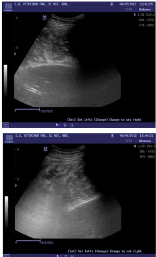
Case history and surgical intervention: The subject of the current case was a 2 years-old Holstein cow. In anamnesis and clinical examination of the animal was revealed the presence of poor appetite, reduced milk production, partial defecation, loss of percussion field for liver, presence of ping and ballottement. Due to detected clinical findings, the animal was diagnosed with a possible
right displaced abomasum. However, the results of the hematological and especially blood gasses (Table 1) did not verify the diagnosis of the right displaced abomasum; similarly, ultrasound examination also did not support our diagnosis. Ultrasound examination indicated the presence of an organ in the right paralumbal area, but this organ did not look like the abomasum (Fig. 3). Conflicting findings regarding the animal necessitated the exploratory laparotomy. After the line block local anesthesia, the right paralumbal area is clipped and prepared surgically for laparotomy. The abdominal cavity was reached by incising the skin, the subcutaneous tissues, external and internal oblique muscles, transversus muscle, and the peritoneum respectively from exterior to interior over the right paralumbal fossa. At first glance, a limited swelling resembling the abomasum was noted (Fig. 2). When the swelling structure was palpated, while its dorsal part was felt to be filled with gas, its distal particularly caudal part was determined to have stiff consistency. It was failed to perform an effective transperitoneal exploration before the decompression of the swelling. The swelling structure was punctured at its highest point with a needle attached to a tube to allow the release of accumulated abnormal gas. Subsequently, to empty the content of this structure, it was partially pulled out with the help of two rumen forceps held in place from the dorsal and ventral edges and a 10 cm in length incision was made. Discharged content contained plenty of hay particles and its smell was similar to that of
Pak Vet J, 2016, 36(3): 382-384. 383
the rumen. It was determined that the mucosa possessed honeycomb appearance fitting to the reticulum after removing about 1.5 kg of the content (Fig. 4A and B). According to the obtained data, the case was diagnosed as ectopic reticulum. Liquid paraffin was administered into the reticulum to soften the content and facilitate its passage, and then the wall of the reticulum was sutured as routine. At last, transperitoneal exploration was performed to understand whether there are any other hidden anomalies. In this examination, it was observed that the rumen was not entirely full; the omasum was stiff, located right under and slightly medial of the rumen (Fig. 2). On the other hand, the abomasum was moderately full and the liver was at its normal situs. The intestines were completely empty. No peritoneal fluids or fibrin masses were noted in the abdominal cavity. The reticulum found residing at the caudo-dorsal aspect of the right abdominal cavity was attempted to relocate it at cranio-ventral direction but effort was failed. Similarly, repositioning of the omasum and abomasum located to be just under and slightly medial to the reticulum also was unsuccessful. The abdominal wall was closed up without additional intra-abdominal manipulations. The animal was hospitalized, and penicillin-streptomycin (5ml/100kg IM; Dipenisol, Bayer Animal Health, Istanbul, Turkey) and flunixin meglumine (2.2 mg/kg IM; Fulimed, Alke, Istanbul, Turkey) were applied for antibiotic and pain therapy during postoperative three days respectively, in addition to intravenous liquid treatment. After the operation, the prognosis of the animal gradually deteriorated and the animal was slaughtered with the consent of the owner to prevent further yield loss. Since the cow, moreover, was insured, her owner refused the autopsy of the animal.
DISCUSSION
There is no literature information concerning the ectopia or sole displacement of the reticulum in cattle. However, sometimes displacement of the reticulum following the omasum displacement (reticulo-omasal-abomasal volvulus) is shown to be seen very rarely (Wittek et al., 2004). In the present report, right displacement abomasum/volvulus was excluded by clinical, ultrasonographic and surgical examination of abdomen. There was no abnormality in results of hematologic and blood gas analyses. Only blood leukocyte count increased compared to reference values. Clinical symptoms are also not specific for ectopia reticulum. In the right flank laparotomy, the reticulum was located in an abnormal anatomical situs where it was right to the rumen, and at the caudal-dorsal aspect of the omasum and abomasum between the right abdominal wall and the intestines. These findings indicated presence of an ectopic reticulum.
The causes for disorders of the forestomach compartments in adult cow are considered to be dietary, inflammatory, and/or mechanical (Chanie and Tesfaye, 2012). However, a published study (Alhendi et al., 1996) reporting the presence of a hypoplastic reticulum emphasizes that one of the potential factors leading to the diseases of the digestive system in adult cow might be congenital. In the case, we speculate that ectopia reticulum may be congenital in the cow. Because displacement of reticulum in cattle is appeared rarely as acquired.
Fig. 1: Schematized image of the gastrointestinal organs in healthy cow.
Fig. 2: Schematized image of the gastrointestinal organs in the current case.
Pak Vet J, 2016, 36(3): 382-384. 384
Table 1: Laboratory test results. Preop: Preoperative data, Postop: Postoperative data (Radostits et al., 2007).
Blood gases Preop Postop Reference Values Hematological data Preop Reference Values
pH 7.43 7.41 7.35-7.50 WBC (m/mm3) 15.02 4-12
CO2 (mmHg) 41 42 34-45 Lym (%) 13.3 45-80
O2 (mmHg) 40 35 >40 Mon (%) 2.0 1-5
Na (mmol/L) 152 145 132-152 Gra (%) 84.7 10-30
K (mmol/L) 2.6 2.4 3.9-5.8 Lym (m/mm3) 1.99 2.0-7.5
HCO3- (mmol/L) 27.2 26.6 20-30 Mon (m/mm3) 0.30 0-0.8
Ca (mmol/L) 0.59 0.95 1.2-1.6 Gra (m/mm3) 12.73 0.4-3.6 Glu (mg/dL) 56 44 45-75 RBC (m/mm3) 5.94 6-11 Lac (mmol/L) 1.3 0.4 0.6-2.2 Hct % 28.9 % 24-46 Hct (%) 28 29 25-50 RDW 14.1 16.7-23.3 BEecf (mmol/L) 2.9 2.0 ±3 Hb (g/dl) 9.6 8-15 BE (B) (mmol/L) 2.6 1.8 ±3 THR (m/mm3) 471 100-800
Fig. 4: A: Image of reticulum observed in the right abdominal cavity during the operation. B: Image of the honeycomb shape of reticulum observed during the operation.
Medical history of the animal indicated that no marked complications with the cow had been noticed until the parturition, implying that the ectopic reticulum did not obstruct the passage of the ruminal content into the lower parts of the alimentary canal. Nonetheless, It was opinion that accumulation of excessive volatile fatty acids in the rumen and abomasum due to feeding of the animal with excessive concentrated food during the dry period, development of the negative energy balance in the postpartum period, suffering from hypocalcaemia, generation of inflammatory mediators induced by other diseases, hypomotility in gastrointestinal system due to vagal nerve hypofunction, and occurrence of sudden enlargement in the abdominal cavity right after the parturition in dairy cows (Zadnik et al., 2001; Van Winden
and Kuiper, 2003) can trigger development of ectopic reticulum-associated transition problems.
Conclusions: We believe that this is the first report to show the presence of an ectopic reticulum in a cow. Ultrasonography examination could be useful technique in diagnosis of congenital anomalies such as ectopic reticulum. However, we could be said that laparotomy is more reliable method in diagnosis of ectopic reticulum in suspicious conditions.
Author’s contribution: SA and YK conceived and designed the study. RY and ME executed clinical and ultrasonographic examination, and analyzed the blood analyses. SA and FA performed the surgical intervention. All authors interpreted the data, critically revised the manuscript for important intellectual contents and approved the final version.
REFERENCES
Akin M, Erginel B, Bilici S, Gedik S, Yıldız A et al., 2014. Crossed testicular ectopia: Report of six cases. Afr J Paediatr Surg, 11: 269-272. Alhendi AB, Gameel AA and Ramadan RO, 1996. Possible hypoplasia of
the reticulum in a cow. Can Vet J, 37: 442-443.
Chanie M and Tesfaye D, 2012. Clinico-pathological findings of metallic and non-metallic foreign bodies in dairy cattle: A review. Acad J Anim Dis, 1: 13-20.
Gökce HI and Bozukluhan K, 2014. Inflammatoric diseases of rumen and reticulum in cattle. Turkiye Klinikleri J Vet Sci, 5: 32-41.
Imran S, Sharma S, and Bhat AA, 2012. Ultrasonographic imaging of normal reticulum and traumatic reticuloperitonitis in crossbred cows. Eurasian J Vet Sci, 28: 214-219.
Koç Y and Alkan F, 2001. A case of extraumbilical ectopia hepatica in a lamb. Eurasian J Vet Sci, 17: 109-111.
Radostits OM, Gay CC, Hinchcliff KW and Constable P, 2007. Reference laboratory values in veterinary medicine: In: A Textbook of the Diseases of Cattle, Sheep, Goats, Pigs and Horses. 10th ed. Saunders Elsevier, Philadelphia, USA, pp: 66, 2047-2049. Shafaey EE, Aoki T, Ishii M, Sasaki M and Yamada K, 2015. A descriptive
study of the bovine stomach using computed tomography. Pak Vet J, 35: 18-22.
Van Winden SCL and Kuiper R, 2003. Left displacement of the abomasum in dairy cattle: Recent developments in epidemiological and etiological aspects. Vet Res, 34: 47-56.
Wittek T, Fürll M, and Constable PD, 2004. Prevalence of endotoxemia in healthy postparturient dairy cows and cows with abomasal volvulus or left displaced abomasum. J Vet Intern Med, 18: 574-580. Zadnik T, Mesaric M, and Reichel P, 2001. A review of abomasal displacement-clinical and laboratory experiences at the clinic for ruminants in Ljubljana. Slov Vet Res, 38: 193-208.
View publication stats View publication stats