alignant glioma is the most common primary tumor of the brain in adults and has a very poor prognosis. Despite advances in surgery, chemotherapy, radiotherapy and imaging techniques no clear trend toward
M
ORĐJĐNAL ARAŞTIRMA / ORIGINAL RESEARCH.
Different Doses of Radiation on Agar Colony
Forming Development in C6 Glioma Cells:
Assessment by Thymidine Labeling Index,
and Bromodeoxyuridine Labeling Index
C6 GLĐOMA HÜCRELERĐNDE DEĞĐŞĐK RADYASYON DOZLARINDA KOLONĐ
OLUŞTURMA ETKĐSĐ: TĐMĐDĐN ĐŞARETLEME ĐNDEKSĐ VE BROMODEOKSĐÜRĐDĐN
ĐŞARETLEME ĐNDEKSĐĐLE DEĞERLENDĐRME
Tolga ÖZMEN, MD,a Gülperi ÖKTEM, MD,b Sevilcan TUNA,c Burcu BĐLTEKĐN,d
Seçnur DENĐR,e Ayhan BĐLĐR, MDc
a
Đstanbul University, Đstanbul Faculty of Medicine, ĐSTANBUL
bDepartment of Histology and Embryology, Ege University School of Medicine, ĐZMĐR cDepartment of Histology and Embryology, Đstanbul University School of Medicine, ĐSTANBUL d
Department of Histology and Embryology, Edirne University School of Medicine, EDĐRNE eDepartment of Biological Science, Balıkesir University Science Faculty, BALIKESĐR
Abstract
Objective: Gliomas are relatively frequent in adults, and are among the most
malignant primary brain tumors. Glioblastoma multiforme, like many other tumors that exhibit radiation sensitivity in vitro, seems to be very resistant to radiation in vivo, thus suggesting that irradiation may not be a rate-limiting factor for malignant glioma tumor growth. In this study, we aimed to determine the optimal dose of radiation in C6 glioma colony forming assay, which is a valuable tool for antitumor treatment screening.
Material and Methods: 105 cell/lamella colony forming cells were radiated with 200 cGy, 400 cGy, 800 cGy and 1600 cGy for 10 minutes. Radiosensitiv-ity was measured systematically 24, 48, 72 and 96 hours after the radia-tion by three methods: soft-agar bilayer assay, thymidinE incorporaradia-tion, and bromodeoxyuridine (BrdU) incorporation.
Results: The soft-agar bilayer assay, which assessed the colony-forming
units, showed that the number of colonies in the control group (609, 3 ± 86.8) were decreased after 200 cGy (8.3 ± 3.6) and 400 cGy (7.2 ± 4.3). No colony was detected in 800 cGy and 1600 cGy irradiated cells [3H] Thymidine incorporation was more prominent with higher doses of radiation. BrdU incorporation revealed that even at low doses (200 cGy) of radiation there was a significant decrease of cell proliferation. On higher doses like 1600 cGy it was more prominent.
Conclusion: Cell survival, doubling time, and cell phases are parameters of
growth kinetics, and the results suggest that C6 glioma cells are radio-sensitive and are virtually affected by all radiation doses in our experi-ment even 200 cGy at 24 hours. Besides, colony forming assay with thymidine labeling index, and BrdU labeling index may be used as new methods for determining radiotherapy doses in clinical applications.
Key Words: Dose fractionation; glioma
Turkiye Klinikleri J Med Sci 2007, 27:321-327
Özet
Amaç: Gliomalar erişkinde oldukça sık görülen, kötü gidişli primer beyin
tümörlerinin büyük bölümünü oluştururlar. Pek çok tümör gibi glioblastoma multiforme de in vitro radyasyona duyarlıdır, ancak in vivo radyasyon direnci gösterir. Bu sebeple malign glioma tümör
geli-şiminde, radyasyon tedavisi sınırlayıcı olmayabilir. Bu çalışmada, anti tümör tedavi taramasında çok uygun bir teknik olabilen “colony forming assay” yöntemi ile C6 glioma hücre hattında en uygun radyas-yon dozunun belirlenmesi amaçlandı.
Gereç ve Yöntemler: 105 hücre/lamel içeren “colony forming” hücreleri 10’ar dk. boyunca 200 cGy, 400 cGy, 800 cGy ve 1600 cGy radyas-yona maruz bırakıldı. Radyasyon alan hücrelerdeki radyosensitivite sistematik olarak 24, 48, 72 ve 96 saatlerde “soft-agar bilayer assay” timidin (TLI) ve BrdU (BLI) işaretleme indeksleriyle değerlendirildi. “Soft-agar bilayer assay” oluşan koloni sayılarını belirlemekteydi ve bu sayı kontrol grubunda (609.3 ± 86.8) iken 200 cGy (8.3 ± 3.6) ve 400 cGy (7.2 ± 4.3)’de belirgin olarak azalmıştı. 800 cGy ve 1600 cGy sonrası koloniye rastlanamadı.
Bulgular: TLI yüksek dozda, BLI ise düşük dozda (200 cGy) radyasyonun
değerlendirilmesinde daha güvenilir bir tekniktir. Bu çalışmada
dü-şük doz (200 cGy) radyasyon sonrasında bile hücre proliferasyonunda belirgin bir azalma olmuştur. Bu 1600 cGy yük-sek dozda çok daha belirgindir.
Sonuç: Hücre yaşam süreleri, ikilenme zamanları ve hücre fazları büyüme
kinetiklerini gösterir. C6 glioma hücreleri radyosensitiftir ve 200 cGY radyasyon dozunda ve 24 saatte bile açıkça etkilenirler. Bu-nunla beraber TLI ve BLI klinik uygulamalarda radyoterapi dozu-nun belirlenmesinde yeni bir metod olarak kullanılabilir.
Anahtar Kelimeler: Radyoterapi doz ayarlama; glioma
Geliş Tarihi/Received: 13.09.2006 Kabul Tarihi/Accepted: 11.12.2006
This research supported by Đstanbul University Research Foundation
Yazışma Adresi/Correspondence: Gülperi ÖKTEM, MD
Ege University School of Medicine, Department of Histology and Embryology, TR-35100, ĐZMĐR
[email protected] Copyright © 2007 by Türkiye Klinikleri
improvement in outcome was achieved for malignant glioma.1-3 Local recurrence occurs in 90% of patients and their survival is extremely low. New molecular targets and new therapeutic moda-lities are essential for further treatments.4 Currently, modern and advanced radiotherapy systems are effective as local treatments of malignant tumors.
While planning for radiotherapy, not only the parameters such as tumor size, tumor invasion and lymphatic involvement but also tumor cell characteristics should be taken into consideration. There are lots of effective methods to define the cell characteristics of tumors. Creating colonies in agar is a method which is called double layer agar colony forming assay.5 With double layer agar colony forming assay, the effects of different antineoplastic drugs and radiotherapy on primer tumor colonies can be comparatively studied. Thymidine labeling index (TLI) and BrdU, have also been used successfully to define tumor cell characteristics.6
Recent studies show that irradiation of C6 glioma cells, decreased their proliferation in vitro in a dose-dependent manner.7 In this study, we treated C6 glioma cells of rats with different doses of radiation. Our aim was to determine the optimal dose of radiation by comparing colony forming assay with TLI and BrdU index. Dose-dependent radiotherapy represents a new therapeutic approach in clinical applications and we aimed to examine the proper dose and method for clinical applica-tions in patients.
Material and Methods
Cell Culture
C6 glioma cells were obtained from American Culture Collection. This tumor cell line is main-tained in culture as adherent cells and cultured in DMEM (Sigma Chemical Co., St Louis, Missouri) plus 10% heat inactivated fetal calf serum (FCS) (Sigma Chemical Co., St Louis, Missouri), sup-plemented with 1% L glutamin (Sigma Chemical Co., St Louis Missouri), 1% nonessential amino acids (Sigma Chemical Co., St Louis, Missouri), 10.000 units/mL penicillin (Sigma Chemical Co., St Louis, Missouri), and 10 mg/mL streptomycin
(Sigma Chemical Co., St Louis, Missouri). Cell line was grown in a humidified atmosphere at 37oC in 5% CO2. When the tumor cell lines were used as
target cells, they were treated with tyripsin-EDTA (Sigma Chemical Co., St Louis, Missouri), washed, and resuspended in complete medium. Dimethylsulfoxide (DMSO) and trypan blue dyes were purchased from Sigma (Sigma Chemical Co., St Louis, Missouri). Stock solutions of the reagents were prepared in PBS, medium or DMSO as appro-priate. A total of 1.500.000 cells were used (500.000 cells for TLI, 500.000 cells for BrdU and 500.000 cells for agar colony development). The cells were seeded in sterile 25 cm2 culture flasks with 5 mL medium as described. This study was performed in
Đstanbul University Medical Faculty Department of Histology and Embryology and passages were con-trolled with invert microscope at least twice a week.
Agar colony development
After the cultures became semiconfluent, flasks were treated with 200, 400, 800 and 1600 cGy radiation. Radiation was applied for 10 minutes for each dose in flasks. After about an hour, the cells were treated with tyripsin and were counted. Then, 1000 cells were added on the upper layer of each well of the culture plates. The plates were incubated for 7 days. After this period, the colonies were evaluated and counted.
Three wells were inoculated for each radiation dose.
The group of cells containing at least 30 cells was accepted as a colony.
The mean values and standard deviations of counted cells were calculated.
TLI of Tumor Cells
After the cultures became semiconfluent the cells in one flask were treated with tyripsin (0.25%) and the resulting cell pellet was inoculated on coverslips placed in sterilized 24 well-culture plates with a final concentration of 1 x 10⁵ cell/coverslip in 2 mL of medium for each experi-ment. The attachment of cells on the coverslips were controlled by invert microscope and the cells were radiated for 10 minutes with 200 cGy, 400
cGy, 800 cGy and 1600 cGy for each plate. Sys-tematically 24, 48, 72 and 96 hours after the radia-tion, cells were treated with 1 mCi/mL 3H TdR for 30 minutes. Then cells were fixed with Carnoy fixative and autoradiography was performed. The percentage of labeled cells over at least 1000 cells from each coverslip was calculated. For each group 3 coverslips were used. The mean values and stan-dard deviations of the results were evaluated.
BrdU Labeling Index of Tumor Cells Cells in the S-phase of the cell cycle (thus syn-thesizing DNA) can be labeled by BrdU incorpora-tion into the DNA strand replacing thymidine and can be subsequently detected by immunocyto-chemical means. C6 glioma cells were incubated with 0.3% H2O2 for 10-15 minutes to inhibit
en-dogenous peroxidase activity. Then, sections were treated with trypsin at 37oC for 30 min and rinsed with water. After buffered phosphate saline incu-bation for 5 minutes at 37oC, sections were treated with 2 N HCl for 30 minutes at 37oC to denature DNA. Sections were incubated in 0.1 M Borax for 10 minutes and then in PBS for 5 minutes at room temperature. Incubation with Ultra V Block for 5 min, primary antibody (Sigma Chemical Co., St Louis, Missouri 1:200 dilution) for 1 hour, secon-dary antibody (biotinylated goat anti-mouse) for 10 minutes, streptavidin peroxidase for 20 minutes and peroxidase-compatible chromogen for 20 minutes in incubation box at room temperature were performed by using Ultravision Large Volume Detection Sys-tem Anti-Mouse HRP Kit (Lab Vision, California, USA) and AEC Substrate System Kit (Lab Vision, California, USA). Sections were rinsed with water and counterstained in Mayer’s Hematoxylin for 3 minutes. BrdU labeled cells had red stained nuclei.
Statistical Analysis
The obtained data were compared between and within groups using ANOVA with Bonferroni test. Differences were considered to be significant at p values less than 0.05. All results are ex-pressed as mean SEM calculated from at least 3 independent experiments. All statistical analyses were performed with GraphPads Instat 3.05
soft-ware (GraphPad Softsoft-ware Inc., San Diego, CA, USA).
Results
Radiotherapy Decreased the Number of Colonies
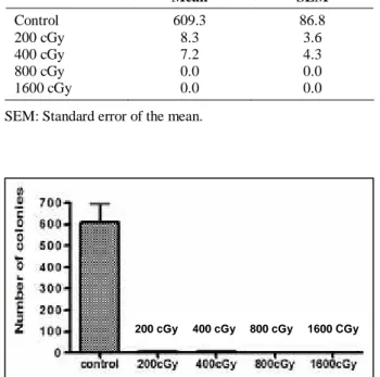
Number of colonies in the control group (609.3 ± 86.8) were decreased after 200 cGy (8.3 ± 3.6) and 400 cGy (7.2 ± 4.3) irradiation. No colony was detected in the 800 cGy and 1600 cGy irradi-ated cells. Data obtained from the controls were significantly different than those of the 200 cGy (p< 0.0001) and 400 cGy (p< 0.0001) irradiated cells (Table 1, Graphic 1). However, ANOVA showed that there was no difference between the 200 cGy and 400 cGy groups (p> 0.05). In addition to the reduction in the number of colonies, smaller colonies were determined in the radiotherapy groups.
Table 1. Number of colonies.
Mean SEM Control 609.3 86.8 200 cGy 8.3 3.6 400 cGy 7.2 4.3 800 cGy 0.0 0.0 1600 cGy 0.0 0.0
SEM: Standard error of the mean.
Graphic 1. Number of colonies in the control group was significantly lower than those in the 200 cGy and 400 cGy cells. There was no colony in the 800 cGy and 1600 cGy cells. Differences between the controls and the 200 cGy (p< 0.0001) and 400 cGy (p< 0.0001) groups were statistically significant.
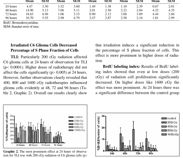
Irradiated C6 Glioma Cells Decreased Percentage of S Phase Fraction of Cells TLI: Particularly 200 cGy radiation affected C6 glioma cells at 24 hours of observation for TLI (p< 0.0001). Higher doses of radiotherapy did not affect the cells significantly (p> 0.005) at 24 hours. However, further observations clearly revealed that 400, 800 and 1600 cGy radiotherapies influenced glioma cells evidently at 48, 72 and 96 hours (Ta-ble 2, Graphic 2). Overall our results clearly show
that irradiation induces a significant reduction in the percentage of S phase fraction of cells. This effect is more prominent in higher doses of radia-tion.
BrdU labeling index: Results of BrdU label-ing index showed that even at low doses (200 cGy) of radiation cell proliferation significantly decreased. On higher doses like 1600 cGy the effect was more prominent. At 24 hours there was a significant difference between the control group
Graphic 3. In all radiotherapy groups, even the 200 cGy, BrdU stained cell numbers were significantly decreased (p< 0.0001) and 400, 800 and 1600 cGy radiotherapies influenced glioma cells evidently at 48, 72 and 96 hours.
Graphic 2. The most prominent effect at 24 hours of observa-tion for TLI was with 200 cGy radiaobserva-tion of C6 glioma cells (p< 0.0001). Higher doses of radiotherapy did not affect the cells significantly (p> 0.005) at 24 hours. However, further observa-tions clearly revealed that 400, 800 and 1600 cGy irradiation influenced glioma cells evidently at 48.72 and 96 hours. Table 2. Evaluation of TLI after irradiation.
Control 200 cGy 400 cGy 800 cGy 1600 cGy
Mean SEM Mean SEM Mean SEM Mean SEM Mean SEM
24 hours 39.50 10.70 13.40 12.80 41.99 10.36 33.25 11.37 31.25 10.14
48 hours 42.40 11.20 41.20 12.40 33.57 12.73 29.22 12.45 34.50 15.62
72 hours 48.60 13.38 40.78 14.11 27.70 14.01 22.70 11.23 27.70 14.01
96 hours 13.51 3.34 17.14 8.76 20.98 8.72 16.25 9.10 1.43 1.22
TLI: Thymidine labeling index, SEM: Standart error of men.
Table 3. Evaluation of BrdU after irradiation.
Control 200 cGy 400 cGy 800 cGy 1600 cGy
Mean SEM Mean SEM Mean SEM Mean SEM Mean SEM
24 hours 4.47 3.30 1.52 3.60 1.49 1.38 1.10 2.39 0.67 2.01
48 hours 14.90 5.13 7.06 5.11 2.01 2.50 2.22 2.04 4.32 4.35
72 hours 18.63 6.96 1.06 3.15 0.90 2.12 3.06 3.89 4.44 2.66
96 hours 10.70 5.55 2.98 4.79 2.47 2.87 2.50 2.16 1.81 2.99
BrdU:Bromodeoxyuridine, SEM: Standart error of men.
and 200cGy radiation group. However, there was statistically no difference between higher doses and 200 cGy at 24 hours of observation. Although at 48 hours of observation there was a statistically significant difference between 200 cGy and 400 cGy, no difference was detected between higher doses and 400 cGy (Table 3, Graphic 3).
Changes on Cell Morphology after Radiotherapy
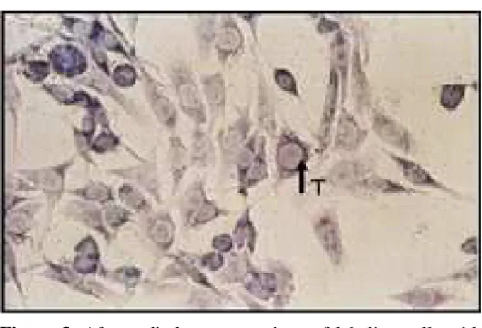
Thymidine and BrdU positive cells were de-tected with immunohistochemical and monoclonal staining techniques (Figure 1, 2). Control cell shapes reflecting regular C6 glioma cell morphol-ogy were used. In the control group, the number of positive stained cells was higher than in the irradi-ated cells. Highly proliferative C6 glioma cells were significantly decreased after irrradiation even low
dose radiation with thymidine and BrdU incorpora-tion (Figure 3, 4). A decrease in the cell number as well as poor staining of DNA was observed in ra-diotherapy groups. Morphological changes indi-cated that radiotherapy affected cells particularly starting from low doses.
Discussion
The aim of this project was to realize a new method-colony forming efficiency with TLI and BrdU labeling index-to determine radiotherapy doses. TLI was one of the first methods utilized to evaluate the proliferative activity of cancer cells. The number of tumor cells undergoing DNA syn-thesis can be measured using in vivo or in vitro assays for 3H-thymidine uptake, which can be visualized by autoradiography.8 The TLI expresses Figure 1. C6 glioma cells with thymidine labeling without
radiotherapy. Arrow shows positive staining cell with dark cytoplasm (x 400).
Figure 3. After radiotherapy, numbers of labeling cells with Thymidine are significantly decreased. Arrow shows positive staining cell (x 400).
Figure 2. BrdU positive cells were detected in the control group. Arrow shows BrdU positive cell (x 400).
Figure 4. Highly proliferative C6 glioma cells were signifi-cantly decreased after radiotherapy even at low dose. Arrow shows positive BrdU incorporation (x 400).
the ratio between thymidine-labeled cells and the total number of tumor cells and was shown to be reliable and stable over time.9 Labeled pyrimidine bases other than (3H) thymidine, such as the halo-genated analogue BrdU, have also been used suc-cessfully.10,11 Of note is that incorporated BrdU may be revealed by immunohistochemistry (IHC). The technique, however, has limitations that have hampered its acceptance as a standard method. In fact, fresh tumor tissue or cultured cells are needed and a complex and time-consuming radioactive assay or in vivo administration of labeled sub-stances is required.12
Our main goal was to combine colony forming assay with both TLI and BrdU indexes. The mod-est benefit seen was thought to be related to the heterogeneous distribution of the halogenated pyrimidines in tumors and the low thymidine re-placement achieved in individual tumor cells. BrdU is a potent radiosensitizer when incorporated into the DNA of target cells; however, the degree of radiosensitization depends on the extent of thymidine replacement by the analog and on the number of cells labeled. The failure of proliferating cells to take up thymidine analogs such as BrdU is attributable to the low availability of the drug to the tumor cell or to the analog being diluted to below useful levels by endogenous nucleotide pre-cursors. The latter results from the activity of en-zymes in the de novo synthesis pathways and/or the enzymes in the alternative pathways for pyrimidine or purine biosynthesis which are so-called salvage pathways. A marked rise in the ac-tivities of enzymes in both categories has been observed in cancer cells in logarithmic growth and in hepatomas of different growth rates.13
Cancers are spreading rapidly among humans and new diagnosis and treatment modalities are essential for the cure of this disease. The optimal treatment for recurrent high-grade glioma remains undefined despite advances in surgery, chemother-apy and radiotherchemother-apy.14-17 Attempts to improve the response of patients to radiotherapy have involved a number of stratagems. Malignant glioma tumors exhibit various behaviors in response to radiother-apy. This is due to the diversity of radiation
sensi-tivity in laboratory conditions and human body.7 C6 glioma cells are quite resistant to irradiation in vitro, even at high doses of X-rays.1 This signifi-cant data was the inspiration of this project. Other tumors, even glioblastoma multiforme cell lines that exhibit radiation sensitivity in vitro, seem to be very resistant to radiation in vivo, thus suggest-ing that irradiation may not be a rate-limitsuggest-ing fac-tor for malignant glioma tumor growth.18
Colony forming assay is based on colony de-velopment of cells which will specialize with stem cells in the tumor cell population. Cancer cells are widely proliferating indefinitely like stem cells. There is increasing evidence that cancers may con-tain their own stem cells. Many cancers, like nor-mal organs, seem to be maintained by a hierarchi-cal organization that includes slowly dividing stem cells, rapidly dividing transit amplifying cells (pre-cursor cells), and differentiated cells.19,20 The pres-ence of a small subpopulation of slowly dividing cancer stem cells may elucidate why so many can-cers recur after treatment with irradiation or cyto-toxic drugs, even when most of the cancer cells seem to be killed by the therapy. Usually, some cancer cells survive the treatment, and these sur-viving cells may be cancer stem cells, which may be not only resistant to the therapy but also essen-tial for the malignancy of the cancer and malignant cell lines, which have been maintained for years in culture, contain a subpopulation of stem cells.21
In this study, we determine the optimal dose of radiation in C6 glioma colony forming assay. Col-ony forming assay represents three dimensional structural forms of tumors in laboratory conditions and many studies suggest that the human tumor colony-forming assay may be a valuable tool for antitumor treatment screening.22 Colony forming assay technique was used to determine the appro-priate dose of radiation therapy and it was investi-gated in terms of feasibility, validity, and potential for discovering new antitumor treatment.23,24 Lu et al showed that clonogenic survival assay offers the potential to study the intrinsic radiosensitivity, repair, long term regeneration, and other radiobio-logical responses of neural stem cells after in vitro or in vivo irradiation.25 Colony forming assay with
TLI and BrdU labeling index may be new methods for determining radiotherapy doses in cancer. In-deed, it is possible to investigate these methods in primary cell cultures to determine the appropriate dose in clinical applications. Either human tumor colony-forming assay or other new remedial meth-ods would possibly depend on advances in basic research.
REFERENCES
1. Kortmann RD, Jeremic B, Weller M, Plasswilm L, Bamberg M. Radiochemotherapy of malignant glioma in adults. Clinical experiences. Strahlenther Onkol 2003;179:219-32.
2. Rave-Frank M, Glomme S, Hertig J, Weiss E, Pradier O, Hess CF, et al. Combined effect of topotecan and irradia-tion on the survival and the inducirradia-tion of chromosome ab-errations in vitro. Strahlenther Onkol 2002;178: 497-503.
3. Schuck A, Muller SB, Kohler A, Konemann S, Wienstroer M, Mosler C, et al. Combined radiochemotherapy with paclitaxel in the treatment of malignant glioma. Strahlenther Onkol 2002;178:486-90.
4. Andratschke N, Grosu AL, Molls M, Nieder C. Perspec-tives in the treatment of malignant gliomas in adults. Anti-cancer Res 2001;21:3541-50.
5. Hornsten P, Granstrom M, Wahren B, Gahrton G. Prog-nostic value of colony-stimulating and colony-forming cells in peripheral blood in acute non-lymphoblastic leu-kemia. Acta Med Scand 1977;201:405-10.
6. Tazzari PL, Gobbi M, Tassi C, Lemoli RM, Dinota A, Visani G, et al. In situ staining of bromodeoxyuridine positive cells in normal and neoplastic colony-forming units grown on plasma clots. J Immunol Methods 1988;113:215-9.
7. Parthymou A, Kardamakis D, Pavlopoulos I, Papadimitriou E. Irradiated C6 glioma cells induce angio-genesis in vivo and activate endothelial cells in vitro. Int J Cancer 2004;110:807-14.
8. Meyer JS, Connor RE. In vitro labeling of solid tissues with tritiated thymidine for autoradiographic detection of S-phase nuclei. Stain Technol 1977;52:185-95.
9. Meyer JS, Friedman E, McCrate MM, Bauer WC. Predic-tion of early course of breast carcinoma by thymidine la-beling. Cancer 1983;51:1879-86.
10. Meyer JS, Province M. Proliferative index of breast car-cinoma by thymidine labeling: prognostic power inde-pendent of stage, estrogen and progesterone receptors. Breast Cancer Res Treat 1988;12:191-204.
11.Tubiana M, Pejovic MH, Koscielny S, Chavaudra N, Malaise E. Growth rate, kinetics of tumor cell proliferation and long-term outcome in human breast cancer. Int J Can-cer 1989;44:17-22.
12.Colozza M, Azambuja E, Cardoso F, Sotiriou C, Larsimont D, Piccart MJ. Proliferative markers as prog-nostic and predictive tools in early breast cancer: where are we now? Ann Oncol 2005;16:1723-39.
13.Weber G. Biochemical strategy of cancer cells and the design of chemotherapy: G. H. A. Clowes Memorial Lec-ture. Cancer Res 1983;43:3466-92.
14.Leibel SA, Sheline GE. Radiation therapy for neoplasms of the brain. J Neurosurg 1987;66:1-22.
15.Glioma Meta-Analysis Trialists (GMT) Group. Chemo-therapy for high-grade glioma. Cochrane Database Syst Rev 2002;4:CD003913.
16.Andratschke N, Grosu AL, Molls M, Nieder C. Perspec-tives in the treatment of malignant gliomas in adults. Anti-cancer Res 2001;21:3541-50.
17.Medical Research Council Brain Tumor Working Party. Randomized trial of procarbazine, lomustine, and vincris-tine in the adjuvant treatment of high-grade astrocytoma: a Medical Research Council trial. J Clin Oncol 2001;19:509-18.
18. Taghian A, DuBois W, Budach W, Baumann M, Freeman J, Suit H. In vivo radiation sensitivity of glioblastoma multiforme. Int J Radiat Oncol Biol Phys 1995;32:99-104. 19.Poste G, Greig R. On the genesis and regulation of cellular heterogeneity in malignant tumors. Invasion Metastasis 1982;2:137-76.
20.Tu SM, Lin SH, Logothetis CJ. Stem-cell origin of metas-tasis and heterogeneity in solid tumours. Lancet Oncol 2002;3:508-13.
21.Kondo T, Setoguchi T, Taga T. Persistence of a small subpopulation of cancer stem-like cells in the C6 glioma cell line. Proc Natl Acad Sci USA 2004;101:781-6. 22.Griffon G, Merlin JL, Parache RM, Martinet N, Marchal
S, Nabet F, et al. Sensitivity to taxoid derivatives of a newly established human endometrioid ovarian adenocar-cinoma radioresistant cell line. Anticancer Res 1996;16:177-87.
23.Kono K, Tsuchida T, Kern DH, Irie R. Ganglioside com-position of human melanoma and response to antitumor treatment. Cancer Invest 1990;8:161-7.
24. Soulieres D, Rousseau A, Tardif M, Larochelle M, Tremblay M, Vaillancourt L, et al. The radiosensitivity of uveal melanoma cells and the cell survival curve. Graefes Arch Clin Exp Ophthalmol 1995;233:85-9.
25.Lu F, Wong CS. A clonogenic survival assay of neural stem cells in rat spinal cord after exposure to ionizing ra-diation. Radiat Res 2005;163:63-71.