Skewed X-Chromosome Inactivation in Scleroderma
Elif Uz&Laurence S. Loubiere&Vijayakrishna K. Gadi&Zeynep Ozbalkan&Jeffrey Stewart&J. Lee Nelson&
Tayfun Ozcelik
Published online: 21 December 2007
# Humana Press Inc. 2007
Abstract Scleroderma is a female-prevalent autoimmune disease of unclear etiology. Two fundamental gender differ-ences, skewed X-chromosome inactivation (XCI) and pregnancy-related microchimerism, have been implicated in scleroderma. We investigated the XCI patterns of female scleroderma patients and the parental origin of the inactive X chromosome in those patients having skewed XCI patterns (>80%). In addition, we investigated whether a correlation exists between XCI patterns and microchimerism in a well-characterized cohort. About 195 female scleroderma patients
and 160 female controls were analyzed for the androgen receptor locus to assess XCI patterns in the DNA extracted from peripheral blood cells. Skewed XCI was observed in 67 (44.9%) of 149 informative patients and in 10 of 124 healthy controls (8.0%) [odds ratio (OR)=9.3 (95% confidence interval (CI) 4.3–20.6, P<0.0001)]. Extremely skewed XCI (>90%) was present in 44 of 149 patients (29.5%) but only in 3 of 124 controls (2.4%; OR=16.9; 95% CI 4.8–70.4, P<0.0001). Parental origin of the inactive X chromosome was investigated for ten patients for whom maternal DNA was informative, and the inactive X chromo-some was of maternal origin in eight patients and of paternal origin in two patients. Skewed XCI mosaicism could be considered as an important risk factor in scleroderma.
Keywords X-inactivation . Microchimerism . Mosaicism . Scleroderma
Introduction
Scleroderma (systemic sclerosis, SSc) is an autoimmune disease characterized by fibrosis and alterations in the microvasculature [1]. Scleroderma is three to ten times more prevalent in women than in men [2]. Most autoim-mune diseases are more prevalent in females than in males [3], and fundamental differences between male and female biology, such as hormone status [4], pregnancy [5], and X-chromosome inactivation [6,7] have been proposed as the underlying pathophysiological mechanisms leading to the female prevalence of autoimmune diseases. Indeed, both pregnancy-related microchimerism [8–11], and skewed (i.e., not the expected 50:50 balance) X-inactivation mosaicism [12] have been shown to be associated with scleroderma.
Clinic Rev Allerg Immunol (2008) 34:352–355 DOI 10.1007/s12016-007-8044-z
E. Uz
:
T. Ozcelik (*)Department of Molecular Biology and Genetics, Faculty of Science, Bilkent University, Bilkent,
Ankara 06800, Turkey
e-mail: [email protected] L. S. Loubiere
:
V. K. Gadi:
J. L. Nelson Clinical Research Division,Fred Hutchinson Cancer Research Center, Seattle, WA, USA
V. K. Gadi
:
J. L. NelsonDepartment of Medicine, University of Washington, Seattle, WA, USA
Z. Ozbalkan
Rheumatology Department,
Ankara Numune Education and Research Hospital, Ankara, Turkey
J. Stewart
Caldera Pharmaceuticals, Los Alamos, NM, USA T. Ozcelik
Institute for Materials Science and Nanotechnology (UNAM), Bilkent University,
This study was performed to investigate the X-inactivation patterns of female scleroderma patients along with the parental origin of the inactive X chromosome in those patients with extremely skewed X-inactivation. In addition, the correlation between skewed X-inactivation and both maternal and fetal microchimerism was examined in a well-characterized cohort of scleroderma patients.
Materials and Method
DNA samples were obtained from 195 scleroderma patients and 160 control women. Clinical characteristics of the patients and controls have been published elsewhere [12, 13]. The ethics review boards at the participating institu-tions approved the study protocol. Informed consent was obtained from all subjects. The X-chromosome inactivation status of the patients and controls was determined by genotyping a highly polymorphic CAG repeat in the first intron of the androgen receptor as previously described [12, 14]. Depending on the definition and quantitative accuracy of the measurement method, a few percent to nearly one fifth of apparently healthy women display skewed (i.e., non-random) patterns of X-inactivation. Whereas ratios in the range of 50–79% are usually regarded as normal variation, deviation from this range in 80–89% of cells is defined as skewed, and above 90% of cells as extremely skewed X-inactivation. [15–17] Fisher’s exact test was used for statistical analyses.
Results
X-Inactivation Ratios
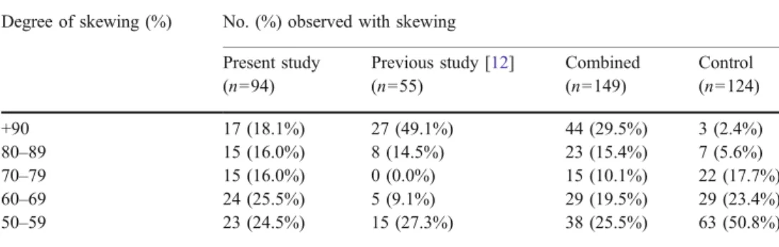
Our data show that skewed X-chromosome inactivation (>80%) was associated with disease. X-chromosome inactivation status was informative in 94 of the 125 scleroderma patients (75%) and in 124 of the 160 controls (78%). Only the individuals whose alleles resolved ade-quately were included in the subsequent densitometric
analysis; thus, some of the heterozygous individuals were considered as uninformative. Skewing in the range of 80– 89% was observed in 15 of the 94 patients (16.0%) but in only 7 of the 124 controls (5.6%; P<0.0001; Table 1). Extremely skewed X-inactivation (>90%) was observed in 17 of the 94 patients (18.1%) but in only 3 of the 124 controls (2.4%;P<0.0001). When the data from the present and the previously published [12] studies were combined (Table1), of the total of 195 patients, 149 were informative for the androgen receptor polymorphism. Skewed X-inactivation was observed in 67 of 149 patients (44.9%) but in only 10 of 124 healthy controls (8.0%). Expressed as a risk factor for scleroderma, the odds ratio (OR) was 9.3 [95% confidence interval (CI) 4.3–20.6; P<.0001]. Numer-ous studies conducted in different control populations indicate that extremely skewed X-inactivation is a rare event not exceeding 3–5% [15–17]. For the current study, extremely skewed X-inactivation was present in 44 of 149 patients (29.5%) but in only 3 controls (2.4%), and the OR was 16.9 (95% CI 4.8–70.4, P<0.0001).
Parental Origin of the Inactive X Chromosome
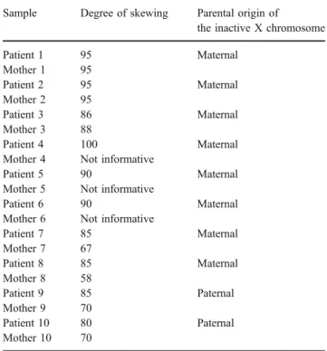
Nineteen patients with skewed X-inactivation for whom maternal DNA was available were further analyzed to determine the parental origin of the inactive X chromosome. For this analysis, DNA samples from mothers of patients were analyzed for the androgen receptor gene polymor-phism, and informative results were obtained for ten patient– mother pairs. For the remaining nine pairs, both the mother and the daughter were heterozygous for the androgen receptor polymorphism, thus providing no difference in allele sizes. Therefore, it is not possible to determine the parental origin of the alleles in the absence of information regarding the paternal genotype. The inactive X chromo-some was of maternal origin in eight patients and of paternal origin in two patients. In three mothers of patients studied, X-inactivation pattern was also skewed (Table2). Interest-ingly, the same allele was skewed in both the mothers and the patients, and one mother had been diagnosed with an autoimmune condition, namely temporal arteritis.
Table 1 Proportions of sclero-derma patients and controls with skewed X-inactivation
For comparison byχ2, P<0.0001(both >80% skewing and >90% skewing)
Degree of skewing (%) No. (%) observed with skewing Present study (n=94) Previous study [12] (n=55) Combined (n=149) Control (n=124) +90 17 (18.1%) 27 (49.1%) 44 (29.5%) 3 (2.4%) 80–89 15 (16.0%) 8 (14.5%) 23 (15.4%) 7 (5.6%) 70–79 15 (16.0%) 0 (0.0%) 15 (10.1%) 22 (17.7%) 60–69 24 (25.5%) 5 (9.1%) 29 (19.5%) 29 (23.4%) 50–59 23 (24.5%) 15 (27.3%) 38 (25.5%) 63 (50.8%)
Evaluation of Skewed X-Inactivation for Correlation with Microchimerism
Maternal and fetal microchimerism was investigated as described previously [18]. We examined X-inactivation ratios for correlation with microchimerism in a limited number of patients for whom microchimerism data was also available. We observed that among patients with more than 80% skewing, 7 of 12 (58.3%) tested positive for maternal microchimerism and 8 of 14 (57.1%) for fetal micro-chimerism (Table 3). In patients with random patterns of X-inactivation, 10 of 15 (66.7%) were positive for maternal microchimerism, and 14 of 25 patients were positive for fetal microchimerism (56%). Neither maternal (P=0.7) nor fetal microchimerism (P=1.0) correlated with skewed X-inactivation.
Discussion
Autoimmune disorders affect more than 5% of the population, and a high female prevalence is characteristic of most autoimmune diseases including scleroderma, autoimmune thyroid diseases, systemic lupus erythematosus, and Sjögren’s syndrome [3]. Pregnancy-related microchimerism [10] and skewed X-inactivation [12] have been proposed as potential contributors to the pathophysiology of scleroderma. In this study, we observed skewed X-inactivation patterns in a significant proportion (34%) of women with scleroderma. Of these women, approximately 18% displayed extreme skew-ing (>90:10). This result is consistent with our previous study [12] and indicates that skewed X-inactivation could be a common finding in different population groups.
We do not know the cause of skewed X inactivation in scleroderma, and probable mechanisms have been discussed in the accompanying manuscript [19]. Among them, X-linked lethal mutations, which would be compatible with life in females because of X-inactivation mosaicism, are appealing causative mechanism. If the cause of skewing is indeed X-linked mutations, these should be inherited from the maternal lineage unless they occur de novo during gametogenesis. We therefore analyzed the parental origin of the inactive X chromosome in 19 patient–mother pairs, and of the 10 pairs with informative genotyping results, inactive X was found to be of maternal origin in 8 pairs. This result is on the border of statistical significance (P=0.055). Assuming this result holds, we will be left with an interesting puzzle. Why are maternally inherited X chromosomes more likely to be inactivated in highly skewed patients? One possibility is that some X chromosomes confer a selective disadvantage to both the organism itself and the peripheral cells within the organism. This precise situation has been documented in X-linked immunodeficiency, where heterozygous (female) carriers have high X-inactivation skew (because of natural selection within the organism), whereas affected males have a survival disadvantage [20]. When an X-encoded genotype is selec-tively disadvantageous to cells, skew is an outcome. When an X-encoded genotype is selectively disadvantageous to an organism, preferential maternal inheritance is an outcome. Together, this leads us to the hypothesis that some highly Table 2 Parental origin of the inactive X chromosome in scleroderma
patients with skewed X-inactivation
Sample Degree of skewing Parental origin of
the inactive X chromosome
Patient 1 95 Maternal Mother 1 95 Patient 2 95 Maternal Mother 2 95 Patient 3 86 Maternal Mother 3 88 Patient 4 100 Maternal
Mother 4 Not informative
Patient 5 90 Maternal
Mother 5 Not informative
Patient 6 90 Maternal
Mother 6 Not informative
Patient 7 85 Maternal Mother 7 67 Patient 8 85 Maternal Mother 8 58 Patient 9 85 Paternal Mother 9 70 Patient 10 80 Paternal Mother 10 70
Table 3 Proportion of mater-nal (MMc) and fetal (FMc) microchimerism in scleroderma patients
MMc FMc
Skewed (>80%) 12 tested 58.3% positive for MMc 14 tested 57.1% positive for FMc
7 positive 8 positive
5 negative 6 negative
Random (<80%) 15 tested 66.7% positive for MMc 25 tested 56.0% positive for FMc
10 positive 14 positive
5 negative 11 negative
skewed patients have an X chromosome that would confer a selective disadvantage on homozygotic women and on males. Because scleroderma is the first disease in which pregnan-cy-related microchimerism has been documented, we investi-gated a subset of patients for whom microchimerism data were also available for correlation of skewed X-inactivation with microchimerism. Neither maternal nor fetal microchimerism was found to be correlated with skewed X-inactivation. In conclusion, the two types of female mosaicism—skewed X-inactivation and maternal/fetal microchimerism—appear to be independent risk factors in scleroderma. One possible expla-nation for these data is that mosaicism itself is an underlying cause of scleroderma and, by extension, female-prevalent autoimmune disease in general.
Acknowledgements We would like to thank Iclal Ozcelik for critical reading of the manuscript. Supported by grants from the Scientific and Technical Research Council of Turkey–TUBITAK-SBAG 3334, Inter-national Centre for Genetic Engineering and Biotechnology –ICGEB-CRP/TUR04-01, and Bilkent University Research Fund (to Dr. Ozcelik).
References
1. Derk CT, Jimenez SA (2003) Systemic sclerosis: current views of its pathogenesis. Autoimmun Rev 2:181–191
2. Silman AJ, Hochberg MC (1993) Scleroderma. In: Silman AJ, Hochberg MC (eds) Epidemiology of the rheumatic diseases. Oxford University Press, Oxford, pp 192–219
3. Whitacre CC (2001) Sex differences in autoimmune disease. Nat Immun 2:777–780
4. Cutolo M, Capellino S, Sulli A, Serioli B, Secchi ME, Villaggio B, Straub RH (2006) Estrogens and autoimmune diseases. Ann N Y Acad Sci 1089:538–547
5. Mullinax F (1993) Chimerism and autoimmunity. In: Feng PH, Boey ML, Chang HH, Fong KY, Howe HS, Leong KH (eds) Proceedings of the 4th ASEAN Congress of Rehumatology. Communication Consultants, Singapore, pp 39–40
6. Kast RE (1977) Predominance of autoimmune and rheumatic diseases in females. J Rheumatol 4:288–292
7. Stewart JJ (1998) The female X-inactivation mosaic in systemic lupus erythematosus. Immunol Today 19:352–357
8. Nelson JL (1996) Maternal-fetal immunology and autoimmune diseases: is some autoimmune disease auto-alloimmune or allo-autoimmune? Arthritis Rheum 39:191–194
9. Mullinax F, Mullinax GL (1996) Pregnancy-induced scleroderma: identification of offspring-derived cells in patients with scleroder-ma [abstract]. Arthritis Rheum 39(Suppl 9):231
10. Nelson JL, Furst DE, Maloney S, Gooley T, Evans PC, Smith A (1998) Microchimerism and HLA-compatible relationships of pregnancy in scleroderma. Lancet 351:559–562
11. Artlett CM, Smith JB, Jimenez SA (1998) Identification of fetal DNA and cells in lesions from women with systemic sclerosis. N Engl J Med 321:1186–1191
12. Ozbalkan Z, Bagislar S, Kiraz S, Akyerli CB, Ozer HT, Yavuz S, Birlik AM, Calguneri M, Ozcelik T (2005) Skewed X chromo-some inactivation in blood cells of women with scleroderma. Arthritis Rheum 52:1564–1570
13. Loubiere LS, Lambert NC, Madeleine MM, Porter AJ, Mullarkey ME, Pang JM, Galloway DA, Furst DE, Nelson JL (2005) HLA allelic variants encoding DR11 in diffuse and limited systemic sclerosis in Caucasian women. Rheumatology (Oxford) 44:318–322 14. Allen RC, Zoghbi HY, Moseley AB, Rosenblatt HM, Belmont JW (1992) Methylation of HpaII and HhaI sites near the polymorphic CAG repeat in the human androgen-receptor gene correlates with X chromosome inactivation. Am J Hum Genet 51:1229–1239 15. Busque L, Mio R, Mattioli J, Brais E, Brais N, Lalonde Y, Maragh M,
Gilliland DG (1996) Non-random X-inactivation patterns in normal females: lyonization ratios vary with age. Blood 88:59–65 16. Sharp A, Robinson D, Jacobs P (2000) Age- and tissue-specific
variation of X chromosome inactivation ratios in normal women. Hum Genet 107:343–349
17. Amos-Landgraf JM, Cottle A, Plenge RM, Friez M, Schwartz CE, Longshore J, Willard HF (2006) X chromosome inactivation patterns of 1,005 phenotypically unaffected females. Am J Hum Genet 79:493–499
18. Lambert NC, Erickson TD, Yan Z, Pang JM, Guthrie KA, Furst DE, Nelson JL (2004) Quantification of maternal microchimerism by HLA-specific real-time polymerase chain reaction: studies of healthy women and women with scleroderma. Arthritis Rheum 50:906–914
19. Ozcelik T (2007) X chromosome inactivation and female predispo-sition to autoimmunity. Clin Rev Alergy Immunol (in press) 20. Puck JM, Willard HF (1998) X inactivation in females with
X-linked disease. N Engl J Med 338:325–328