ONCOLOGY REPORTS 11: 1337-1341, 2004
Expression of TAP73 and ANP73 in malignant gliomas
Abstract. The p73 gene is able to encode transcriptionaly
active TAp73, as well as a dominant-negatively acting ANp73 transcript isoforms. We studied differential expression of these forms in normal brain as well as glial tumors, by semi quantitative RT-PCR. The expression of p73 was low or undetectable in normal brain tissues. Most of the tumors and non-tumor brain tissues also lacked significant expression of p73 in patients with low-grade astrocytomas. In contrast, most high-grade glial tumors displayed strong up-regulation of TAp73, whereas only a few displayed ANp73 expression. These aberrations may reflect the inactivation of retinoblastoma pathway in these tumors which result in the activation of E2F transcription factors, since TAp73 is a known target of E2F1 gene. The study of TAp73 expression in brain tumors may serve as a means to evaluate the retinoblastoma pathway-dependent tumor progression.
Introduction
Malignant gliomas are the most prevalent primary neuro ectodermal tumors, accounting for 15-23% of intracranial tumors and about 50% of all primary brain tumors in adults (1). The World Health Organization classifies these tumors, which arise from supportive glial cells of the central nervous system, into four grades of malignancy in the increasing order from I to IV (2). Despite technological improvements in neurodiagnostics, neurosurgery and adjuvant therapy, there has been little improvement in the survival rates of the patients with malignant gliomas. For example, the mean survival rate for astrocytoma grade IV (glioblastoma multi forme) is less than one year (3). Current therapies have limited impact on the mortality rate of malignant gliomas for several reasons. First, these tumors diffusely infiltrate the brain to
Correspondence to: Dr Mehmet Ozturk, Department of Molecular
Biology and Genetics, Bilkent University, 06800 Ankara, Turkey E-mail: ozturk@fen.bilkent.edu.tr
Key words: p73, transcriptionally active p73, dominant negative p73,
glial tumor, brain tumor, retinoblastoma pathway, tumor progression
render complete resection impossible. Second, they are resistant to conventional chemotherapy because of blood-brain barrier that limits the penetration of drugs into the brain. Finally, radiotherapy is limited to low dose irradiations because of its harmful effects to the normal brain tissue at high doses (4). Thus, it is necessary to develop new therapeutic strategies for these incurable tumors and this will only be possible once a thorough understanding as to their behavior and biology have been achieved.
The p53 gene is the most frequently mutated tumor suppressor gene in human cancers (5). Germline mutations of p53 gene are known to lead to Li-Fraumeni syndrome, which harbors the carcinoma of brain in the tumor spectrum of these patients (6). Moreover, allelic loss of p53 locus was described as an early event in gliomagenesis and p53 gene mutations were shown to be present at 30-50% of astrocytic tumors (7,8). Thus, inactivation of p53 is a common event in glial tumor formation. The p53 protein, together with p63 and p73, forms a family of transcription factors. The p73 gene was identified as the first p53 homologue (9). The structural and functional similarities of p53 and p73 proteins confer a probable tumor suppressor function for the new family member (10,11). However, in contrast to p53, p73 mutations are rare if not absent in different cancers including the tumors of the brain (10). Nevertheless, p73 gene display aberrant changes in its expression patterns in tumors suggesting its involvement in malignancy in an unusual manner. The p73 gene has an unusual ability to express multiple transcript forms resulting from either alternative splicing or alternative promoter usage. In addition to TAp73 and ANp73 isoforms generated by alternative promoter usage, 10 different isoforms generated by alternative splicing have been identified so far (11,12). All these isoforms encode different protein products that are able to form hetero-oligomers and tetramers as they all contain an oligomerization domain (9). The transcriptionally active full-length TAp73 and the dominant-negative ANp73 display opposite activities. TAp73 induces cell-cycle arrest and apoptosis, whereas ANp73 inhibits both TAp73- and p53-induced apoptosis. Furthermore, ANp73 is p53-induced by TAp73 and p53, creating a dominant-negative feedback loop that regulates p53 and p73 function (12-14).
The ANp73 isoform is generated by the usage of an internal promoter and an additional exon within the intron 3 of human p73 gene (13,15,16). The putative protein product of this novel HASAN UGUR1'2, A. EMRE SAYAN1, SUKRU O. OZDAMAR3,
YUCEL KANPOLAT2 and MEHMET OZTURK1
Department of Molecular Biology and Genetics, Bilkent University, 06800 Ankara; Department of Neurosurgery, Faculty of Medicine, Ankara University;" Department of Pathology,
Faculty of Medicine, Hacettepe University, 06100 Ankara, Turkey Received December 16, 2003; Accepted February 25, 2004
transcript (ANp73) lacks the transactivation (TA) domain that is present in TAp73. This truncation confers to the ANp73 the ability to inhibit the transcriptional activities of both wild-type p53 and TAp73 proteins (13,16-18). The ANp73 transcripts were shown to be the major p73 gene product in mouse tissues during development and adulthood (17). In vitro overexpression of ANp73 was shown to inhibit p53-dependent apoptosis in nerve cells following NGF withdrawal (18). There are no functional studies describing a probable function for ANp73 in glial cells. However, p53 and ANp73 complexes were shown to be present in the mouse brain (18).
A large number of p73 expression studies in different tumors have been conducted, but there are very few reports about the relative expressions of TAp73 and ANp73 transcripts/proteins in human malignancies. In primary hepatocellular carcinomas (HCCs) and HCC-derived cell lines, ANp73 levels were shown to be in comparable amounts with that of normal liver, whereas TAp73 expression was up-regulated (15). A recent report describes an overexpression of ANp73 transcript and protein in primary breast carcinoma samples (19). The status of p73 gene in neuroblastomas is of particular interest, since the loss of chromosomal locus lp36 where p73 gene resides and N-myc amplification is strongly associated with poor prognosis in these tumors (20). In addition, the expression of ANp73 transcript was described in 30% of neuroblastoma samples which display a poor prognosis (21). In another study both TAp73 and ANp73 transcripts were defined to be present in neuroblastic tumors (22). However, to our knowledge, the relative expression of TAp73 and ANp73 transcripts in glial tumors has not been reported yet.
In this study, we analyzed the relative expression of TAp73, in comparison with ANp73 isoform in normal brain tissues, as well as in glial tumors using semi-quantitative RT-PCR methods. Our data show that these p73 transcipts are low or undetectable in normal human brain tissues as well as early stage glial tumors, whereas most advanced glial tumors express the TAp73 form, and occasionally the ANp73 transcripts.
Materials and methods
Tumor and tissue samples. All tumor and tissue samples
were obtained at surgery. Freshly collected samples were immediately frozen and stored in liquid nitrogen prior to analysis. Normal brain samples (n=3) were obtained as discarded materials from anterior temporal lobectomies in amygdalohippocampectomy surgeries applied for surgical therapy of epilepsy. A total of 20 tumor samples were also collected together with 9 available corresponding non-tumor tissues (Table I). Tumor and tissue samples were sliced using a microtome, stained with hematoxylin-eosin and were evaluated by two different pathologists. Tumor tissues containing more than 80% tumor positive nuclei and non-tumor tissues with no tumor contamination were selected for further studies.
RNA extraction. Total RNA from tumor and normal tissues
were extracted using TriPure reagent (Boehringer Mannheim, IN, USA), as described by the supplier. Following column purification and DNase treatment (MN RNA Quickspin II, Duren, Germany), 5 u.g total RNA was used to prepare cDNA
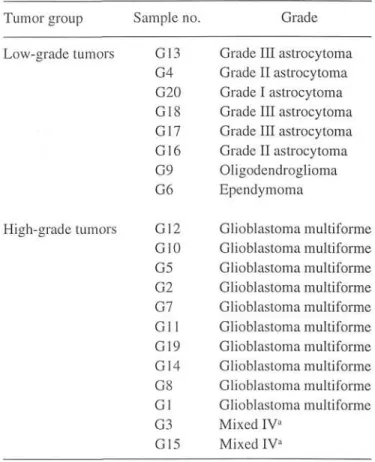
Table I. The tumors studied for p73 gene expression. Tumor group Low-grade tumors High-grade tumors Sample no. G13 G4 G20 G18 G17 G16 G9 G6 G12 G10 G5 G2 G7 Gil G19 G14 G8 Gl G3 G15 'Tumors containing astroglial and oli
Grade Grade III astrocytoma Grade II astrocytoma Grade I astrocytoma Grade III astrocytoma Grade III astrocytoma Grade II astrocytoma Oligodendroglioma Ependymoma Glioblastoma multiforme Glioblastoma multiforme Glioblastoma multiforme Glioblastoma multiforme Glioblastoma multiforme Glioblastoma multiforme Glioblastoma multiforme Glioblastoma multiforme Glioblastoma multiforme Glioblastoma multiforme Mixed IVa Mixed IV" godendroglial cells.
using RevertAid First Strand cDNA Synthesis Kit (MBI-Fermentas, Vilnius, Lithuania).
Semi-quantitative PCR. The quality and the quantity of cDNA
were initially tested by GAPDH RT-PCR amplification with primer pair GAPDH-F (5'-GGCTGAGAACGGGAAGCT TGTCAT-3') and GAPDH-R (5'-CAGCCTTCTCCATGG TGGTGAAGA-3'), using 1/20 and 1/40 volume of cDNA preparation. Further PCR studies were performed with cDNA preparations yielding equal amounts of GAPDH amplification products. For the semi-quantitative PCR analysis of each transcript (TAp73 and ANp73), the optimum number of PCR cycles have been defined following an initial study at 22, 26, 30, 34 and 37 cycles, in order to remain in the logarithmic phase of amplification. All RT-PCR results have been repeated several times from different batches of cDNA preparations and TAp73 and ANp73 transcripts were tested with 2 different primer sets. Appropriate positive and negative controls were used in all experiments. For the detection of TAp73 and potential N-terminal alternatively spliced forms (13,23), two sets of primers were used. Primers and PCR conditions described by Fillippovich et al were used initially (23). Later, the same set of tissue and tumor samples were tested using another primer set composed of a forward primer from exon 1 (P73X1F: CCAGGCCAGCCGGGACGGA) and a reverse primer from exon 5 (p73X5R: CTTGGCGATCTGG CAGTAGA). The expression profile of ANp73 transcript was also assayed by using 2 different primer pairs, one using the
ONCOLOGY REPORTS 11: 1337-1341, 2004 1339
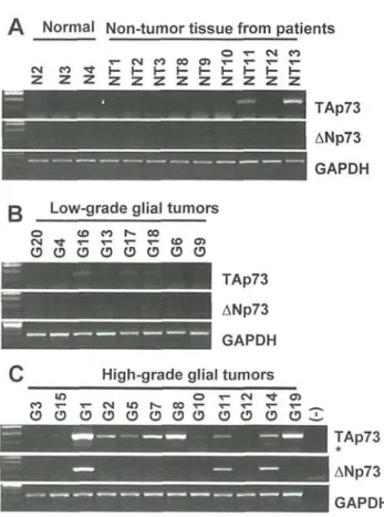
Figure 1. Differential expression of TAp73 and ANp73 in normal brain and malignant gliomas. The expression of TAp73 and ANp73 in (A), normal brain and non-tumor tissues; (B), low-grade tumor samples and (C), high-grade tumor samples. The expression of p73 transcript isoforms was performed semi-quantitatively, using GAPDH as a control. Asterisk denotes the PCR band corresponding to a minor p73 transcript isoform (A2) that lacks exon 2 (see text).
same PCR conditions and primers described at Sayan et al (15) and another using a forward primer at exon 3 (p73-NDNF: GCTGTACGTCGGTGACCCC) and a reverse at exon 5 (p73X5R). The PCR conditions and additional information is available upon request.
Densitometric analysis of transcripts. The ethidium
bromide-stained agarose gel pictures were captured by Bio-Rad Gel Doc 2000 system and analyzed by NIH-Scion Image analysis program. Following a background reduction, the crude absorption of the bands was calculated. The samples were normalized with the corresponding GAPDH values. Next, the maximum values registered with normal brain samples for each class of transcripts were calculated. Then, we calculated the ratio of each sample value to that maximum value observed in normal brain. The ratio of 3.0 or higher was chosen arbitrarily as a significant increase.
Results
The differential expression of TAp73 and ANp73 transcript isoforms in normal and diseased brain tissues was analyzed using two separate primer pairs for each isoform, as described in Materials and methods. The results obtained by these alternative primer pair combinations were essentially similar for both TAp73 and ANp73 isoforms. The data obtained with
primer pairs described in Fillippovich et al (23) for TAp73 isoform and Sayan et al (15) for ANp73 isoform are presented in Fig. 1, as an example.
The expression ofTAp73 and ANp73 gene in non-tumor brain tissues. We used 12 tissues for this purpose. Three samples
from non-cancer patients were used as normal brain tissues. As shown in Fig. 1A, these normal tissues did not express detectable levels of p73 transcripts under semi-quantitative PCR conditions (see Materials and methods). Under these conditions, 9 additional non-tumor brain tissues were also negative, except for 2 tissues that showed weak TAp73 expression (Fig. 1 A). In order to quantify the changes in the levels of TAp73 and ANp73 under different conditions, we compared the densitometric intensities of the PCR bands as explained in Material and methods. Using the normal brain as a background reference, a ratio of 3.0-fold or higher was set arbitrarily as a significant increase. Accordingly, the TAp73 levels were determined to be positive in non-tumor samples NT11 and NT13, with ratios 3.0 and 5.2 respectively (Fig. 1A).
The expression of TAp73 and ANp73 gene in low-grade versus high-grade glial tumors. A total of 20 brain tumors
were tested. Low-grade tumor group consisted of 8 samples (Table I). Similar to non-tumor brain samples, these low-grade tumors did not display a significant expression of TAp73 or ANp73 transcripts, with the exception of one tumor (G16) that displayed a ratio of 3.1 for TAp73 (Fig. IB). In sharp contrast with these low-grade tumors, most of the high-grade glial tumors displayed increased levels of TAp73 (Fig. 1C). A significant increase (ratio 3.0 or higher) of TAp73 was detectable in eight out of 12 tumors (67%). In some tumors (i.e., Gl, G8, G14), this increase was more than 10-fold, when compared to normal brain (data not shown). The ANp73 levels were also increased, but only in 3/12 (25%) of these high-grade glial tumors (Fig. 1C). It was noted that a faster migrating PCR band was also detectable with TAp73 in some high-grade tumors (shown with asterisk in Fig. 1C). Nucleic acid sequencing revealed that this weakly positive band represents the N-terminal alternatively spliced D2 form lacking the exon 2 sequence (11) co-amplifying with TAp73 sequences (data not shown).
Discussion
We report here that p73 gene expression in normal human brains is undetectable under semi-quantitative PCR conditions. Similarly, most brain tissues from patients with low-grade glial tumors also lack significant p73 expression. In contrast, the expression of p73 is induced in most high-grade glial tumors that display increased levels of TAp73 transcripts. This increase in TAp73 levels is accompanied with a concomitant increase in the levels of ANp73, in some but not all tumors. Although the differential expression of p73 isoform in these cancers has not been reported previously, Loiseau et al (24) defined a grade-dependent increase of total p73 transcripts in astrocytic tumors as compared to non-tumor samples. Dong et al (25) have also reported that total p73 transcript levels were increased in some astrocytic tumors, although they also reported a decrease in some other
High-grade glial tumors Low-grade glial tumors
tumors. Kamiya and Nakazoto studied the expression of p73 by immunohistochemistry and reported an increase in p73 p r o t e i n l a b e l i n g i n d e x in g r a d e IV a s t r o c y t o m a s when compared to grade I, II and III astrocytomas (26). Thus, in a g r e e m e n t with these studies, our observations p r o v i d e additional evidence that the expression of p73 gene is indeed induced in advanced glial tumors.
More importantly, we demonstrate that the induction of p73 gene expression in advanced glial tumors results in the production of the transcriptionally active TAp73 isoform in most cases, although additional forms including ANp73 and D2 isoforms are also detected in some of these tumors. As stated earlier, the protein encoded by TAp73 is a pro-apoptotic and growth-inhibitory molecule, whereas ANp73 transcripts encode oppositely-acting anti-TAp73 and anti-p53 molecules (11,13,16). Therefore, it may appear quite surprising that it is the TAp73 rather than ANp73 that is up-regulated in glial tumors, as they evolve from low- to high-grade tumors. We believe that this pattern of p73 expression may reflect the implication of a well-described oncogenic pathway in glioma-genesis. One of the dysregulations during gliomagenesis, especially in astrocytoma-derived tumors, is the disruption of the retinoblastoma pathway. It has been clearly established that the transformation from low to i n t e r m e d i a t e - g r a d e gliomas (anaplastic astrocytomas) is accompanied by allelic losses on chromosomes 9p and 13q, and less frequently by 12q amplification. The allelic losses involve the inactivation of R B I and pl6I N K 4 A genes, whereas the 12q amplification
reflects the amplification of cyclin-dependent kinase 4 (27). Experimental evidence demonstrates that the inactivation of the retinoblastoma pathway causes an abnormal activation of E2F transcription factors acting as key positive regulators of cell cycle progression from G l to S phase (28). Oncogenic activation of E2F1 also results in the induction of TA-p73, by the positive regulation of the first promoter of the p73 gene (29). The alternative p r o m o t e r of p73 which drives the expression of ANp73 is not controlled by E2F1, although it is responsive to TAp73 protein (12-14). Thus, the induction of TAp73 in advanced gliomas correlates with the inactivation of retinoblastoma pathway which occurs in intermediate-grade tumors. In support of this hypothesis, we observed in a retinoblastoma-deficient cell line (Hep3B) that ectopically-introduced E 2 F 1 induces the expression of e n d o g e n o u s TAp73, followed by a weaker induction of ANp73 expression (Karakuzu O et al, unpublished data).
Although we do not know whether the TAp73 expression affects the behavior of malignant gliomas, it is noteworthy that the inactivation of p53 tumor suppressor gene is an early event in gliomagenesis. In human gliomas, p53 mutations are primarily missense mutations and target the evolutionarily conserved domains in exons 5, 7, and 8, thus affecting residues that are crucial to DNA binding (27). As some mutant p53 molecules were shown to effectively inhibit the activity of TAp73 protein, malignant glioma cells could use mutant p53 protein to antagonize tumor suppressor functions of TAp73 protein (30).
Taken together our observations provide indirect evidence that, glioma tumors up-regulate the expression of TAp73 in relation with the inactivation of the retinoblastoma pathway in these tumors. Thus, the study of TAp73 expression in these
malignancies can serve as a marker for tumor progression. This could be particularly important for the selection of patients for new therapeutic trials aiming to kill selectively gliomas displaying retinoblastoma pathway inactivation (31). It is not clear whether the increase of TAp73 transcripts is associated with a c o n c o m i t a n t a c c u m u l a t i o n of T A p 7 3 p r o t e i n in advanced gliomas. However, it appears that, even if it is present in these tumors, it must be in a dormant/inactive state, probably by mutant p53 proteins and/or dominant negative p73 gene derived proteins. However, TAp73 protein was shown to be activated by some chemical drugs. Therefore, it would be interesting to test whether the TAp73 protein could be used as a target for glioma therapy by drug-induced activation of its pro-apoptotic functions (32).
Acknowledgements
This work was supported by grants from Bilkent University and TUBA and partially by TUBITAK (Turkey).
References
1. Vandenberg SR: Current diagnostic concepts of astrocytic tumors. J Neuropathol Exp Neurol 51: 644-657, 1992.
2. Kleihues P, Louis DN, Scheithauer BW, et al: The WHO classification of tumors of the nervous system. J Neuropathol Exp Neurol 61: 215-225, 2002.
3. Salcman MS, Scholtz H, Kaplan RS and Kulic S: Long-term survival in patients with malignant astrocytoma. Neurosurgery 34: 213-220, 1994.
4. Sasaki M and Plate KH: Gene therapy of malignant glioma: recent advances in experimental and clinical studies. Ann Oncol 9: 1155-1166, 1998.
5. Levine AJ, Chang A, Dittmer D, Notterman DA, Silver A, Thorn K, Welsh D and Wu M: The p53 tumor suppressor gene. J Lab Clin Med 123: 817-823, 1994.
6. Malkin D: p53 and the Li-Fraumeni syndrome. Cancer Genet Cytogenet66: 83-92, 1993.
7. Fueyo J, Gomez-Manzano C, Yung WK and Kyritsis AP: The functional role of tumor suppressor genes in gliomas: clues for future therapeutic strategies. Neurology 51: 1250-1255, 1998. 8. Nieder C, Petersen S, Petersen C and Thames HD: The challenge
of p53 as prognostic and predictive factor in gliomas. Cancer Treat Rev 26: 67-73, 2000.
9. Kaghad M, Bonnet H, Yang A, et al: Monoallelically expressed gene related to p53 at 1 p36, a region frequently deleted in neuro blastoma and other human cancers. Cell 90: 809-819, 1997. 10. Stiewe T and Putzer BM: Role of p73 in malignancy: tumor
suppressor or oncogene? Cell Death Differ 9: 237-245, 2002. 11. Melino G, De Laurenzi V and Vousden KH: p73: friend or foe
in tumorigenesis. Nat Rev Cancer 2: 605-615, 2002.
12. Zaika Al, Kovalev S, Marchenko ND and Moll UM: Over-expression of the wild type p73 gene in breast cancer tissues and cell lines. Cancer Res 59: 3257-3263, 1999.
13. Stiewe T, Theseling CC and Putzer BM: Transactivation-deficient Delta TAp73 inhibits p53 by direct competition for DNA binding: implications for tumorigenesis. J Biol Chem 277: 14177-14185,2002.
14. De Laurenzi V, Costanzo A, Barcaroli D, et al: Two new p73 splice variants, gamma and delta, with different transcriptional activity. J Exp Med 188: 1763-1768, 1998.
15. Sayan AE, Sayan BS, Findikli N and Ozturk M: Acquired expression of transcriptionally active p73 in hepatocellular carcinoma cells. Oncogene 20: 5111-5117, 2001.
16. Grob TJ, Novak U, Maisse C, et al: Human delta Np73 regulates a dominant negative feedback loop for TAp73 and p53. Cell Death Differ 8: 1213-1223, 2001.
17. Yang A, Walker N, Bronson R, et al: p73-deficient mice have neurological, pheromonal and inflammatory defects but lack spontaneous tumours. Nature 404: 99-103, 2000.
18. Pozniak CD, Radinovic S, Yang A, McKeon F, Kaplan DR and Miller FD: An anti-apoptotic role for the p53 family member, p73, during developmental neuron death. Science 289: 304-306, 2000.
ONCOLOGY REPORTS 11: 1337-1341,2004 1341
19. Zaika AI, Slade N, Erster SH, et at. DeltaNp73, a dominant-negative inhibitor of wild-type p53 and TAp73, is up-regulated in human tumors. J Exp Med 196: 765-780, 2002.
20. Maris JM and Matthay KK: Molecular biology of neuroblastoma. J Clin Oncol 17: 2264-2279, 1999.
21. Casciano I, Mazzocco K, Boni L, et at. Expression of DeltaNp73 is a molecular marker for adverse outcome in neuroblastoma patients. Cell Death Differ 9: 246-251, 2002.
22. Douc-Rasy S, Barrois M, Echeynne M, et at DeltaN-p73alpha accumulates in human neuroblastic tumors. Am J Pathol 160: 631-639,2002.
23. Fillippovich I, Sorokina N, Gatei M, et at Transactivation-deficient p73alpha (p73Deltaexon2) inhibits apoptosis and competes with p53. Oncogene 20: 514-522, 2001.
24. Loiseau H, Arsaut J and Demotes-Mainard J: p73 gene transcripts in human brain tumors: overexpression and altered splicing in ependymomas. Neurosci Lett 263: 173-176, 1999.
25. Dong S, Pang JC, Hu J, Zhou LF and Ng HK: Transcriptional inactivation of TP73 expression in oligodendroglial tumors. Int J Cancer 98: 370-375, 2002.
26. Kamiya M and Nakazato Y: The expression of p73, p21 and MDM2 proteins in gliomas. J Neurooncol 59: 143-149, 2002. 27. Maher EA, Furnari FB, Bachoo RM, et at. Malignant glioma:
genetics and biology of a grave matter. Genes Dev 15: 1311-1333, 2001.
28. Muller H and Helin K: The E2F transcription factors: key regulators of cell proliferation. Biochim Biophys Acta 1470: M1-M12, 2000.
29. Stiewe T and Putzer BM: Role of the p53-homologue p73 in E2F1 -induced apoptosis. Nat Genet 26: 464-469, 2000.
30. Bergamaschi D, Gasco M, Hiller L, et at p53 polymorphism influences response in cancer chemotherapy via modulation of p73-dependent apoptosis. Cancer Cell 3: 387-402, 2003. 31. Fueyo J, Alemany R, Gomez-Manzano C, et at. Preclinical
characterization of the antiglioma activity of a tropism-enhanced adenovirus targeted to the retinoblastoma pathway. J Natl Cancer Inst 95: 652-660, 2003.
32. Gong JG, Costanzo A, Yang HQ, et at The tyrosine kinase c-Abl regulates p73 in apoptotic response to cisplatin-induced DNA damage. Nature 399: 806-809, 1999.