A S A N O v a APOPTOSIS M ARKER
A T iilS IS SUBM TTEO TO
'
8W L06Y AHD SENETtCS
A R S THE iNSTirUTE Or Q ie M E E R IW n U IO S № R C E O F
firnAt FU U % tM a iT O F TH E RESUiREM EHTS
£ B EiM E OF D
ÑAPO
AS A NOVEL APOPTOSIS MARKER
A THESIS SUBMITTED TO
THE DEPARTMENT OF MOLECULAR BIOLOGY AND GENETICS AND THE INSTITUTE OF ENGINEERING AND SCIENCE OF
BILKENT UNIVERSITY
IN PARTIAL FULFILLMENT OF THE REQUIREMENTS FOR THE DEGREE OF DOCTOR OF PHILOSOPHY
BY
BERNA S. SAYAN AUGUST, 2002
I certify That I read this thesis and in my opinion it is fully adequate, in scope and quality, as thesis for the degree of Doctor of Philosophy
Prof Dr. Mehmet Öztürk
I certify That I read this thesis and in my opinion it is fully adequate, in scope and quality, as thesis for the degree of Doctor of Philosophy
Prof Dr. Ahmet Koman
I certify That I read this thesis and in my opinion it is fully adequate, in scope and quality, as thesis for the degree of Doctor of Philosophy
Prof DkiCuyaş Buğra
I certify That I read this thesis and in my opinion it is fully adequate, in scope and quality, as thesis for the degree of Doctor of Philosophy
...
Assist, ^ ro f Tamor Yağcı
I certify That I read this thesis and in my opinion it is fully adequate, in scope and quality, as thesis for the degree of Doctor of Philosophy
Assisi) P rof Uygar Tazebay
Approved for the Institute of Engineering and Science
ABSTRACT
ÑAPO as a novel apoptosis marker Berna S. Sayan
Ph.D. in Molecular Biology and Genetics Supervisor: Prof. Dr. Mehmet Oztiirk
2002, 128 pages
Apoptosis or programmed cell death plays a pivotal role in embryonic development and maintenance of homeostasis. It is also involved in the etiology and of pathophysiological conditions such as cancer, neurodegenerative, autoimmune, infectious and heart diseases. Consequently, the study of apoptosis is now at center of both basic and clinical research applications. Therefore sensitive and simple apoptosis detection techniques are required. This study involves identification and characterization of a monoclonal antibody-defined novel antigen, namely NAPO (negative in apoptosis), which is specifically lost during apoptosis. The anti-NAPO antibody recognizes two nuclear polypeptides of 60 kD and 70 kD. The antigen is maintained in quiescent and senescent cells, as well as in different phases· of the cell cycle including mitosis. Thus, immunodetection of NAPO antigen provides a specific, sensitive and easy method for differential identification of apoptotic and non-apoptotic cells.
ÖZET
Yeni bir apoptoz belirteci: NAPO
Berna S. Sayan
Doktora Tezi, Moleküler Biyoloji ve Genetik Bölümü Tez yöneticisi: Prof. Dr. Mehmet Öztürk
2002, 128 sayfa
Apoptoz veya programlanmış hücre ölümü embriyonik gelişimde ve homeostazm sağlanmasında çok önemli bir role sahiptir. Ayrıca kanser, nörodejeneratif hastalıklar, otoimmün hastalıklar ve kalp hastalıklarının patofızyolojilerinde de rol alır. Buna bağlı olarak, apoptozun tayini ve çalışılması hem temel hem de klinik araştırmalar açısından son derece önemlidir. Bu sebeple apoptoz tayininde kullanılacak hassas ve basit metodların geliştirilmesi gerekmektedir. Bu çalışma NAPO (apoptozda negatif) adlı, spesifik olarak apoptoz sırasında yok olan yeni bir antijenin bulunması ve karakterizasyonunu içermektedir. NAPO’ya karşı olan anti-NAPO antikoru 60 ve 70 kDa civarında yürüyen iki adet polipeptidi tanımaktadır. Bu antijen kuisens, senesens ve mitoz da dahil olmak üzere hücre siklüsünün tüm safhalarında varlığını sürdürmektedir. Yani NAPO antijeninin immünodeteksiyonu apoptotik olan ve olmayan hücrelerin teşhisi için kullanılabilinecek spesifik, hassas ve kolay bir metoddur.
a c k n o w l e d g e m f:n t s
First, I would like to thank my supervisor Prof. Dr. Mehmet Ozturk for giving me the chance to enter the splendid world of Molecular Biology and Genetics in Turkey and particularly for letting me enter and discover this world with him, under the light of his extraordinary experience and knowledge, admirable brilliance and forethought, and amazing patience. I know, I would not be able to succeed, if I had worked with someone else. I have not only learned scientific thinking from him, but he has also influenced my scope of living.
1 also would like to thank my husband Emre, for being such a perfect person and being with me in all my good and bad days. I do appreciate him for all the things we have shared in the last seven years. He is one of the very few people who has encouraged me and take me up whenever I was down, and make me smile whenever I was sad. He is not only a great spouse but also a great working partner. I truly owe him the point where I am at the moment. I thank him for all his support, motivation and love. I feel very lucky to have him in my life.
I would like to thank my father Enver Özçelik and my mother Keriman Özçelik for raising me as the person I am. I thank my father for teaching me to struggle with difficulties and to be a diligent person. He showed me that honesty, idealism, intellectuality and hard-working will always open the doors of success. His principles will always guide my life. I am deeply missing you... I also want to thank my mother for her encouragement, support, love and most importantly for being my best friend. She has always guided me with her tenderness, tolerance, deep love and understanding. I thank my sister Ayşin for being with me whenever I needed her. She will always be a support in every step I take in my life.
I also want to express my deepest gratitude to Esin and Erol Sayan, for their indispensable support and encouragement and to Eser and Korean for their friendship.
Very special thanks to my best friends Abdullah Yalcin and Ahmet Ucar. The times I spent with them were the best times 1 have spent in Bilkent. They made my time here memorable and unforgettable.
I also would like to thank all the faculty of the department for their guidence and helps.
Special thanks to the members of the “Molecular Oncology Group”: Esra, Tolga, Nuri and Ozgur for their helps and friendship. Also thanks to Tuba G., Banu, Ebru, Cemaliye, Belhaj, Hani and Ai'zu. Biggest thanks to Tulay, for her “always good mood” and snailing face. Also to Fusun for her friendship and supply of reagents at maximum speed and I also want to thank Sevim Baran, Yavuz Ceylan and Abdullah Unlu.
TABLE OF CONTENTS PAGE ABSTRACT...i ÖZET...ii ACKNOWLEDGEMENT... iii TABLE OF CONTENTS...v LIST OF TABLES...xi LIST OF FIGURES...xii ABBREVIATIONS...xv CHAPTER I INTRODUCTION 1 -1 General Introduction... I 1-2 C.Elegans As The Model Organism...7
1-3 Caspases... 9
1-3.1 Structure Of Caspases...9
1-3.2 Regulation Of Caspase Activation... 13
1-3.3 Inhibitor Of Apoptosis Proteins (lAPs)... 14
1-3.4 Targets Of Active Caspa.ses...15
1 -4 The BCL-2 Family...16
1 -4.1 General Structure... 16
1-4.2 Bcl-2 Family Function In Apoptosis... 19
1-4.3 Regulation Of Bcl-2 Family Members... 19
1-4.3.1 Post-Translational Modifications... 19
1-4.3.2 Cellular Localization And... 21
Dimerization 1-5 Death Receptor Pathway... 22
1-5.1 CD95/Fas/Apo-l... 23
1-5.2 TNF Receptors... 25
1-5.3 TRAIL Receptors...26
1-6 Mitochondria...27
1-6.1 General Structure...28
1-6.2 Cytochrome C Release From The... 29
Mitochondria And Apoptosis 1-6.3 Other Factors Released From The Mitochondria... 32
During Apoptosis 1-6.3.1 SMAC/DIABLO... 32
1-6.3.2 Apoptosis Inducing Factor (AIF)... 32
1-6.3.3 Pro-Caspases...33
1 -7 Regulation Of Apoptosis...33
1-8 Role Of Apoptosis In Human Disease... 34
CHAPTER II DETECTION OF APOPTOSIS IN CELLS AND TISSUES 2-1 Analysis of morphological features of apoptosis... 40
2-2 DNA fragmentation assays... 41
2-3 Analysis of free DNA ends... 43
2-4 Use of Annexin V... 44
2-5 Detection of caspase activity...44
CHAPTER III AIM OF THE STUDY...46
CHAPTER IV MATERIALS AND METHODS 4-1 Production of unti-NAPO antibody from hybridomas... 48
4-1.1 Production of anti-A^APO antibody producing... 48
hybridomas 4-1.2 Production of anti-NAPO ascites in Balb/c mice... 49
4-1.3 Characterization of Ig subtype of the anti-NAPO... 49
antibody 4-2 Screening of À,TripIEx expression library with the anti-... 50
NAPO antibody 4-2.1 Bacterial strains... 50
4-2.2 Solid and liquid mediums... 50
4-2.3 Antibiotics...51
4-2.4 Growth and maintenance of primary and working... 51
bacterial stoeks 4-2.5 Titration of A-TriplEx expression library... 51
4-2.6 Transfer of plaques to nitrocellulose filters... 53
4-2.7 Immunodetection... 54
4-2.7.1 Optimization of the secondary... 54
antibody concentration 4-2.7.2 Immunodetection... 54
4-3 Western Blotting with the anti-NAPO antibody... 54
4-3.1 Tissue culture studies... 54
4-3.1.1 Defrosting cells... 55
4-3.1.2 Subculturing of cells... 57
4-3.2 Protein extraction from cells... 57
4-3.3 Bradford assay for protein quantitation... 57
4-3.4 SDS-Polyacrylamide gel electrophoresis of... 58
proteins 4-3.5 Transfer of proteins from SDS-polyacrylamide... 63
gels to solid supports 4-3.6 Staining proteins immobilized on solid... 65
surfaces with Ponceau S 4-3.7 Dénaturation and renaturation of proteins on...65
nitrocellulose membranes 4-3.8 Immunological detection of immobilized... 66
proteins (Western Blotting) 4-3.9 Detection of proteins immobilized on membranes... 66
4-4 Immunoprécipitation with the anti-NAPO antibody... 67
4-6 Induction of apoptosis in different cell lines... 68
4-6.1 Induction of apoptosis by serum starvation... 68
4-6.2 Induction of apoptosis by oxidative stress and cisplatin.. 69
4-6.3 Induction of apoptosis by UV-C treatment... 69
4-6.4 Induction of apoptosis by activation of... 69
death-receptor mediated apoptosis 4-6.4.1 Induction of apoptosis by TN F-a... 69
treatment 4-6.4.2 Induction of apoptosis by anti-Fas... 70
antibody treatment 4-7 p53 staining of apoptotic versus non-apoptotic Huh7 cells... 70
4-8 TUNEL staining of apoptotic cells... 70
4-9 Synchronization of Huh7 and MRC5 cells... 71
4-9.1 Synchronization of MRC5 cells by serum withdrawal.... 71
4-9.2 Synchronization of Huh7 cells by nocodazole treatment. 71 4-9.2.1 Optimization of nocodazole concentration 72 4-9.2.2 Mitotic .shake-off... 72
4-10 BrdU labeling and identification of S phase cells... 72
4-11 Induction of quiescence in MRC5 cells... 73
4-12 Senescence associated P-Galactosidase Assay... 73
CHAPTER V RESULTS 5-1 Introduction... 74
5-2 Production of the anti-NAPO monoclonal antibody... 75
5-2.1 Production of anti-NAPO antibody producing... 75
hybridomas 5-2.2 Characterization of Ig subtype of the anti-... 75
NAPO antibody 5-3 Screening of A,TripIEx expression library with the... 75
5-3.1 Titration of À,TripIEx expression library... 75
5-3.2 Optimization of the secondary antibody concentration... 76
5-3.3 Immunodetection...76
5-4 Biochemical characterization of the NAPO antigen... 76
5-4.1 Western Blotting with the anti-NAPO antibody... 76
5-4.2 Immunoprécipitation with the anti-NAPO antibody... 77
5-4.3 Immunofluorescence with the anti-NAPO antibody... 77
5-4.3.1 Staining of HOC cell lines with... 77
the anti-NAPO antibody 5-4.3.2 Species specific expression of the... 78
NAPO antigen 5-5 Identification of NAPO as an apoptotic marker... 79
5-5.1 NAPO immunoreactivity of apoptotic... 80
SNU 398 and Huh7 cells 5-5.2 p53 immunoreactivity of apoptotic Huh7 cells.... 82
5-5.3 NAPO immunostaining in cells treated with... 83
various apoptotic stimuli 5-5.3.1 NAPO immunostaining in death-... 83
receptor mediated apoptosis 5-5.3.2 NAPO immunostaining in UV-C... 84
irradiation induced apoptosis 5-5.4 NAPO immunostaining in tumorous versus non-... 85
tumorous cells exposed to various apoptotic stimuli 5-5.4.1 NAPO immunostaining in apoptotic... 86
tumorous cell lines versus viable counterparts 5-5.4.2 NAPO immunostaining in apoptotic... 86
non-tumorous celt lines versus viable counterparts 5-6 Expression of the NAPO antigen in quiescent cells... 89
5-7 Expression of the NAPO antigen during cell cycle... 91
5-7.1 Expression of NAPO in synchronized Huh7 cells... 92
mitotic shake-off
5-7.1.2 Determination of S phase cells by... 93 BrdU incorporation assay
5-7.1.3 NAPO immunostaining at different... 94 phases of the cell cycle
5-7.2 Expression of NAPO in synchronized MRC-5 cells... 96 5-7.1.1 Synchronization of Huh7 cells by... 92
5-7.2.1 Synchronization of MRC-5 cells... by serum starvation
.... 96
5-7.2.2 Determination of S phase cells... by BrdU incorporation assay
... 96
5-7.2.3 NAPO immunostaining at different...
phases of the cell cycle
.... 98
5-8 Expression of the NAPO antigen in senescent cells... 100
CHAPTER VI
DISCUSSION AND PERSPECTIVES. .103
CHAPTER VII
REFERENCES. 12
L IST O F TA BL E S
NUMBER/NAME PAGE
Table 1-1: Differences between apoptosis and necrosis... 3
Table 1-2: The main sub-groups of mammalian caspases... 9
Table 1-3: Dysrégulation of apoptosis in some autoimmune... 35
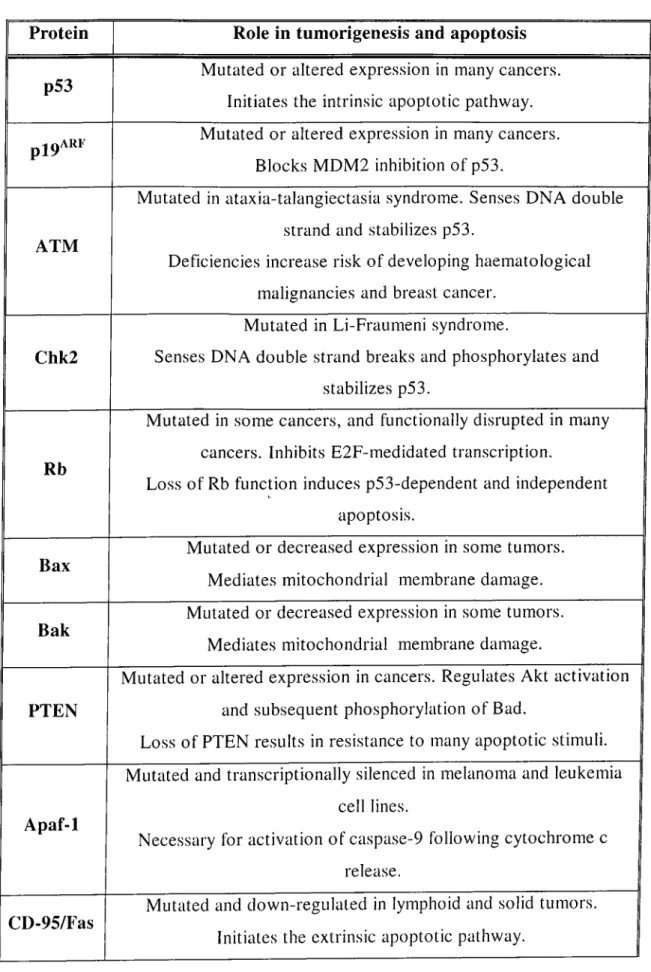
diseases. Table 1-4: Summary of the Roles of Apoptotic Initiators,... 38
Regulators or Executioners in Tumorigenesis and Apoptosis Table 4-1: Concentrations of the antibiotics used in this study... 51
Table 4-2: Dilution chart of A,TripIEx for titration... 52
Table 4-3: The cell lines used for characterization of NAPO... 56
Table 4-4: Effective range of separation of SDS-PAGE gels... 59
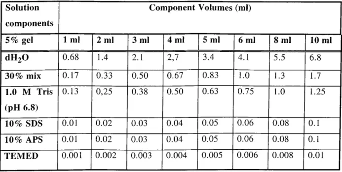
Table 4-5: Solution of preparing resolving gels for Tris-glycine... 61
SDS-PAGE. Table 4-6: Solution of preparing 5% stacking gels for... 63
Tris-glycine SDS-PAGE. Table 5-1: Expression of the NAPO antigen in cell lines of... 79
different species. Table 5-2: List of cell lines tested for loss of NAPO... 80
immunoreactivity after induction of apoptosis by various stimuli.
L IST OF FIG URES
NUMBER/NAME PAGE
Figure 1-1: Schematic representation of mechanisms leading... 2 to apoptotic cell death.
Figure 1-2: The apoptotic pathway is conserved in metazoans... 4 Figure 1-3: Activation of death-receptor mediated apoptosis... 5
through binding of the cognate receptor to its ligand.
Figure 1-4: Induction of apoptosis through cytochrome c ...6 release from the mitochondria.
Figure 1-5: Activation of apoptosis machinery in the nematode... 8 C. elegans.
Figure 1-6: Activation of caspases... 11 Figure 1-7: Structure of some of the initiator and... 12
effector caspases.
Figure 1-8: Release of inhibitory effects of lAPs on caspases... 15 by the SMAC/DIABLO protein.
Figure 1-9: Structure of some pro- and anti-apoptotic Bcl-2... 18
family members.
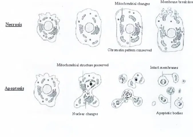
Figure 2-1: Basic morphological features of apoptosis,... 41
differing from necrosis used in the detection of apoptosis.
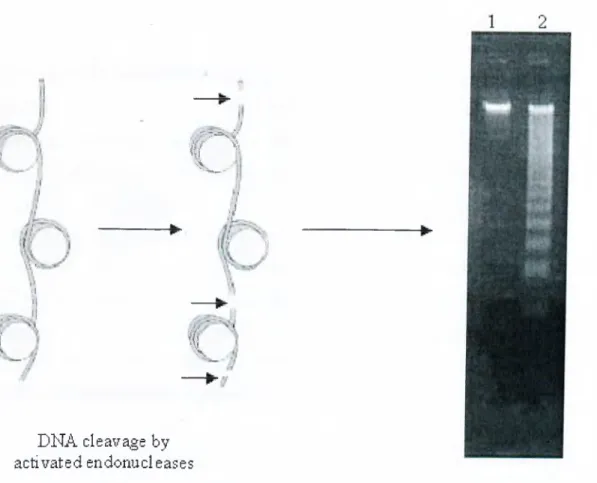
Figure 2-2: Principle of DNA laddering assay and a typical... 42
gel appearance of apoptotic cell DNA.
Figure 2-3: Labelling principle of terminal polymerase... 43 and terminal transferase
Figure 2-4: Use of PARP cleavage for the detection of... 45
Huh7 cell lysate.
Figure 5-2: Nuclear localization of the NAPO antigen in Huh7... 78 and SNU 398 cell lines.
Figure 5-3: Loss of i\\t NAPO antigen in serum-starved... 81 SNU 398 HCC cells.
Figure 5-4: NAPO immunoreactivity of apoptotic Huh7 cells... 82 in comparison with TUNEL assay
Figure 5-5: p53 staining of apoptotic Huh7 cells... 83
Figure 5-6: NAPO immunostaining in death receptor... 84 mediated apoptosis in MCF-7 and Jurkat cells.
Figure 5-7: NAPO staining of UV-C induced apoptotic... 85 MCF-7 and Jurkat cells.
Figure 5-8: Expression of NAPO in cells induced to undergo... 87
apoptosis by UV-C irradiation.
Figure 5-9: Expression of NAPO in non-tumorous cells... 88 induced to undergo apoptosis.
Figure 5-10: Brd-U incorporation test to serum starved and... 90
non-starved p l8 MRC-5 cells.
Figure 5-11: VAPO immunostaining of serum starved and... 91
non-starved p l8 MRC-5 cells.
Figure 5-12: Optimization of the most efficient nocodazole... 93
concentration required to arrest Huh7 cells in M phase of the cell cycle with the least cytotoxic effect.
Figure 5-13: BrdU incorporation index of M phase arrested... 94
Huh7 cells after mitotic shake-off.
Figure 5-14: VAPO immunostaining of synchronized Huh7 cells... 95 Figure 5-15: BrdU incorporation assay of synchronized... 97
MRC-5 cells.
Figure 5-16: BrdU incorporation index of serum starved MRC-5... 98
cells after release from the quiescent state.
Figure 5-17: NAPO immunostaining of synchronized MRC-5 cells.... 99
Figure 5-1: Immunoprécipitation of the antigen from... 77
Figure 5-18: SA-β Galactosidase and ÑAPO immunostaining of... 101
ABBREVIATIONS
AIDS Acquired Immunodeficiency Virus
AIF Apoptosis Inducing Factor
ANT Adenine Nucleotide Translocator
Apaf-1 Apoptotic Protease Activating Factor
ASK Apoptosis Signal Regulating Kinase
ATP Adenosine triphosphate
BH Domain Bcl-2 Homology Domain
BIR Baculovirus lAP Repeats
Bisacrylamide N, N, methylene bis-acrylamide
Bp Base
Pail-BSA Bovine Serum Albumin
C. e le gems Caenorhabditis elegcins
CAD Caspase Activated DNase
Caspase Cysteine Aspartyl Protease
CARD Caspase Recruitment Domain
CD Cluster of Differentiation
ced Cell Death Defective
CO2 Carbon Dioxide
crmA Cytokine Response Modifier A
D. melcinogaster Drosophila melanogaster
Da Dalton
Daxx Death Domain Associated Protein
DcR Decoy Receptor
DD Death Domain
DED Death Effector Domain
DISC Death Inducing Signaling Complex
DMSO Dimethyl Sulfoxide
DNA deoxyribonucleic acid
dUTP deoxyuridine triphosphate
EOF Epithelial Growth Factor
ERK Extracellular Signal-Related Kinase
EtOH Ethanol
FADD Fas Associated DD
PCS Fetal Calf Serum
FIST Fas Interacting Seine/Threonine Kinase
FLIP Fas Associated DD like ICE Inhibitory Protein
HCC Hepatocellular Carcinoma
HIPK3 Homeodomain Interacting Protein Kinase
HIV Human immunodeficiency virus
HRP Horse Redish Peroxidase
lAP Inhibitor of Apoptosis proteins
ICAD Inhibitor of CAD
ICE Interleukin-1P Converting Enzyme
IkB Inhibitor of NF-kB
IF Interleukin
IPTG isopropylthio-P-D-galactoside
JNK/SAPK c-Jun N-terminal Kinase/Stress-activated protein
kinase
Kan kanamycin
Kb Kilo base
kDa kilo daltons
LB Luria-Bertani media
MAPK Mitogen Activated Protein Kinase
MgS04 Magnesium Sulfate
ml Mililiter
mg Miligram
MQ MilliQ water
N-terminus amino terminus
NaCl Sodium Chloride
NaOH Sodium Hydroxide
NAPO Negative in Apoptosis
NIK NF-kB Inducing Kinase
Nm nanometer (1/109 of a meter)
0/N Over Night
PAGE polyacrylamide gel electrophoresis
PARP Poly (ADP-ribose) polymerase
PBS Phosphate Buffered Saline
PBS-T Phosphate Buffered Saline with Tween-20
Pfu Plaque Forming Unite
PKA Protein Kinase A
PKB/Akt Protein kinase B
PKC Protein Kinase C
PTP Permeability Transition Pore
RAIDD RIP Associated ICH-I/Ced3 Homologous Protein
with a Death Domain
RIP Receptor Interacting Protein
RPM Revolutions per minute
SDS Sodium Dodecyl Sulfate
SDS-PAGE SDS- Polyacrylamide Gel Electrophoresis
Ser Serine
VDAC Voltage dependent Anion Channel
tBid Truncated Bid
TBS Tris.Buffered Saline
TBS-T Tris Buffered Saline with Tween-20
TEMED N,N,N,N-tetramethyl-1,2 diaminoethane
Tris tris (hydroxymethyl)-methylamine
TRADD TNF-R-associated DD
TRAE TNF Receptor Associated Factor
TRAIL TNF-related Apoptosis-Inducing Ligand
TRIP TRAF-Interacting Protein
TUNEL TdT-mediated dUTP nick end labeling
UV Ultraviolet
X-Gal 5-bromo-4-chloro-3-indolyl-p-D-galactoside
CHAPTER I
INTRODUCTION
1-1 GENERAL INTRODUCTION
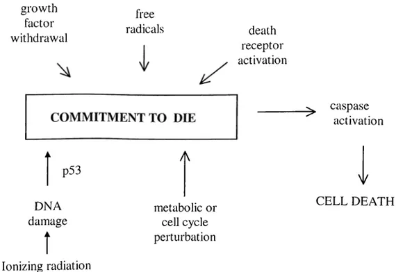
Apoptosis (programmed cell death) is the most common physiological form of cell death. It is essential for the precise regulation of cellular homeostasis and development as it serves to remove unwanted (excess, damaged or infected) cells at critical and appropriate times (Vaux and Korsmeyer, 1999; Wyllie and Golstein, 2001). Apoptosis is a tightly regulated process and can be activated by various mechanisms as summarized in figure 1-1. Many morphogenetic abnormalities and diseases are linked to dysrégulation of this process thus it is critical for development and survival of all metazoans (Reed, 2000; Mattson, 2000; Chervonsky, 1999; Roulston et al., 1999; Narula et al., 2000). For example in many different types of cancer, pro-apoptotic factors are inactivated and pro survival (anti-apoptotic) factors are upregulated. This leads to accumulation of apoptosis-resistant cells and since effective chemotherapy depends on the induction of programmed cell death, these types of cancers are hard to treat (Lowe and Lin, 2000).
Apoptotic cell death is characterized by a series of morphological changes including cell shrinkage, nuclear condensation, chromatin segregation, membrane blebbing, formation of membrane-bound apoptotic bodies and internucleosomal DNA cleavage. In more detail, chromatin condenses and forms aggregates near the nuclear membrane, the nucleolus becomes enlarged and appears abnormally granular. Chromatin is then degraded first into 300-350 kb, then into 180 bp fragments by the actions of different activated endonucleases (Brown and Rose, 1992; Oberhammer et al., 1993; Wyllie AH., 1980). Then the cells shrink and
form apoptotic bodies. These apoptotic bodies are rapidly phagocytosed and digested by neighboring cells or macrophages thus inflammation does not occur during apoptosis (Saraste and Pulkki, 2000). These features of apoptosis make it a unique mechanism of cell death. In the other form of cell death, which is necrosis, cells swell and inflammation occurs. The major differences between apoptosis and necrosis are summarized in table 1-1.
growth factor withdrawal free radicals death receptor activation p53 DNA damage
Î
Ionizing radiation A metabolic or cell cycle perturbation ^ caspase activation y CELL DEATHFigure 1-1: Schematic representation of mechanisms leading to apoptotic cell death.
T able 1-1: D ifferences betw een apoptosis and necrosis.
Features Necrosis Apoptosis
Stimuli
Toxins, severe hypoxia, massive insult and conditions of ATP
depletion
Physiological and pathological conditions without ATP depletion
Energy Requirement None ATP dependent
Histology
Cellular swelling, disruption of organelles, death of patches of tissue
Chromatin condensation, apoptotic bodies, death of
single isolated cells
DNA breakdown pattern Randomly sized fragments Ladder of fragments in internucleosomal multiples of 180 bp
Plasma membrane Lysed Intact, blebbed, with
molecular alterations
Phagocytosis of dead cells
Immigrant phagocytes Neighboring cells
Tissue reaction Inflammation No inflammation
Components of the apoptotic cell death mechanism are conserved in all metazoans, from nematodes to humans (figure 1-2). Thus, study of programmed cell death in the nematode “Caenorhabditis elegans” (C. elegans) has revealed both the mechanisms underlying the highly regulated apoptotic processes and the genes that are involved in the regulation of this process. As nomenclature, the genes whose protein products induce apoptosis are called “pro-apoptotic” and the ones whose protein products inhibit apoptosis are called “pro-survival” or “anti- apoptotic”.
In metazoans, apoptosis can be activated by one of the two pathways. The first is the activation of death receptors by binding of a death inducing ligand to its cognate receptor (summarized in figure 1-3). In the other pathway, apoptosis
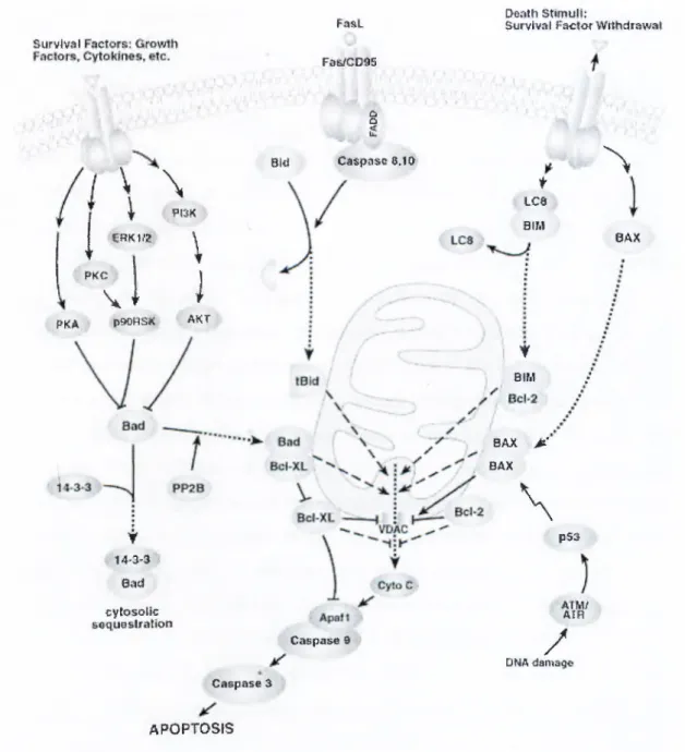
is induced by the release of cytochrome c from the mitochondria due to activation of apoptosis inducing factors such as the pro-apoptotic members of the bcl-2 family (summarized in figure 1-4). In both cases the final outcome is the activation of the caspase cascade.
C, Blegans M aftim als
E G L - 1 ' B ik i a a k l i i a x j
1
iCED^1
C E D - 4 :i
D: nmiaaogBstBf mm1
•k s vC^4 ' i· Smac^ RsBpe*' A p a f-1 i CSriffl: D A R K , OmiJ ни : I -T i I T C E b - a - C a s p a s e ;9 , J_ J_ f p R O N C ; ifte c to r К ^ Effector 1 ::^ s p a s e s ; ) easpa ses Ii ... ... 1....
Ap-Qptosis A p o p to sis Apoptosis
TH f TNFB1 AP0^3i AFO-2L CdS|>.1;S«3' 3
M / /
Ci&ll shririkagi» Membraiie bobbing CAD OisiA iragmentasioiii\
DNA fii pali
V y
APOPTOSIS
Figure 1-3: Activation of death-receptor mediated apoptosis through binding of the cognate receptor to its ligand, (taken from cellsignaling.com)
Survivíll
Fíís.1 fii^ctm
Peíith StimuH:
Survi^val Factor WílNcíir^w?*í
a D PKA i \
\
1 1 nm ««Km:. \ f \ PKC- ' 1 \ JÍ f P90RSÍI AKT ¥ i4^3-s;v 8,ntl cytosoíío seqyeslraslloti B\4 ClliS|>iaSO 6,1 íí f Lca BIM f eiM QAX jÉt' BAX\
^
p$3 ÁTftriíAIR Caspas« t/
DíáA damaig^ Caspas« 3 APOPTOSIS 0AXFigure 1-4: Induction of apoptosis through cytochrome c release from the mitochondria, (taken from cellsignaling.com)
1-2 C M egans AS THE MODEL ORGANISM
One of the organisms that have been used extensively for the study of apoptosis is the nematode C. elegans. During development 131 cells among 1090 cells die by apoptosis to leave a final total of 959 in the adult. The analysis of apoptosis in C. elegans has identified four steps in apoptosis. These are: (1) determination step, (2) execution step, (3) enguliment of apoptotic bodies, (4) degradation of engulfed cell DNA.
In the execution steps, three genes have been identified that play critical roles. These are named as “ced (cell death defective)” genes as they were first identified by analysis of mutant nematodes, which had abnormalities in their apoptotic procedures (Ellis and Horvitz., 1986). Two of these ced-3 and ced-4 are required for cell death. When a mutation occurs hitting one of these genes, none of the 131 cells dies. The third gene ced-9 acts as an antagonist of the other two ced genes. When a “gain of function” mutation occurs in ced-9 gene, cells become resistant to apoptosis and mutations leading to “loss of function” result in extensive apoptosis (Hengartner et al., 1992). Therefore, ced-3 and ced-4 are regarded as pro-apoptotic, whereas ced-9 is regarded as anti-apoptotic. Biochemical analysis revealed that these proteins function as a ternary complex, in which activation of the protease CED-3 by the adapter protein CED-4 is normally repressed by the anti-apoptotic CED-9. In cells destined to die, expression of EGL-1 results in displacement of CED-4, activation of CED-3, and cellular demise. In more detail, the protein encoded by ced-3; Ced-3 is a cysteine aspartyl protease (caspase), which cleaves after aspartate residues. Caspases are synthesized as inactive pro-enzymes and upon activation their N-terminal pro domain is cleaved resulting in the formation of an active caspase. The protein encoded by ced-4; Ced-4 can physically interact with both Ced-3 and Ced-9, acting as an adaptor molecule (Chinnaiyan et al., 1997). It contains a putative ATPase domain, which is required for its ability to activate Ced-3. The human homologue of Ced-4 is the Apaf-1 protein. The protein encoded by ced-9; Ced-9 prevents apoptosis by blocking the activation of Ced-3. It has been shown that Ced-9 utilizes two different mechanisms to inhibit Ced-3 mediated apoptotic pathways (Xue and Horvitz., 1997). It may interact directly with Ced-3 inhibiting the cleavage of pro-domain of Ced-3 or it may compete with Ced-3 and inhibit
apoptosis indirectly. For the latter mechanism first Ced-9 is cleaved by Ced-3 to form a Ced-3 like Ced-9 protein fragment. A schematic representation of the activation of apoptosis machinery in the nematode C. elegans is shown in figure 1-5, where egl-1 gene product acts as a pro-apoptotic protein for the inhibition of the anti-apoptotic Ced-9 protein.
EGL-1 (BH3 dornainl ol isomerization 4 3 r i- 3 . a Ced3 crocessine
i
artive Ced 3Caspases are cysteine proteases that cleave substrates after a conserved aspartate residue and are known to be the executers of apoptosis in metazoans. They are found as inactive zymogens and are activated when cleaved appropriately. The activated caspases initiate a caspase cascade by cleaving each other and the initially activated caspases are called “initiator ca.spases”. The caspases activated as a result of this cascade and have functions in the execution phase of apoptosis are called “effector caspases”. The first caspase; caspase-1 (ICE: Interleukin-1P converting enzyme) was identified in 1993 as the human homologue of ced-3 (Yuan et al., 1993). Today 14 different caspases are known. These caspases are divided into three main sub-groups according to their phylogenetic background as shown in table 1-2.
1-3 CASPASES
Table 1-2: The main sub-groups of mammalian caspases
Name Components
ICE-subfamily Caspa.se-1, -4, -5, -11, -12, -13, -14
Ced-3/CPP32 subfamily Caspase-3, -6, -7
ICH-1/Nedd2 subfamily Caspase-2, -8, -9,-10
1-3.1 STRUCTURE OF CASPASES
Caspases share structural and catalytic homologies. In all caspases the active site is composed of a pentapeptide sequence with the general structure QACXG (X=G, Q or R), where the cysteine in the middle, together with a distant histidine, is directly involved in the catalysis. Caspases recognize a tetrameric primary sequence in their targets, which contains an aspartic acid in the first position (Thornberry and Lazebnik. 1998).
The inactive caspases are called pro-caspases, and they contain a N- terminal prodomain, which is cleaved during activation. Caspases can be activated by one of the three mechanisms. They can cleave themselves (autoactivation), can be cleaved by previously activated caspases or they can be activated by non-caspase activators such as granzyme-B. After cleavage the cleaved subunits (large subunit and small subunit) form a heterodimer and two heterodimers form the active caspase as shown in figure 1-6.
Caspases differ in the length of their prodomains. Caspases -3, -6, -7 and - 10 contain short prodomains (10-40 residues). The caspa.ses with long prodomains contain recognizable domains in their prodomains involved in signal transduction via protein-protein interaction. Thus these prodomains play important roles in caspase regulation and function (Earnshaw et al., 1999). For example the prodomain of caspase-8 and -10 contain “death effector domain” in their prodomain, which mediate signal transduction between the death receptor and the downstream caspases through adaptor molecules. This allows death receptor-induced activation of downstream caspases in response to ligand binding to the receptor. The caspa.ses -1, -2, -4 and -9 contain CARD domain (caspase recruitment domain) in their prodomain. CARD domain is also present in the adaptor molecule Apaf-1. The CARD domain is thought to mediate specific intermolecular interactions that regulate caspase activation. Structures of some of the caspases are shown in figure 1-7.
prodomain ’ large subunit ^ sniallsnbunit
precursor
active enzyme
O
“ active siteFigure 1-6: Activation of caspases due to cleavage of the prodoniain region and cleavage of the remaining part into two subunits namely small and large subunits.
-Casp-3 ■Ca5p-7 ■Casp-6 ■Casp-8 -Casp-10 ■Casp-d •C89p-2 *Casp-4 •Casp-13 ■Casp-5 ’Casp-11 -fj30
21^
f D E D j f D E D
1
1521I I
j
CARO L card j II J 1 Lf -p1C 4 ’: 5 i . 31 & I 1331 , L J i; 3i«| umi l
2731 1200 r ~ n 311^ 331 r --- i r ¿ 7 7 1 1 1 1 n i ^ a i J&D3 1 1 l! 1II r ~ ■ I! 2 1 H | • ir yu
h21 416HI
3 7 7 TT 37/ 4lfl M73 11« C asp-12 n s f f i i n 1 I 111 m 11«!^ “ 3 »!/ Cusp-1 f CARD 1 1 II 2A2 C asp -l-l _ L _ _____ L L2 L3 L4Figure 1-7: Structure of some of the initiator and effector caspases.
All listed caspases are o f human origin except easpase-11 (mouse), -12 (mouse), and -13 (bovine). The initiator and effector caspases are labeled in purple and red, respectively. The position o f the first activation cleavage (between the large and small subunits) is highlighted with a large arrow while additional sites of cleavage are represented by medium and small arrows. L1-L4 represent the regions required for the formation o f the catalytic groove. The catalytic residue Cys is shown as a red line at the beginning o f L2.
The availability and activation of caspases are crucial steps in the commitment of a cell to die; therefore their regulation is strictly controlled and maintained by several cellular processes. For example, caspases are subject to transcriptional regulation and posttranslational modification (Earnshaw et al., 1999). In addition, active caspases can be permanently eliminated by the ubiquitination-mediated proteasome degradation pathway (Huang et al., 2000; Suzuki et al., 2001).
Being the executers of apoptosis, caspases are targets of various caspase inhibitory proteins. This inhibition plays a very important role in pathogenesis as well as tumorigenesis. For example, as infected cells commit suicide and undergo apoptosis for the benefit of the organism, some viruses have developed mechanisms to inhibit caspase function in order to overcome the cellular apoptosis response generated due to the viral infection.
Baculoviruses express the “caspase inhibitory protein p35”, which competes with the substrates of the caspase-8 for binding to the enzyme. After binding of p35 to the caspase, caspase-8 cleaves p35 and the N-terminal fragment of p35 protein blocks the active site of the caspase in an irreversible manner (Xu et al., 2001). In mammals a homologue of p35 has not yet been identified.
A cowpox protein “cr.mA” (cytokine response modifier A) has been shown to be the inhibitor of caspase-1 and -8. Like the baculovirus protein p35, crmA is also cleaved by the caspase that it inhibits, but the inhibitory effects results from a conformational change in the active site of the ca.spase due to crmA binding (Renatus et al., 2000).
Baculoviruses also encode for another type of proteins named “lAPs” (inhibitor of apoptosis proteins). The enzymatic activity of caspases is subject to inhibition by the conserved lAP (inhibitor of apoptosis) family of proteins (Deveraux and Reed, 1999).
Some chemicals are also known to inhibit caspase activation. These are aldehydes or fluoromethyl ketone-derivatized synthetic peptide inhibitors such as (ZVAD-fmk).
1-3.3 INHIBITOR OF APOPTOSIS PROTEINS (lAPs)
lAPs have been identified in many different organisms ftom viruses to mammals due to the presence of a 65 amino acid homology domain named “BIR” (bacLilovirus lAP repeats). These repeats are typically found in the N-terminal region of the lAPs and mediate various types of protein-protein interactions. Certain lAPs also contain a C-terminal ring finger, which is presumed to mediate other specific protein-protein interactions. Eight distinct mammalian lAPs, including XIAP, c-IA Pl, C-IAP2, and ML-IAP/Livin (Ashhab et al., 2001; Kasof and Gomes, 2001; Vucic et al., 2000), have been identified, and they target the initiator caspase, caspase-9, and the effector caspases, caspase-3 and -7 (Deveraux and Reed, 1999).
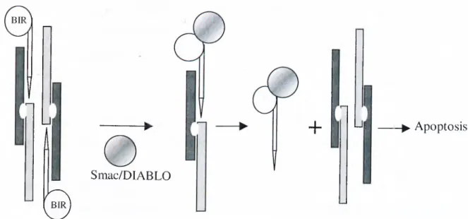
Recent structural analyses revealed that a linker segment at the N-terminal region between BİRİ and BIR2 of XIAP occupies the active site of caspases directly which results in a blockade of substrate entry. This makes lAPs unique inhibitors of caspases that function by directly binding to and inhibiting caspase function, rather then targeting the enzyme indirectly and affecting their activation. (Chai et al., 2001; Huang et al., 2001; Riedl et al., 2001). It is also noteworthy to mention that only processed caspase-9 is subject to inhibition by lAPs. It has been shown that, BIR3 of XIAP binds to the N terminus of the small subunit of caspase-9, which becomes exposed after proteolytic processing (Srinivasula et al., 2001).
However induction of apoptosis requires elimination of the caspase inhibition enforced by lAPs. Thus when apoptosis is induced, lAPs are inhibited by binding of a mitochondrial apoptosis promoting factor Smac/DIABLO to the BIR domains of lAPs (Green DR., 2000) (Figure 1-8).
n
Lit
Smac/DIABLO
0
+ Ui -► Apoptosis
lAP-mediated Smac/DIABLO destabilizes lAP is released from caspase
caspase inhibition caspase-IAP complex
Figure 1-8: Release of inhibitory effects of lAPs on caspases by the SMAC/DIABLO protein.
it has been shown that deregulation of lAPs contribute to human disease. For example a single BIR domain containing lAP “Survivin’’ has been shown to be upregulated in many human tumours.
1-3.4 TARGETS OF ACTIVE CASPASES
When activated, initiator caspases activate downstream caspases and when the effector caspases are activated they cleave critical cellular protein substrates in order to advance apoptosis. (Thornberry and Lazebnik, 1998). The targets of active caspases can be grouped in 4 groups.
1- Pro- and anti-apoptotic proteins:
a) Pro-caspases; The already activated caspases transactivate other caspases. This allows the generation of a caspase activation cascade.
b) Anti-apoptotic proteins: Bcl-2 and bcl-XL are cleaved by caspase-3. This way the anti-apoptotic activities of these proteins are eliminated.
c) Cleavage of the pro-apoptotic protein Bid by caspase-8: The cleavage product t-Bid translocates to the mitochondria and induces the release of cytochrome c.
2- Components of apoptotic machinery:
The inhibitor of CAD (caspase activated DNAase) ICAD is cleaved by caspase-3, allowing CAD to translocate to nucleus and cleave DNA.
3- vStructural proteins:
The structural proteins gelsolin (which regulates actin dynamics), lamins (which are the major structural proteins of nuclear envelope) and P-catenin (which plays important roles in cell-cell adhesion) are degraded by caspases.
4- Homeostatic proteins:
Poly (ADP-ribose) polymerase (PARP) is a DNA double-break repair enzyme that is cleaved to facilitate the DNA degradation during apoptosis.
1-4 THE BCL-2 FAMILY
1-4.1 GENERAL STRUCTURE
Bcl-2 family proteins play a pivotal role in deciding whether a cell will die or survive. Bcl-2 was first identified as a proto-oncogene in follicular B-cell lymphoma (Tsujimoto et al., 1985). In 1993 Oltvai et al discovered the first pro- apoptotic member of the Bcl-2 fomily namely Bax (Oltvai et al., 1993).
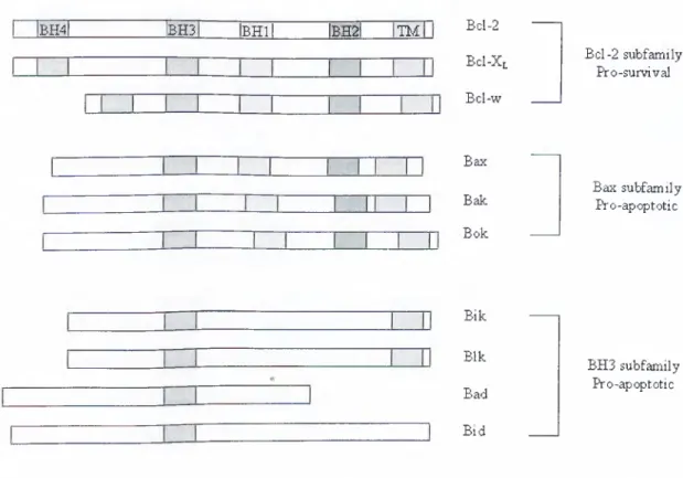
The growing Bcl-2 femily consists of ‘multidomain’ death antagonists (e.g. Bcl-2, Bcl-XL, Bcl-w, Mcl-l, A l) and death agonists (e.g. Bax, Bak, Bok), which function primarily to protect or disrupt the integrity of mitochondrial membranes, respectively. The homology between the Bcl-2 family members is restricted to four Bcl-2 homology (BH) domains (Figure 1-9). Through these homology domains they can form homo- or heterodimers with each other. Thus the ratio of pro- versus anti-apoptotic members is very critical for making the decision of cell death. All of the anti-apoptotic members have four of the BH
domains whereas the pro-apoptotic members do not have the BH4 domain except for Bcl-Xs. There are also “BH3 only” pro-apoptotic members, which carry only the BH3 homology domain. These proteins serve as ligands to activate multidomain pro-apoptotic Bcl-2 family members or inactivate anti-apoptotic Bcl-2 family members (Huang and Strasser, 2000). BH3-only proteins include Bim, Bid, Bad, Bik/Nbk, BNIP3, Blk, Noxa, Puma, and Hrk. Another characteristic of these proteins is that they can become integral membrane proteins.
Protein crystallography and liposome reconstitution analysis with purified Bcl-2 family members suggest that, these proteins regulate the mitochondrial membrane barrier by forming channels themselves or influencing pre-existing channels in the membrane (Martinou and Green, 2001).
Alterations in expression, subcellular localization, phosphorylation status, and proteolytic processing of Bcl-2 family molecules determine whether the death program will be activated.
The anti-apoptotic members can dimerize with pro-apoptotic members to
inhibit apoptosis. Among these, BHl and BH2 are impoi'tant for
heterodimerization of the anti-apoptotic members Bcl-2 and Bc1-Xl with Bax for the suppression of apoptosis (Borner et al., 1994). But anti-apoptotic members can perform their inhibitory effects on apoptosis without the need to bind and inactivate a pro-apoptotic member. It has been shown that a mutant form of Bcl- Xl, which cannot bind the anti-apoptotic members Bax or Bad can still preserve the 70-80% of its anti-apoptotic ability (Cheng et al., 1996).
BH3 domain is the most critical domain for pro-apoptotic activity. This domain has been termed as “suicide domain” because when the BH3 domain of Bax was inserted into Bcl-2, the anti-apoptotic protein became a killer protein (Hunter et al., 1996). Also although the “BH3 only” pro-apoptotic members (Bid, Bik, Bim, Blk and Bad) carry the BH3 domain, they induce apoptosis through heterodimerizing with pro- or anti-apoptotic proteins. Crystal structure of Bcl-Xi. and Bid showed the presence of a region named the “flexible region” at the N- terminal of the BH3 domain, which serves as a potential phosphorylation site (Muchmore et al., 1996).
BH4 domain can be regarded as the anti-apoptotic domain, as its presence is enough to confer anti-apoptotic activity to Bcl-2 family members. BcI-2
mutants lacking BH4 domain loose their anti-apoptotic activity and become killer proteins (Grandgirard et al., 1998). It has also been shown that during apoptosis, the BH4 domain of Bcl-2 is cleaved to accelerate the apoptotic procedure (Cheng et al., 1997, Grandgirard et al., 1998)
Another important domain found in some Bcl-2 family members is the C- terminal hydrophobic domain, which mediates the localization of these proteins to membranes.
IbH4| IbH3I iBHlI IBM It mI Bd-2
Bc1-Xl Bcl-w Bcl-2 subfamily Pro-survival Bax Bak Bok Bax subfamily Pro-apoptotic Bik Blk Bad Bid BH3 subfamily Pro-apoptotic
Figure 1-9: Structure of some pro- and anti-apoptotic Bcl-2 family members.
(TM: transmembrane domain)
1-4.2 BCL-2 FAMILY FUNCTION IN APOPTOSIS
Currently, three (non-exclusive) models are used to explain Bcl-2 function. In the first model, it is proposed that Bcl-2 proteins can act as ion channels due to their structural resemblance to the diphtheria toxin and colicin (Muchmore et al., 1998). The second model suggests that Bcl-2 proteins function in the modulation of caspase activation for example by inhibition of the apical caspase-9 by Bcl-2/Bcl-xL. According to this model, Bcl-2/Bcl-xL should displace Bcl-2/Bcl-xL from the Apaf-1/ cytochrome c/caspase-9 complex and so trigger caspase-9 autoactivation just as in the case of C. elegans (Chinnaiyan et al., 1997). Finally, the third model proposes that these proteins act as inhibitors of cytochrome c export from mitochondria. Very recently it has been shown that Bax and Bak serve as the major mitochondrial sensors of upstream apoptosis signaling, and cells missing both factors are resistant at the mitochondrial level to killing by a wide range of stimuli (Wei et al., 2001).
Another possible mechanism by which Bcl-2 might function in the suppression of apoptosis arose from observations that Bcl-2 expression can affect intracellular Ca^"^ homeostasis. Alterations in intracellular Ca^"^ concentrations are known to influence apoptosis so it remains possible that Bcl-2 either directly modulates calcium channels or acts to protect lipid membranes from damage by peroxide radicals which is known to disrupt Ca""^ homeostasis (Pintón et al., 2002).
1-4.3 REGULATION OF Bcl-2 FAMILY MEMBERS
Regulation and activity of the family members is regulated by phosphorylation, proteolysis, dimerization and by their cellular localization.
1-4.3.1 POST-TRANSLATIONAL MODIFICATIONS
Members of the Bcl-2 family can be regulated by phosphorylation or proteolysis. The anti-apoptotic activity of Bcl-2 can be either inhibited or
amplified by phosphorylation. Phosphorylation of Bcl-2 at residues serine 70 and 87 and threonine 56 and 74, has been shown to inactivate its anti-apoptotic activity (Chang et ah, 1997). Bcl-2 was found to be phosphorylated and thus inactivated in many tumor cell lines that are treated with chemotherapeutic agents such as taxol (Haidar et al., 1995 and 1996). One of the kinases responsible for Bcl-2 phosphorylation is a МАРК (mitogen-activated protein kinase) family member JNK/SAPK (c-Jun N-terminal Kinase/Stress-activated protein kinase) (Maundrell et al., 1997). JNK/SAPK is known to be activated by stimuli favouring apoptosis. The anti-apoptotic function of bcl-2 has been shown to be amplified by phosphorylation at serine 70 by the activation of a classic protein kinase C (PKC) isoform. This phosphorylation is required for suppression of apoptosis by Bcl-2 in murine growth factor-dependent celt lines (Ruvolo et al.,
1998). Another МАРК family member ERK (extracellular signal-related kinase), is also involved in the regulation of apoptotic proteins. While JNK/SAPK functions to promote apoptosis by inhibiting Bcl-2, ERK can be activated to inhibit apoptosis (Xia et al., 1995). When activated ERK phosphorylâtes Bad at residues serine-112 and serine-136. Phosphorylated Bad associates with 14-3-3 protein and becomes sequestered in the cytosol (Zha et ah, 1996). To inhibit this inhibitory function of Erk, during apoptosis this kinase is inactivated by the actions of caspase 3 (Widmann et al., 1998).
Similar to Bcl-2, the pro-apoptotic “BH3 only” protein Bad is a target for kinases. In the presence of survival factors BAD is phosphorylated on two serine residues (Ser-112 and Ser-136) and sequestered in the cytosol by the 14-3-3 protein (Zha et al., 1996). The kinases responsible for Bad phosphorylation are PKA (Protein kinase A) and PKB/Akt (Protein kinase B) (Harada et ah, 1999, del Peso et ah, 1997). PKA phosphorylâtes Bad at ser-112 and PKB/Act from serine- 136. Thus when cells are stimulated with survival factors. Bad is phosphorylated and its anti-apoptotic activity diminishes. Following a death signal BAD is de- phosphorylated and binds to the anti-apoptotic molecules Bcl-2 and Bc1-Xl·
Cleavage of pro- or anti-apoptotic proteins is also an important factor, affecting the apoptotic process. By cleavage anti-apoptotic factors may become pro-apoptotic and pro-apoptotic members become more apoptotic. For example, the pro-apoptotic Bcl-2 family member Bid has been shown to be cleaved during Fas-mediated apoptosis by caspase 8 (Li et al., 1998). After activation, caspase 8
cleaves Bid, generating a C-terminal fragment of the protein that is “truncated Bid” (tBid). While full-length p22 Bid is localized in cytosol, p l5 tBid translocates to mitochondria and thus transduces apoptotic signals from cytoplasmic membrane to mitochondria. Cleavage of Bcl-2 by activated caspases results in formation of a C-terminal cleavage product which has a pro-apoptotic function (Cheng et al., 1998). It has also been demonstrated that Bax is a substrate for both caspases and the calcium-activated protease calpain (Wood et al., 1998).
1-4.3.2 CELLULAR LOCALIZATION AND DIMERIZATION
The Bcl-2 members have been shown to be both cytosolic and membrane associated. A considerable portion of pro-apoptotic members are localized to cytosol in the absence of an apoptotic signal in contrast to anti-apoptotic members which are localized to membranes (Hsu et al., 1997, Gross et al., 1998, Puthalakath et al., 1999). Following a death signal the pro-apoptotic members undergo a conformational change and this alteration enables them to target and integrate into membranes, especially the outer membrane of the mitochondria. Activation of Bax requires both dimerization and cellular localization.
In viable cells Bax is monomeric and localized to cytosol. Following a death stimulus Bax is translocated to mitochondria where it becomes an integral membrane protein and forms a homo or heterodimer with Bcl-2/Bcl-xL (Wolter et al., 1997, Gross et al., 1998). However in contrast to Bax, Bak is usually resides in the outer mitochondrial membrane.
The anti-apoptotic protein Bcl-2 resides in the outer mitochondrial membrane, endoplasmic reticulum and the nuclear membrane (Krajewski et al., 1993; Lithgow et al., 1994). The three dimensional structures of Bc1-Xl and Bid demonstrated that they share structural homology with the pore-forming domain of certain bacterial toxins especially diphtheria toxin and colicins A and El (Muchmore et al., 1996). It has been shown that Bc1-Xl, Bcl-2 and Bax can form channels in synthetic lipid membranes (Minn et al., 1997, Schendel et al., 1997, Antonsson et al., 1997).
Thus localization of Bcl-2 family members to mitochondria and their pore-forming activities play a very important role during apoptosis.
1-5 DEATH RECEPTOR PATHWAY
Activation of a specific group of transmembrane receptors also induce activation of caspases hence apoptosis. These receptors belong to the “tumor necrosis factor (TNF)” superfamily. Mammalian TNF-R (TNF receptor) family members are type I membrane proteins with conserved extracellular cysteine-rich domains. The TNF-R superfamily members include: TNF-Rl, TNF-R2, TNF-R3, CD95, LT-aR, LT-^R, Ox 40, CD27, CD 28, CD 30, CD 40, 4-1 BB, p75 NGFR, GIT-R, Rank, DR6 and the TRAIL receptors. When a receptor is activated it typically forms trimeric or multimeric complexes stabilized by disulfide bonds. Some of these receptors such as CD95, TNF-Rl and TNF-R2 can also exist in soluble forms (Hughes DPM. and Crispe IN., 1995).
The ligands of these receptors also comprise a family. Some members of this family are; TNF, CD 95 ligand (FasL/CD95L), LT-a (lymphotoxin-a), LT-P, Ox 40L, CD27L, CD 28L, CD 30L, CD 40L and 4-1 BBL. These ligands share a characteristic 150 amino acid region at their C-terminus. This C-terminal region is responsible for .specific interaction of the ligand with its cognate receptor. Although TNF and CD95L can exist as soluble proteins, others are usually found as membrane-bound trimeric or multimeric complexes.
Some members of the TNF superhimily have been shown to induce apoptosis when activated by binding of their cognate ligand and therefore are called as “death-receptors”. These include CD95, TNF-Rl, DR3, DR4, DR5 and DR6. These receptors share a homology region at their cytoplasmic region, which is called “death domain”. This domain is responsible for transduction of death signals to intracellular proteins by initiating a protein-protein interaction and activation cascade.
Upon ligand binding to the receptor, intracellular adapter proteins such as FADD/MORTl, TRADD and RAIDD are recruited to the cytoplasmic regions of the receptors through homotypic death-domain (DD) interactions to form a death- inducing signaling complex (DISC) (Ashkenazi and Dixit, 1998). In turn, FADD
recruits pro-caspase- 8 via interactions between death-effector-domains (DEDs) present in both proteins, thereby stimulating caspase-8 autoproteolytic activation and initiating a caspase cascade leading to cell death. Caspase-2 can also associate with the cytoplasmic regions of death receptors, and this association requires the interaction of caspase-2 with the molecular adapter RAIDD/CRADD (Duan and Dixit, 1997; Ahmad et al., 1997)
1-5.1 CD95/Fas/Apo-l
CD95 is expressed in activated lymphocytes and in all other tissues including liver, lung and heart. The ligand of the receptor CD95L is expres.sed in activated lymphocytes, natural killer cells, erythroblasts, immune privileged tissues and in some tumors. The main function of CD95/CD95L signaling is deletion of autoreactive lymphocytes and maintenance of peripheral tolerance.
CD95 does not have a catalytic domain. Thus in order to activate downstream signaling effector molecules it has to interact with an adaptor molecule. This interaction is mediated by a 65 amino acid intracellular domain of CD95, which is called “death domain (DD)”. Through this domain CD95 can recruit downstream effector molecules and induce apoptosis (Itoh and Nagata, 1993). These adaptor/effector znolecules include FADD/MORT (Fas-associated death domain), RIP (receptor interacting protein), Daxx (death domain associated protein) and FIST/HIPK3 (Fas interacting serine/threonine kinase/Homeodomain interacting protein kinase).
One of the adaptor molecules that interact with DD of CD95 is FADD/MORT. FADD contains a DD at its C-terminus and a death effector domain at its N-terminus. Through this DED, the adaptor protein can interact with the DED of procaspase-8 leading to autocleavage and activation of caspase- 8. (Boldin et al., 1996; Muzio et al., 1996). This protein complex generated upon activation of CD95 is called “DISC (death inducing signaling complex)”. Thus, recruitment of DISC to the activated receptor provides a direct link between external apoptotic signals and the basal apoptotic machinery of the cell (Kischkel et al.,1995). When caspase-8 is activated it can either directly activate downstream caspases such as caspa,se-3 or it can cleave the pro-apoptotic
molecule Bid to generate a Bid fragment namely t-Bid (truncated Bid). t-Bid then translocates to mitochondria where it can induce cytochrome c release (Li et al., 1998; Lluo et al., 1998).
Another receptor-associated signal transducer molecule is RIP (Stanger et al., 1995). RIP contains a DD at its C-terminal and a tyrosine kinase-like domain at its N-terminal. It has been shown that blockers of tyrosine kinase activity also inhibit the CD-95 mediated apoptotic signal transduction (Eischen et al., 1994). RIP can also activate caspase-2 through an interaction with an adaptor molecule named “RAIDD (RIPP associated ICH-l/Ced-3 homologous protein with a death domain)” (Wallach D., 1997). This DD-DD interaction recruits procaspase-2, which interacts with RAIDD through the ICH-l/Ced-3 homology region. Another molecule activated by RIP is the anti-apoptotic molecule NF-kB. Thus RIP can also be an intermediate for inhibition of apoptosis. Therefore caspase-8 can also cleave RIP in order to inhibit the NF-kB activation (Lin et al., 1999).
Daxx is an inducer of “c-Jun N-terminal kinase” INK and has been shown to bind to DD of CD95 although it lacks a DD (Yang et al., 1997). Therefore Daxx functions as an intermediate protein that couples CD95 to activation of JNK hence apoptosis. CD95 mediated JNK activation can be inhibited by FIST/HIPK3 (Rochat-Steiner et al., 2000). FIST/HIPK3 can also interact with CD95 as well as FADD. Binding of FIST/HIPK3 to FADD induces FADD phosphorylation and inhibits CD95 mediated JNK activation.
There are two main inhibitors of CD95 mediated apoptotic signaling. One of these inhibitors is FLIPs (Fas-associated death domain like ICE inhibitory proteins) and the other is the “decoy receptors”. FLIPs exert their inhibitory effect by interfering with recruitment of caspases to CD95 signaling complex. Some viruses synthesize FLIPs (v-FLIP) in order to evade host immune system (Thome et al., 1997). The cellular homologue of v-FLIP is c-FLIP and been identified by several groups in 1997 (Srinivasula et al., 1997; Irmler et al., 1997; Goltsev et al., 1997; Shu et al., 1997; Inohara et al., 1997; Hu et al., 1997; Han et al., 1997; Rasper et al., 1998). c-FLIP shows homology to caspase-8 containing two DEDs. It also contains an inactive caspase-like domain without the conserved functional cysteine domain. Therefore c-FLIP can block caspase-8 activation at the DISC and can inhibit CD95-mediated apoptosis (Scaffidi et al.,
1999).
1-5.2 TNF RECEPTORS
TNF is a cytokine produced by activated T-cells and macrophages that influences the proliferation, differentiation and apoptosis of cells involved in inflammation. Thus TNF plays a pivotal role in the regulation the host inflammatory response.
There are two receptors for TNF: TNF-Rl and TNF-R2. TNF- R1 is able to mediate most of the biological responses initiated by TNF, whereas TNF-R2 provides an auxiliary function in cooperating in the binding of TNF to TNF-Rl (Tartaglia et al., 1993). Although TNF-Rl alone can trigger apoptosis TNF-R2 mainly seems to promote cell survival. Nevertheless TNF-R2 was also shown to kill certain cells when over-expressed (Grell et al., 1999). Both TNF-Rl and TNF-R2 can activate the pleiotropic transcription factor NF-icB (Rothe et al.,
1994).
Binding of the ligand to TNF-Rl activates the proteolytic caspa.se cascade by recruiting caspase-8 via FADD/MORT. However FADD/MORT adapter molecule cannot directly bind to TNF-Rl, therefore another cytoplasmic adapter molecule with a death domain called “TRADD” (TNF-R-associated death domain) is recruited to the activated receptor. TRADD can also bind to RIP, thereby linking TNF-Rl to caspa.se-2 activation via RAIDD and CRADD.
Another class of signaling adapter proteins recruited to the TNF receptor is the “TRAFs” (TNFR-associated factors) of which six are currently identified (Inoue et al., 2000). TRAF proteins are signal transduction adapter proteins. All TRAFs share a conserved 230 amino acid “TRAF domain” which mediates their homo- or hetero-oligomerization with other TRAFs, their interaction with the cytoplasmic tails of members of the TNF-R superfamily, and interactions with downstream signal transducers (Lee et al., 1997). TRAFs are held in abeyance in the cytoplasm through their association in oligomeric complexes with I-TRAF [177].
TRAF-2, -5, and -6 mediate activation of SAPK/JNK or NF-kB (Lee et al., 1997), the latter by interaction with the downstream signaling kinase NIK. NIK, in turn, activates the IkB kinases, which phosphorylate and inactivate IkB, the endogenous cellular inhibitor of NF-kB (Malinin et al., 1997; Ye et al..
1999). It is believed that TRAFs’ anti-apoptotic effect is due to the activation of NF-kB. TRAF proteins can also interact with the death domain kinase RIP and the serine/threonine kinase IRAK. On the other hand, apoptosis signal-regulating kinase ASK-1, a TRAF-interacting kinase, was recently demonstrated to be a downstream target of TRAF-2, TRAF-5, and TRAF-6 in the INK signaling pathway.
TRAFs can also interact with “TRIP” (TRAF-interacting protein) (Lee et al., 1997). TRIP associates with TNF receptor family members through its interaction with TRAF proteins. This binding inhibits TRAF-mediated activation of the apoptosis suppressor NF-kB. However TRAFs can also interact with the lAP proteins. Thus, TRAF interactions with cIAPs would suppress apoptosis whilst interactions with TRIP would promote it. The availability of TRAF interacting proteins, therefore influence the outcome of TNF-R activation.
In another report it has been shown that in the presence of RIP (required for NF-kB activation by TNF-R 1), TNF-R2 triggers cell death in T-cells whereas in the absence of RIP, TNF-R2 activates NF-kB (Pimentel-Muinos and Seed, 1999). RIP is induced during interleukin (IL)-2-driven T-cell proliferation, and its inhibition reduces susceptibility to TNF-dependent apoptosis.
1-5.3 TRAIL RECEPTORS
Another subfamily of TNF receptors is the “TRAIL receptors” (TNF- related apoptosis-inducing ligand receptors) (Golstein, 1997; Gura, 1997; Ashkenazi and Dixit, 1998). The ligand TRAIL (is also called as apo-2L) is synthesized as a membraire-bound protein and cleaved to generate a soluble ligand, which does not to bind either CD95 or TNF-R 1.
The receptors for TRAIL are: DR4 (TRAIL-Rl), DR5 (TRAIL- R2/KILLER), decoy receptor l(DcRl/TRID/LIT/TRAIL-R3), decoy receptor 2 (DcR2/TRUNDD) and osteoprotegerin. The latter three are called “decoy receptors” because although they can bind to TRAIL they cannot transduce signals as they lack a death domain. TRAIL induces apoptosis through binding to DR4 and DR5 requiring FADD and caspase-8 (Bodmer et al., 2000).