Summary
The aim of this study was to estimate the number of ependymal cells in the spinal cord of Leghorn chicks using optical fractionator technique. Forty weeks old six female and six male Leghorn were used. Animals were anesthetized by administration of xylazine-ketamine combination and euthanasia was carried out. Saline solution was administered to the animals for draining blood completely from the body. Ten percent formalin saline solution was used as a fixative. Decalcification was performed on dissected vertebral columns using Trichloroacetic acid. Spinal cords were removed from vertebral columns and measured. All cords were cut 1 cm in length, 10 and 11 systematic random sampled tissue samples with a sampling ratio of 1/3 were taken from females and males respectively. One transversal section was taken from each of tissue samples at the thickness of 10 µm. Optical fractionator was performed on sections to estimate the total number of ependymal cells in the cord. It was found that male Leghorns had more ependymal cells than female Leghorns. The maximum number of ependymal cells was found in the caudal part whereas the minimum number of cells was observed in cervical part of the spinal cord in female. Male animals showed a homogeneous distribution of the ependymal cells. It was thought that sex difference must be considered in studies on spinal cord of Leghorn.
Keywords: Ependymal cell, Spinal cord, Optical fractionator, Leghorn, Stereology
Leghorn Irkı Kanatlılarda Medulla Spinalis’te Yer Alan Ependim
Hücre Miktarının Optik Parçalama Yöntemi Kullanılarak
Belirlenmesi
Özet
Bu çalışmanın amacı, Leghorn ırkı kanatlılarda medulla spinalis’in içerdiği ependim hücrelerinin sayısının optik parçalama yöntemi ile belirlenmesidir. Kırk haftalık altı dişi, altı erkek Leghorn kullanıldı. Hayvanlar ksilazin-ketamin kombinasyonu ile anesteziye alındı ve ötenazi gerçekleştirildi. Kanın vücuttan tamamen uzaklaştırılması için fizyolojik tuzlu su kullanıldı. Kadavralara yüzde onluk tuzlu formol solusyonu uygulandı. Triklor asetik asit kullanarak diseke edilen omurgalar dekalsifiye edildi. Medulla spinalis’ler canalis vertebralis’ten dışarı alındı ve uzunlukları ölçüldü. Tüm medulla spinalisler 1 cm uzunluğunda parçalara ayrıldı ve her bir parçadan 10 µm kalınlığında bir kesit alındı. Sistematik rastgele örnekleme ile 1/3 örnekleme oranı kullanılarak dişilerde 10 erkeklerde 11 kesit elde edildi. Medulla spinalis’te yer alan ependim hücrelerinin saysını hesaplamak için optik parçalama yöntemi kullanıldı. Erkek medulla spinalis’lerinin dişilere nazaran daha fazla ependim hücresine sahip oldukları tespit edildi. Ependim hücre sayılarının medulla spinalis’in farklı bölümlerinden alınan kesitlerdeki dağılımına bakıldığında dişilerde caudal bölgede en fazla, cervical dördüncü kesit düzeyinde ise en az olduğu, erkek hayvanlarda ise medulla spinalis’in genelinde homojen bir dağılımın olduğu görüldü. Leghorn ırkı kanatlılarda cinsiyet farkının ependim hücre sayısı bakımından önemli olduğu sonucuna varıldı.
Anahtar sözcükler: Ependim hücresi, Medulla spinalis, Optik parçalama, Leghorn, Stereoloji
The Use of the Optical Fractionator to Estimate the Total Number
of Ependymal Cells of the Spinal Cord in Leghorn
[1]Durmus BOLAT *
Sadettin TIPIRDAMAZ **
[1] * **
This study is a part of PhD thesis supported by SUBAPK (Project no: 09102043)
Kirikkale University, Faculty of Veterinary Medicine, Department of Anatomy, TR-71451 Campus, Kırıkkale - TÜRKİYE Selcuk University, Faculty of Veterinary Medicine, Department of Anatomy, TR-42000 Campus, Konya - TÜRKİYE
Makale Kodu (Article Code): KVFD-2011-5167
Quantitative data of biological structures obtained
using appropriate methods are valuable for decisive diagnosis or monitoring the disease progress and are used to detection of variation among biological species.
INTRODUCTION
İletişim (Correspondence)
+90 318 3574242/3328Quantitative measurements can be stored and analyzed more easily than subjective measurements 1. New approaches such as design-based stereological methods have been used to investigate quantitative morphology of the biological structures besides the central nervous system 2. Stereological methods have many unbiased approaches for avoiding subjective measurements. For example, systematic random sampling provides more precise and closer results to the true values than random sampling 3. In systematic random sampling, each part of the object must have equal opportunity to be sampled once and should contribute at the same rate to the results of the research 4. Another unbiased three-dimensional sampling method is disector developed by Sterio 5 to calculate the number of particles in a known volume of an object. This method is applied to the biological structures in various ways as physical and optical disector 6. However, the most widely used method to calculate the number of object is optical fractionator 7. Optical fractionator is comprised sampling steps and optical disector. The results obtained using optical fractionator cannot be affected by any assumption namely, object size and shape and tissue shrinkage or swelling since it does not depend on reference volume 8,9. This method can be used to determine the number of ependymal cells of the spinal cord because of its unbiased property.
Ependymal cells known as neural stem cells of central nervous system are important for homeostasis of cerebro- spinal fluid 10,11. They also have ability of proliferation because of the spinal cord injuries 10,12. In mammals, ependymal cells compose of simple epithelial lining of the ventricles and central canal of the central nervous system and lack an external lamina 13. In avian, there are three types of ependymal cells; typical ependymal cells, astrocytic ependymal cells, and lamellate ependymal cells. Typical ependymal cells are placed in the fourth ventricle and central canal of the CNS and their nuclei are round or oval shaped with a diameter of 6-8 µm 14. To the best our knowledge, there is not an obvious method in the literature about how to cut and sample entire spinal cord to calculate the number of ependymal cells.
The aim of this study was to demonstrate cutting and sampling procedure of the spinal cord to estimate the number of ependymal cells surrounding the central canal in both genders of Leghorn using optical fractionator and to investigate possible sex differences.
MATERIAL and METHODS
Material
In this study, forty weeks-old six female (1500±0.063 g) and six male (1766±0.082 g) Leghorn chicken obtained from Ankara Poultry Research Institute were used. The research was approved by the Ethical Committee of College
of Veterinary Medicine, Selcuk University (2009/007, 2011/002).
Methods
Preparation of Cadavers
Animals were anesthetized by administration of xylazine (0.5 mg/kg, IM) plus ketamine (25 mg/kg, IM). Ketamine hydrochloride (15 mg/kg, IM) was used as maintenance dose 15. Abdominal cavity of the animals in the supine position was entered made an incision along abdominal wall behind the sternum. The left ventricle of the heart was reached displacing liver and other surrounding structures. Heparin (5.000 IU ml/kg, intracardiac) administration was performed to the animals through a cannula placed in the left ventricle and then euthanasia was carried out by an incision made on the right atrium. Saline solution (0.9%) was administered to the animals for draining blood completely from the body. Ten percent formalin saline solution (100 ml per animal, pH: 7.4) was used as a fixative. Cadavers were kept in a container of 10% formalin solution for 15 days at room temperature. After this period, vertebral column with the cranium was dissected out from the body. Vertebral columns with cranium were washed in distilled water for 20 min and decalcification was performed using 6% Trichloroacetic acid 16. After decalcification, occipital bone and atlas were opened dorsally. The midpoint between first cervical spinal nerve which lacks a dorsal root and hypoglossal nerve was accepted as the border of spinal cord and medulla oblongata.
Spinal cord was dissected out from medulla oblongata at this point and laminectomy of the entire cord was performed to uropygium. Spinal cords were removed from vertebral canals cutting the spinal nerve roots at the level of the intervertebral foramen and the lengths of the cords were measured.
Tissue Sampling for Counting
Spinal cords were cut in 1 cm long, 10 and 11 systematic random sampled tissue samples with a sampling ratio of 1/3 were taken from females and males, respectively. First sample was taken randomly as a rule of systematic random sampling 17. Following routine histological procedure, paraffin blocks were prepared and cut with a rotary microtome into 10 µm sections as well as one transversal section was taken randomly from each of tissue samples. Section sampling fraction (ssf) was calculated as 1/3.000 9. All tissue samples were stained with Hematoxylin & Eosin.
Counting Procedure of Ependymal Cells Using Optical Fractionator Technique
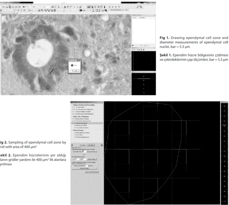
Before applying optical fractionator technique, dia- meters of the ependymal cells were reckoned using quick line tool and recorded (Fig.1). Optical fractionator technique was performed to estimate the number of ependymal
cells in the cord. To do this, optical fractionator workflow of Stereo Investigator (MicroBrightField Inc., VT, USA) software was used. The perimeter of the ependymal cell zone was drawn under 40X objective (Fig.1). Area fraction sampling was used on the region of interest after switching 100X objective (Leica, Plan Apo, 1.40 Na Oil) (Fig. 2). The areas of sampling grid and unbiased counting frame were set 400 µm2 (20 µm X 20 µm) and 100 µm2 (10 µm X 10 µm), respectively. Area sampling fraction (asf) value was equal to ¼ and the area of interest was sampled randomly with unbiased counting frame. Mean mounted thickness of sections (distance between upper and lower surface of section) were calculated as 7 µm using Heidenhein microcator. Guard zone was set 10% and disector height was adjusted for 5 µm. The calculation of height sampling fractions (hsf) of the sections was performed using following formula, hsf = typical mounted section thickness
(t)/disector height (h) and hsf 1.4 9,18. All sampled areas were visited using unbiased counting frame and calculation
of ependymal cells were performed according to the counting rules 19 (Fig. 3).
The number of ependymal cells was estimated as N = x x x ,
Where, is equal to counted cells in the sampled areas 9, and N refers to the number of ependymal cells in the spinal cord. Coefficient of Error (CE) value was calculated using the formula 1/ 8. Numerical density of ependymal cells of the spinal cord in female and male leghorn chicks were calculated according to Gundersen 20.
Statistical Analysis
Two sample t test was used to evaluate difference between the length of the spinal cord and numerical density of ependymal cells in two gender. Mann-Whitney
Fig 1. Drawing ependymal cell zone and
diameter measurements of ependymal cell nuclei, bar = 5.3 µm
Şekil 1. Ependim hücre bölgesinin çizilmesi
ve çekirdeklerinin çap ölçümleri, bar = 5.3 µm
Fig 2. Sampling of ependymal cell zone by
grid with area of 400 µm2
Şekil 2. Ependim hücrelerinin yer aldığı
alanın gridler yardımı ile 400 µm2 lik alanlara ayrılması
U test was used in the statistical evaluation of the counted ependymal cells (SPSS 17.0). The results were presented as Mean±SEM, Median and P<0.05 was considered statistically significant.
RESULTS
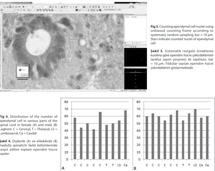
The last sections of the spinal cord of female Leghorns were destroyed during histological process because it was so thin in the caudal part of the cord. Therefore, tenth section could not be counted. The length of the spinal cord was found to be 31.0±0.89 cm in female and 33.3±0.52 cm in male Leghorns (P<0.01). It was observed that spinal cord ends in uropygium. The number of ependymal cells of the female was found to be 1.957.200 (CE = 0.091) and of male 2.755.200 (CE = 0.078) using optical fractionator. Statistical difference found between two values (P<0.05). The number of counted cells on the cross-section of the spinal cord of female and male is given in Fig. 4.
When considering distribution of ependymal cells on the sections taken from various parts of the spinal cord, the most number of ependymal cells were found in the caudal
part, whereas the fewest number of cells observed in the fourth section from cervical part of the spinal cord in female (P<0.05) (Fig. 4). The spinal cord in the male animals showed a homogeneous distribution of the ependymal cells (Fig, 4).
The distribution of the ependymal cells in the entire spinal cord was evaluated and it was observed that 52 cells could be counted in female although 60 cells in male with the thickness of 10 µm. Although the difference between two genders were found statistically important (P<0.02), numerical density of ependymal cells of the spinal cord in female and male leghorns were found to be 0.030±0.00013 and 0.024±0.00006 respectively and there was no difference between two (P>0.05).
Ependymal cells were observed as cubic or flattened prismatic-shaped and were located basal membrane as well as their nuclei were rounded or oval-shaped with a diameter of 5-7.5 µm (Fig. 1). The wall of the central canal was lined by ependymal cells in all spinal cord segments. Ependymal cell zone was supported by plenty of blood vessels. There were observed microvilli and kinocilium on the apical surface of the ependymal cells.
Fig 4. Distribution of the number of
ependymal cell in various parts of the spinal cord in female (A) and male (B) Leghorn. C = Cervical, T = Thoracal, LS = Lumbosacral, Ca = Caudal
Şekil 4. Dişilerde (A) ve erkeklerde (B)
medulla spinalis’in farklı bölümlerinde tespit edilen toplam ependim hücre sayıları
Fig 3. Counting ependymal cell nuclei using
unbiased counting frame according to systematic random sampling, bar = 10 µm. Stars indicate counted nuclei of ependymal cell
Şekil 3. Sistematik rastgele örnekleme
kuralına göre ependim hücre çekirdeklerinin tarafsız sayım çerçevesi ile sayılması, bar = 10 µm. Yıldızlar sayılan ependim hücre çekirdeklerini göstermektedir
DISCUSSION
Ependymal cells lying the brain ventricles play essential roles in the transport of cerebro-spinal fluid and brain homeostasis. Ependymal cells known as neural stem cell of central nervous system constitute a thin epithelial membrane lining the spinal cord 10,18. These cells have ability to response spinal cord injuries and also olfactory bulb neurons are derived from ependymal cells 10,11.
The average number of ependymal cells in the spinal cord was found to be 1.957.200 for female and 2.755.200 for male Leghorn chicks using optical fractionator technique. Comparison of the results could not be performed since there was not enough relevant information to ependymal cells of the avian species in the literature.
In a previous study conducted on spinal cord of female mice using physical disector, the number of ependymal cells has been found being 163.000 along entire spinal cord 21. However, the volume of spinal cord has been measured by applying Archimedes’ principle and counting cells has been performed using physical disector after histological procedure 21. Tissue shrinkage occurs during or after histological process 22. Therefore, assuming the volume measurement before histological procedure as a reference value for determining the number of cells in the unit volume ignoring factors such as tissue shrinkage is not a proper approach. Because of this problem, multi correction factors have been used to correct the results of the mentioned study.
In the current study, the number of ependymal cells was counted using optical fractionator regardless of volume. Volume parameter is not a key factor for this method, thus it is not affected by volume changes which may occur to any of the tissues caused by tissue shrinkage or swelling. Ependymal cells nuclei were found rounded or oval shaped with a diameter of 5-7.5 µm (Fig. 1). These findings were similar to the results of previous study carried out by Hodges 14.
In conclusion, the number of ependymal cells was evaluated using the most unbiased method, optical fractionator, in Leghorn chicks for both genders. Sex difference was found an important factor for Leghorn chicks and it was advised that this factor must be considered even as conducting studies on the spinal cord of poultry. In the light of the present results, important information was acquired about the number of ependymal cells in avian species. Therefore, the results of this study may contribute to evaluate severity of degeneration or proliferation process in experimental studies which conduct on ependymal cells of the spinal cord.
REFERENCES
1. James NT: Morphometry and stereology in biology and medicine
using confocal microscopy. New Microscopies in Medicine and Biology, IEE
Colloquium on,1-4, 1994.
2. Schmitz C, Hof PR: Design-based stereology in neuroscience. Neuroscience, 130, 813-831, 2005.
3.Gundersen H, Jensen E, Kieu K, Nielsen J: The efficiency of systematic
sampling in stereology-reconsidered. J Microsc, 193, 199-211, 1999.
4. Cruz-Orive LM: Precision of Cavalieri sections and slices with local
errors. J Microsc, 193, 182-198, 1999.
5. Sterio DC: The unbiased estimation of number and sizes of arbitrary
particles using the disector. J Microsc, 134, 127-136, 1984.
6. Gundersen HJ: Stereology of arbitrary particles. A review of unbiased
number and size estimators and the presentation of some new ones, in memory of William R. Thompson. J Microsc, 143, 3-45, 1986.
7. Howard V, Reed MG: Unbiased Stereology. 2nd ed., QTP Publications,
Liverpool,2010.
8. Bonthius DJ, McKim R, Koele L, Harb H, Karacay B, Mahoney J, Pantazis NJ: Use of frozen sections to determine neuronal number in
the murine hippocampus and neocortex using the optical disector and optical fractionator. Brain Res Protoc, 14, 45-57, 2004.
9. West MJ, Slomianka L, Gundersen HJ: Unbiased stereological
estimation of the total number of neurons in the subdivisions of the rat hippocampus using the optical fractionator. Anat Rec, 231, 482-497, 1991.
10. Johansson CB, Momma S, Clarke DL, Risling M, Lendahl U, Frisen J: Identification of a neural stem cell in the adult mammalian central
nervous system. Cell, 96, 25-34, 1999.
11. Radojicic M, Nistor G, Keirstead HS: Ascending central canal
dilation and progressive ependymal disruption in a contusion model of rodent chronic spinal cord injury. BMC Neurology, 7, 30-42, 2007.
12. Frisén J, Johansson CB, Török C, Risling M, Lendahl U: Rapid,
wide-spread, and longlasting induction of nestin contributes to the generation of glial scar tissue after CNS injury. J Cell Biol, 131, 453-464, 1995.
13. Ross MH, Reith EJ, Romrell LJ: Histology: A Text and Atlas. Williams
& Wilkins, Baltimore, MD, 1989.
14. Hodges RD: The Histology of the Fowl. Academic Press, London1974.
15. Cowen S: Wild Birds. In, Gosden C (Ed): Exotics and Wildlife: A Manual
of Veterinary Nursing Care. Butterworth-Heinemann, London, 2004.
16. Begum F, Zhu W, Namaka MP, Frost EE: A novel decalcification
method for adult rodent bone for histological analysis of peripheral-central nervous system connections. J Neurosci Methods, 187, 59-66, 2010.
17. Mayhew TM: The new stereological methods for interpreting
functional morphology from slices of cells and organs. Exp Physiol, 76, 639-665, 1991.
18. Spassky N, Merkle FT, Flames N, Tramontin AD, García-Verdugo JM, Alvarez-Buylla A: Adult ependymal cells are postmitotic and are
derived from radial glial cells during embryogenesis. J Neurosci, 25, 10-18, 2005.
19. Gundersen HJ: Notes on the estimation of the numerical density of
arbitrary profiles: The edge effect. J Microsc, 111, 219-223, 1977.
20. Gundersen H, Bendtsen T, Korbo L, Marcussen N, Møller A, Nielsen K, Nyengaard J, Pakkenberg B, Sørensen F, Vesterby A. Some new,
simple and efficient stereological methods and their use in pathological research and diagnosis. APMIS, 96, 379-394, 1988.
21. Bjugn R, Bøe R, Haugland H: A stereological study of the ependyma
of the mouse spinal cord. With a comparative note on the choroid plexus ependyma. J Anat, 166, 171, 1989.
22. Dorph-Petersen KA, Nyengaard JR, Gundersen HJ: Tissue shrinkage
and unbiased stereological estimation of particle number and size. J Microsc, 204, 232-246, 2001.