doi:10.30569.adiyamansaglik.649416
Bu eser, Creative Commons Atıf-GayriTicari 4.0 Uluslararası Lisansı ile lisanslanmıştır. Telif Hakkı © 2020 Adıyaman Üniversitesi Rektörlüğü
Research Article/Özgün Araştırma
Comparison of two types of surgery for lumbar spinal stenosis in elderly:
decompressive laminectomy with and without fusion
Yaşlılarda lomber spinal stenoz için iki tip cerrahinin karşılaştırılması: füzyonlu
ve füzyonsuz dekompresif laminektomi
Tayfun ÇAKIR1 , Şeyho Cem YÜCETAŞ2
1Erzincan Binali Yıldırım University, Medical Faculty, Department of Neurosurgery, 24100, Erzincan-Turkey 2Adıyaman University, Medical Faculty,Department of Neurosurgery, 02040, Adıyaman-Turkey
Atıf gösterme/Cite this article as: Çakır T, Yücetaş ŞC. Comparison of two types of surgery for lumbar spinal stenosis
in elderly: decompressive laminectomy with and without fusion. ADYÜ Sağlık Bilimleri Derg. 2020;6(1):54-58. doi:10.30569.adiyamansaglik.649416
Abstract
Aim: To compare decompressive total laminectomy
with and without instrumented fusion.
Materials and Methods: 54 patients who underwent
decompression formed the group 1, and 48 patients who underwent decompression plus fusion formed group 2. Patients were compared in terms of age, sex, body mass index, duration of surgery, length of hospital stay, visual analog scale (VAS) and oswestry disability index (ODI) scores and complications.
Results: No significantly differences about age, sex,
body mass index (BMI), length of hospital stay were found between the groups. There was significant difference between the groups about mean operative times (p<0.05). In both of the groups VAS scores improved significantly. In both groups there was statistically significant decrease in ODI values. And second surgery requirement was significantly higher in the group 2 (p<0.01).
Conclusion: Facet protective decompression surgery
performed up to two levels can be successfully terminated without fusion.
Keywords: Spinal; Stenosis; Laminectomy; Fusion;
Elderly.
Öz
Amaç: Lomber spinal stenoz cerrahisinde füzyonlu ve
füzyonsuz dekompresif laminektomi yapılan hastaların 3 yıl sonundaki klinik bulgularını ve komplikasyon oranlarını karşılaştırmak.
Gereç ve Yöntem: Grup 1'de tek başına dekompresyon
uygulanan 54 hasta, grup 2'de ise dekompresyon ve posterior transpediküler füzyon uygulanan 48 hasta vardı. Ameliyat süresi, hastanede kalış süresi, yaş, cinsiyet, vücut kitle indeksi gibi özellikler değerlendirildi. Hastaların vizüel ağrı skalası (VAS), oswestry disability index (ODI) skorlarında değişim ve komşu segment sendromu gibi komplikasyon oranları karşılaştırıldı.
Bulgular: Yaş, cinsiyet, vücut kitle indeksi (VKİ) ve
hastanede kalış süresi açısından anlamlı bir farklılık yoktu ama ameliyat süresi açısından anlamlı fark vardı (p<0,05). İki grupta da VAS ve ODI açısından anlamlı şekilde iyileşme gözlendi. Grup 2'de 3 yıllık süreç içerisinde ikinci cerrahi gereksinimi daha yüksekti (p<0,01).
Sonuç: İki seviyeye kadar yapılan dekompresyon
cerrahisinin füzyon gerektirmeden başarıyla yapılabileceği sonucuna vardık.
Anahtar Kelimeler: Spinal; Stenoz; Laminektomi;
Füzyon;Yaşlılık.
Yazışma Adresi/Address for Correspondence: Dr. Tayfun ÇAKIR, Erzincan Binali Yıldırım University, Mengücek Gazi Training
and Research Hospital, Brain and Nerve Surgery Clinic,24100, Erzincan-Turkey, E-mail: [email protected]
Geliş Tarihi/Received:21.11.2019 Kabul Tarihi/Accepted:22.03.2020 Yayım Tarihi/Published online:23.04.2020 https://dergipark.org.tr/tr/pub/adiyamansaglik
55
Introduction
Just about 80% of the society complain of low back pain in any part of their lives.1,2
Lumbar spinal canal stenosis (LSCS) due to degenerative changes is one of the causes of persistent low back and leg pain and a serious disorder mainly affects elderly people.3,4 Also neurogenic claudication caused by LSCS greatly reduces the quality of life. Despite the advanced age, the majority of these patients were generally decided to have surgical intervention.5-7 Recent studies stated that rates of surgery have been increasing dramatically in the world.8,9 Although there have been
studies reporting surgical intervention is not superior to conservative treatment at long-term follow-ups, most studies have reported that satisfactory results have been obtained after decompression surgery. As a sample Deyo et al. suggest that surgical intervention was more successful than conservative treatment for selected patients according to the clinical outcomes and radiological findings.10 But the need of fusion after
decompression is still unclear. Thomas et al. stated in his study consisting 309 patients that, posterior fusion after decompression did not provide any advantages in degenerative LSCS patients.11 In the same way Forsth et al. concluded that adding fusion after decompression did not result in better clinical outcomes than decompression alone.12
Our primary aim was to compare the clinical outcomes and complications between the patients underwent decompressive total laminectomy with and without instrumented posterolateral spinal fusionat 3 years follow-up. Our hypothesis was that satisfactory clinical results can be obtained in LSCS patients who underwent only decompressive laminectomy without fusion, and, naturally, there will be no complications due to instrumentation use.
Materials and Methods
This retrospective study was conducted at the Department of Neurosurgery of the Adıyaman University Education and Research Hospital (Adıyaman–Turkey) between 2011 and 2016 years.
Patients
A total of 102 patients operated due to one or two level LSCS were enrolled in this study. Spinal canal less than 10 mm in MRI axial sectional images was considered as stenosis. Exclusion criteria were previous lumbar spinal surgery, history of severe lumbar spinal trauma, spinal metastasis, concomitant scoliosis, spinal infection, diabetes mellitus, and diagnosis of spondylolisthesis or deformity.
The patients were categorized into two groups: Group 1 consisted of 54 patients who underwent decompression alone, and group 2 consisted of 48 patients who underwent decompression plus posterior transpedicular enstrumanted fusion. Operative time, length of hospital stay, patient characteristics such as age and sex in addition to clinical characteristics such as body mass index were recorded. And change in visual analog scale (VAS), oswestry disability index (ODI) values and presence of adjacent segment syndrome requiring second surgery were compared.
Surgical Technique
After the midline skin incision and subperiosteal dissection of the erector spinae muscles, a standard fenestration laminectomy was performed. Then the ligamentum flavum was resected and the dura was exposed. For cases of severe stenosis or lateral recess stenosis, extensive decompression was performed by a partial undercutting facetectomy but the medial border of the superior facet was usually preserved. And for the instrumanted patients pedicle screws were placed transpedicularly without destruction of the facet joints, and the position of the screws was confirmed by the C-arm. Postoperatively, patients wore a soft lumbar brace for three months. After three months, patients were allowed to return to their normal activities without restriction.
Statistical analysis
Pearson’s chi-squared and t-test were used for statistical analysis. Statistical significiance was defined as a p value of less than 0,05. Results were reported as means and standard deviations for numerical variables, and as percentages for categorical data.
56
Results
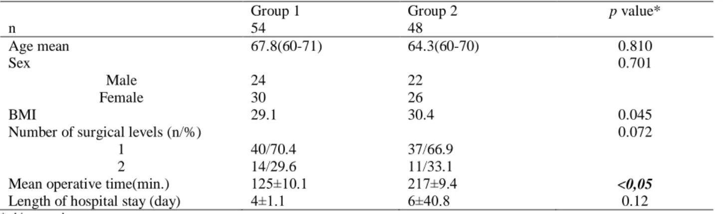
As seen in table 1 no significiant differences were seen between the groups about number of surgical levels, demographic findings such as age, sex, body mass index
(BMI) and length of hospital stay. But there was a significant difference between the groups in terms of mean operation time (125±10.1 min, 217±9 min, respectiveley, p<0.05).
Table 1. Patient characteristics.
Group 1 Group 2 p value*
n 54 48 Age mean 67.8(60-71) 64.3(60-70) 0.810 Sex 0.701 Male 24 22 Female 30 26 BMI 29.1 30.4 0.045
Number of surgical levels (n/%) 0.072
1 40/70.4 37/66.9
2 14/29.6 11/33.1
Mean operative time(min.) 125±10.1 217±9.4 <0,05
Length of hospital stay (day) 4±1.1 6±40.8 0.12
* chi-squared test
And in both of the groups VAS and ODI scores improved significantly within 3 years. In the group 1 VAS decreased from 9.4 (range from 10 to 7) to 3.02 (range from 4 to 1) and in the group 2 it decreased from 8.7 (range from 10 to 6) to 4.99 (range from 5 to 1). There was no significant difference between the groups in terms of improvement in VAS values (p=0.41).
In the group 1 ODI decreased from 60.9±2.4 to 30.02±5.1 and in the group 2 it decreased from 64.1±1.9 to 35.2±3.2. There was no significant difference between the
groups in terms of improvement in VAS values (p=0.770).
Second surgery requirement during 3 years follow-up was significantly higher in the group 2 (p<0.01) (Table 2). In the group 1 spondylolisthesis was observed in 2 patients (3.7%) and underwent posterior stabilization (4.1%). In the group 2, 3 patients (6.25%) underwent second surgery due to malposition of the instruments. Also adjacent segment syndrom was observed in 6 patients (12.5%) and these underwent second surgery.
Table 2. Comparison of second surgery requirement of the groups.
Group 1 Group 2 pvalue*
Second surgery requirement (n/%) 4.1% 9/18.7% <0.01
*chi-squared test
Discussion
This study compared the two types of surgery for LSCS in elderly patients. Patients who underwent only decompression and who underwent posterior transpedicular fusion in addition to decompression were compared for radiological findings and clinical outcomes. The need for surgical intervention in LSCS is still controversial but many studies reported satisfactory recovery in LSCS patients after surgical intervention compared with conservative treatment.10,13 According to our results, patient satisfaction increased after surgery with or without fusion at the end of the third year. Although there was no difference between the two groups, we
observed more decrease in VAS and ODI scores in the non-fusion group. Similar to our results in many studies greater back pain was found in the patients underwent decompression plus fusion surgery than patients without fusion.11,14 Also in group 2 we found significantly longer operative time and longer hospital stay although not statistically significant. Thomas et al. found the ratio of undesirable conditions due to surgery and longer operative time in the instrumented group operated due to LSCS.11 Also in their study, Forsth et al. found longer operative time, longer hospital stayand higher complication rate in the instrumented patients.12 And we found second surgery
57
requirement during 3 years follow-up significantly higher in the group 2. Most of them were due to adjacent segment syndrome. Probably posterior fusion causes changes in the mechanical balance of the spine, eliminates motion so increases the load at the adjacent segments.15-17 Review studies on this subject reported that adjacent segment syndrome could be seen radiologically up to 100%.18,19 Some of them become
symptomatic and require second surgery so the rate of complications increases in patients and the length of hospitalization increases.20
According to the previous studies the incidence of symptomatic adjacent segment syndrome requiring second surgery was ranging from 5 to 18% and most commonly seen on the cranial side.19,21 This difference between results may be because of differences among patient populations and differing methodologies. Some researchers have reported the increased incidence of adjacent segment syndrome according to advancing age.22,23 But, in the many previous studies it
was reported that there was no association between adjacent segment syndrome and age.24,25 In our study no significantly
difference was found between age and adjacent segment syndrome. Also Zhong et al. stated that simultaneous decompression at the adjacent segment was one of the risk factors for adjacent segment syndrome.19 So damage to the posterior elements appeared to be the most obvious cause.
In this present study there are some limitations. As a sample. The fact that no distinction was made between the two diagnoses; central canal stenosis and lateral recess stenosis. It was a limitation of this study. In addition, the fact that the amount of decompression was not measured postoperatively is another important limitation of this study.
Conclusion
We obtained more satisfactory results for both surgeon and patient in patients without fusion. If there is no evidence of serious listesis, total decompressive laminectomy can be performed safely in elderly patients.This successful result may be related to the type of surgery not demographic findings.
Conventional techniques through a minimally traumatic surgical corridorby preservation of spinal stability, optimal visualization, safe well-defined osteotomy lines and adequate decompression of stenotic pathology will ultimately build on achieving improved clinical outcomes. Sodecompression surgery performed up to two levels can be successfully terminated without fusion in elderly population.
Ethics Committee Approval
This study was approved by the Erzincan Binali Yıldırım University Medical Faculty ethics committee (2018-9/17) and continued in accordance with the Helsinki Principles Declaration.
Informed Consent
All participants signed the Informed Consent Form and their consent was obtained. Author Contributions
Conceptualization, Methodology and writing–original draft: T.Ç., Ş.C.Y. Data curation and investigation: Ş.C.Y. Resources, software, visualization and editing: T.Ç. Conflict of Interest
In this article, there is no conflict of interest between the authors
Financial Disclosure
No financial support is received. References
1. Çelik AA, Coşkun S, Kılınç O, Yıldırım A. Bel ağrısı nedeniyle
polikliniğimize başvuran hastalarda skolyoz sıklığı ve Cobb açısı değerlerinin yaş ve cinsiyet ile ilişkisi. Adıyaman
Üniversitesi Sağlık Bilimleri Dergisi,2017;3(3), 551-564.
2. Dönmez YC, Van Giersbergen MY, Başlı AA, Yıldız MD,
Yıldız E. Lomber Disk Hernisi Olan Hastaların Sağlıklı Yaşam
Biçimi Davranışlarının ve Öz-Bakım Gücünün
Belirlenmesi. Adıyaman Üniversitesi Sağlık Bilimleri Dergisi, 2019;5(2), 1628-1641.
3. Jönsson B, Annertz M, Sjöberg C, Strömqvist B. A prospective
and consecutive study of surgically treated lumbar spinal stenosis: part I: clinical features related to radiographic findings. Spine, 1997;22(24), 2932-2937.
4. Truumees E. Spinal stenosis: pathophysiology, clinical and radiologic classification. Instructional Course Lectures, 2005;54, 287-302.
5. Ježek J, Waldauf P, Krbec M, Douša P, Skála-Rosenbaum J.
Outcomes and Complications of Surgical Treatment for LSS at
1-Year Follow-up-Prospective Study. Actachirurgiae
orthopaedicae et traumatologiae Cechoslovaca, 2019;86(4),
256-263.
6. Kalbarczyk A, Lukes A, Seiler RW. Surgical treatment of
lumbar spinal stenosis in the elderly. Acta Neurochirurgica, 1998;140(7), 637-641.
58 7. Machado GC, Ferreira PH, Yoo RIJ, Harris IA, Pinheiro MB,
Koes BW, Ferreira ML. Surgical options for lumbar spinal stenosis. Cochrane Database Syst Rev 2016;11:CD012421. 8. Ciol MA, Deyo RA, Howell E, Kreif, S. An assessment of
surgery for spinal stenosis: time trends, geographic variations, complications, and reoperations. J Am Geriatr Soc 1996;44:285–90
9. Taylor VM, Deyo RA, Cherkin DC, et al. Low back pain hospitalization: recent United States trends and regional variations. Spine 1994;19:1207–13.
10. Deyo RA, Martin BI, Kreuter W, Jarvik JG, Angier H, Mirza SK. Revision surgery following operations for lumbar stenosis.
J Bone Joint Surg Am 2011;93:1979–86.
11. Thomas K, Faris P, McIntosh G, Manners S, Abraham E,
Bailey CS, Manson NA. Decompression Alone vs Decompression plus Fusion for Claudication Secondary to
Lumbar Spinal Stenosis. The Spine Journal
2019;(10):1633-1639.
12. Forsth P, Olafsson G, Carlsson T, Frost A, Borgstr€om F, Fritzell P, Sandén, B. A randomized, controlled trial of fusion surgery for lumbar spinal stenosis. N Engl J Med 2016;374:1413–23.
13. Herkowitz HN, Kurz LT. Degenerative lumbar spondylolisthesis with spinal stenosis. A prospective study comparing decompression with decompression and intertransverse process arthrodesis. J Bone Joint Surg
Am 1991;73:802–8.
14. Demiröz S, Bayram S, Coskun T, Cirakli A, Yanik HS, Atici Y, Erdem Ş. Evaluation of the distal adjacent segment after longsegment posterior instrumentation and fusion for adolescent idiopathic scoliosis. Annals of Medical Research 2019;26(10):2134-8.
15. Amundsen T, Weber H, Nordal HJ, Magnaes B, Abdelnoor M, Lilleas F. Lumbar spinal stenosis: conservative or surgical management? A prospective 10‐year study. Spine 2000;25(11):1424‐35.
16. Weinstein JN, Tosteson TD, Lurie JD, Tosteson AN, Blood E, Hanscom B, Hilibrand, A. SPORT Investigators. Surgical versus nonsurgical therapy for lumbar spinal stenosis. New
England Journal of Medicine 2008;358(8):794‐810.
17. Çakir T, Çakir M, Okay HÖ, Yolaş C, Tanriverdi O, Ömeroğlu
M, Arslan YK. Single level discectomy with and without disc prosthesis: A comparative study of 114 patients. Medicine 2018;97(52).
18. Ilharreborde B, Morel E, Mazda K, Dekutoski MB. Adjacent
segment disease after instrumented fusion for idiopathic scoliosis: review of current trends and controversies. Clinical
Spine Surgery 2009;22(7), 530-539.
19. Zhong ZM, Deviren V, Tay B, Burch S, Berven SH. Adjacent
segment disease after instrumented fusion for adult lumbar
spondylolisthesis: incidence and risk factors. Clinical
Neurology and Neurosurgery 2017;156, 29-34.
20. Aydin A, Çilingir D. Yeniden Ameliyat Olma (Reoperasyon)
ve Hemşirelik Bakımı. Koç Üniversitesi Hemşirelikte Eğitim ve
Araştırma Dergisi 2017;14(3), 218-222.
21. Park P, Garton HJ, Gala VC, Hoff JT, McGillicuddy JE.
Adjacent segment disease after lumbar or lumbosacral fusion: review of the literature. Spine 2004;29(17), 1938-1944.
22. Kizilay Z, Topcu A, Aydin YS, Berber O, Ozturk, H. Short and
Medium Term Results of Posterior Segmental Instrumentation
and Posterolateral Fusion in Female Patients with
Spondylolisthesis: A Clinical Trial/Spondilolistezli Kadin
Hastalarda Posterior Segmental Enstrumantasyon ve
Posterolateral Fuzyonun Kisa ve Orta Vadeli Sonuclari: Klinik Calisma. Meandros Medical and Dental Journal 2018;19(4), 328-336.
23. Min JH, Jang JS, joo Jung B, Lee HY, Choi WC, Shim CS, Lee
SH. The clinical characteristics and risk factors for the adjacent segment degeneration in instrumented lumbar fusion. Clinical
Spine Surgery 2008;21(5), 305-309.
24. Ghiselli G, Wang JC, Bhatia NN, Hsu WK, Dawson EG.
Adjacent segment degeneration in the lumbar spine. The
Journal of Bone and Joint Surgery 2004;86(7), 1497-1503.
25. Lee CS, Hwang CJ, Lee SW, Ahn YJ, Kim YT, Lee DH, Lee
MY. Risk factors for adjacent segment disease after lumbar fusion. European Spine Journal2009; 18(11), 1637.