402 Indian Journal of Ophthalmology Vol. 64 No. 5 spectrum in familial exudative vitreoretinopathy and Norrie
disease with identification of 21 novel variants in FZD4, LRP5, and NDP. Hum Mutat 2010;31:656‑66.
9. Wong N, Lasko D, Rabelo R, Pinsky L, Gordon PH, Foulkes W. Genetic counseling and interpretation of genetic tests in familial adenomatous polyposis and hereditary nonpolyposis colorectal cancer. Dis Colon Rectum 2001;44:271‑9.
10. Maguire AM, Simonelli F, Pierce EA, Pugh EN Jr., Mingozzi F, Bennicelli J, et al. Safety and efficacy of gene transfer for Leber’s congenital amaurosis. N Engl J Med 2008;358:2240‑8.
11. Bainbridge JW, Smith AJ, Barker SS, Robbie S, Henderson R, Balaggan K, et al. Effect of gene therapy on visual function in Leber’s congenital amaurosis. N Engl J Med 2008;358:2231‑9. 12. Jacobson SG, Cideciyan AV, Ratnakaram R, Heon E, Schwartz SB,
Roman AJ, et al. Gene therapy for leber congenital amaurosis caused by RPE65 mutations: Safety and efficacy in 15 children and adults followed up to 3 years. Arch Ophthalmol 2012;130:9‑24. 13. Xia CH, Liu H, Cheung D, Wang M, Cheng C, Du X, et al. A model
for familial exudative vitreoretinopathy caused by LPR5 mutations. Hum Mol Genet 2008;17:1605‑12.
Departments of Ophthalmology, and 1Pathology, Baskent University Faculty of Medicine, Ankara, Turkey
Correspondence to: Dr. Muge‑Coban Karatas, Department of Ophthalmology, Baskent University Faculty of Medicine, Ankara, Turkey. E‑mail: [email protected]
Manuscript received: 14.10.15; Revision accepted: 14.03.16
Spindle cell carcinoma of the
conjunctiva: A rare entity
Muge Coban‑Karatas, Nebil Bal
1,
Rana Altan-Yaycioğlu, Aysen Terzi
1An 85‑year‑old male presented with painless bulging lesion over the cornea. Clinical history, diagnostic imaging studies, and histopathologic sections were evaluated. The patient clinically displayed an vascularized conjunctival lesion located at the superior bulbar conjunctiva with extension onto cornea covering 2/3 of his pupillary aperture superiorly. His visual acuity was counting fingers at 4 m. The patient underwent a total excision of the lesion including conjunctival and corneal parts. Histopathologic evaluation revealed spindle cell carcinoma which involves the whole conjunctival squamous epithelium with significant polarity loss, nuclear enlargement with hyperchromasia and pleomorphism, and mitotic activity. Diagnosis of spindle cell carcinoma is challenging because of overlapping histopathological features with other spindle cell tumors. The detailed pathologic examination is very important for the decision of proper treatment.
Key words: Conjunctiva, spindle cell carcinoma, squamous epithelium
Spindle cell carcinoma is a rare and unusual biphasic malignant tumor, which involves sarcomatoid proliferation of pleomorphic spindle cells and squamous cell carcinoma (SCC). SCC, with the spindle cell component, is an uncommon phenomenon and a rare type of malignant tumor.[1,2] Spindle
cell carcinoma is a poorly differentiated variant of SCC that rarely occurs in the conjunctiva.[3‑7]
We aimed to present a case with conjunctival spindle cell carcinoma to emphasize the importance of detailed pathologic examination to differentiate the cell type for the decision of proper treatment. Informed consent was obtained from the patient.
Case Report
An 85‑year‑old male referred to our clinic with decreased vision in the right eye. He did not have any history of trauma and did not complain about pain. He described a pedunculated lesion without any ulceration, which grew slowly over 3 months. In his ophthalmologic examination, best‑corrected visual acuity was counting fingers at 4 m in his right eye and 0.1 in his left eye. His intraocular pressure was 18 mmHg in both eyes. Anterior segment examination revealed a large vascularized lesion located in the superior bulbar conjunctiva with extension onto cornea closing 2/3 of the pupillary area [Fig. 1]. The left eye revealed no pathology in the anterior segment of the eye. Fundoscopic examination of the right eye could not be performed; on the left eye, optic nerve was pale. The patient underwent an excisional biopsy of the lesion removing the whole tumor in the conjunctiva as well as on the cornea and cryotherapy to the conjunctival margins.
Histopathologic evaluation revealed in situ carcinoma which holds the whole conjunctival squamous epithelium with significant polarity loss, nuclear enlargement with hyperchromasia and pleomorphism, and mitotic activity. The stroma was rich in atypical cells forming herds and bundles of spindle or epithelioid cells, with hyperchromatic nuclei and pleomorphism mixed with inflammatory cells [Fig. 2]. In immunohistochemical staining, atypical stromal cells were stained positive with vimentin, pancytokeratin (cytokeratin AE1/ AE3), epithelial membrane antigen (EMA), smooth muscle actin (SMA), CD99, p63, and calponin and were stained negative with caldesmon and MyoD1. Positivity of EMA and pancytokeratin
Cite this article as: Coban-Karatas M, Bal N, Altan-Yaycioğlu R, Terzi A.
Spindle cell carcinoma of the conjunctiva: A rare entity. Indian J Ophthalmol 2016;64:402-4.
This is an open access article distributed under the terms of the Creative Commons Attribution‑NonCommercial‑ShareAlike 3.0 License, which allows others to remix, tweak, and build upon the work non‑commercially, as long as the author is credited and the new creations are licensed under the identical terms.
For reprints contact: [email protected]
Access this article online
Quick Response Code: Website:
www.ijo.in
DOI:
10.4103/0301-4738.185630 PMID:
***
May 2016 Brief Communications 403
revealed the epithelial origin of the tumor [Fig. 3]. In addition, p63 positivity is indicated the squamous differentiation. Vimentin, SMA, and calponin positivity proved the sarcomatous component with myoepithelial differentiation [Fig. 4].
Because of the malignant potential of the lesion, an orbital magnetic resonance imaging was taken, which showed no posterior and orbital extension.
In his medical history, our patient had a nonmetastatic carcinomatous gingival lesion for which he had received radiotherapy 4 years ago. However, his conjunctival lesion was accepted as primary because the new lesion was far from the radiotherapy treatment area and in situ carcinoma was detected in the superficial epithelium.
After surgical excision, his vision improved to 0.2. He was referred to the oncology department for further evaluation. Unfortunately, the patient lost to follow‑up. Contact with a relative revealed that he was taking home care because of severe senile health problems.
Discussion
SCC of the conjunctiva is a rare malignancy; however, it is reported to be the most common malignant tumor of the ocular surface.[8] SCC has the potential to penetrate the corneoscleral
lamella into the anterior chamber and can breach the orbital septum to invade the soft tissues of the orbit, sinuses, and brain. These tumors may metastasize via lymphatics or blood during the disease.[9] Surgical excision with or without cryotherapy
and radiotherapy remains the widely accepted treatment for SCC of the conjunctiva.[9,10]
Spindle cell carcinoma of the conjunctiva is relatively rare, with only a few cases reported in literature.[3‑7] Cervantes et al.
reported a total of 287 cases of SCC of the conjunctiva, in which only two cases were documented as spindle cell carcinoma.[11]
Spindle cell carcinoma is a poorly differentiated variant of SCC which is considered to be more aggressive and can also affect the progress and outcome of the disease.
Figure 2: The stroma was rich in atypical cells forming herds and bundles of spindle or epithelioid cells with hyperchromatic nuclei and pleomorphism (H and E, × 40)
Figure 3: Pancytokeratin positivity in atypical spindle‑shaped and
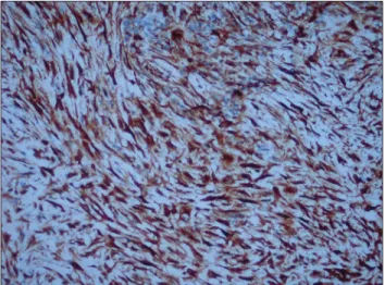
epithelioid cells infiltrating the stroma (pancytokeratin, × 400) Figure 4: Calponin positivity in atypical spindle‑shaped and epithelioid cells infiltrating the stroma (calponin, × 200)
Figure 1: Clinical picture of a large vascularized lesion located in superior bulbar conjunctiva with extension onto the cornea
404 Indian Journal of Ophthalmology Vol. 64 No. 5
Conclusion
Because of their possible aggressive behavior, conjunctival malignancies are known to be sight‑ and life‑threatening.[9] The
detailed pathologic examination is very important to differentiate the cell type for the decision of proper treatment.
Financial support and sponsorship Nil.
Conflicts of interest
There are no conflicts of interest.
References
1. Zheng Y, Xiao M, Tang J. Clinicopathological and immunohistochemical analysis of spindle cell carcinoma of the larynx or hypopharynx: A report of three cases. Oncol Lett 2014;8:748‑52.
2. Torenbeek R, Hermsen MA, Meijer GA, Baak JP, Meijer CJ. Analysis by comparative genomic hybridization of epithelial and spindle cell components in sarcomatoid carcinoma and carcinosarcoma: Histogenetic aspects. J Pathol 1999;189:338‑43.
3. Cohen BH, Green WR, Iliff NT, Taxy JB, Schwab LT,
de la Cruz Z. Spindle cell carcinoma of the conjunctiva. Arch Ophthalmol 1980;98:1809‑13.
4. Huntington AC, Langloss JM, Hidayat AA. Spindle cell carcinoma of the conjunctiva. An immunohistochemical and ultrastructural study of six cases. Ophthalmology 1990;97:711‑7.
5. Ni C, Guo BK. Histological types of spindle cell carcinoma of the cornea and conjunctiva. A clinicopathologic report of 8 patients with ultrastructural and immunohistochemical findings in three tumors. Chin Med J (Engl) 1990;103:915‑20.
6. Schubert HD, Farris RL, Green WR. Spindle cell carcinoma of the conjunctiva. Graefes Arch Clin Exp Ophthalmol 1995;233:52‑3. 7. Slusker‑Shternfeld I, Syed NA, Sires BA. Invasive spindle cell
carcinoma of the conjunctiva. Arch Ophthalmol 1997;115:288‑9. 8. Sun EC, Fears TR, Goedert JJ. Epidemiology of squamous cell
conjunctival cancer. Cancer Epidemiol Biomarkers Prev 1997;6:73‑7. 9. Shields CL, Shields JA. Tumors of the conjunctiva and cornea. Surv
Ophthalmol 2004;49:3‑24.
10. Miller CV, Wolf A, Klingenstein A, Decker C, Garip A, Kampik A, et al. Clinical outcome of advanced squamous cell carcinoma of the conjunctiva. Eye (Lond) 2014;28:962‑7.
11. Cervantes G, Rodríguez AA Jr., Leal AG. Squamous cell carcinoma of the conjunctiva: Clinicopathological features in 287 cases. Can J Ophthalmol 2002;37:14‑9.