Omer Ozel, Emre Demircay
MINIREVIEWS
Review of management of unstable elbow fractures
Omer Ozel, Emre Demircay, Department of Orthopedic Surgery, Baskent University, 34662 Istanbul, Turkey
Author contributions: All the authors contributed to this paper. Conflict-of-interest statement: All authors declare that they have no conflict of interest.
Open-Access: This article is an open-access article which was selected by an in-house editor and fully peer-reviewed by external reviewers. It is distributed in accordance with the Creative Commons Attribution Non Commercial (CC BY-NC 4.0) license, which permits others to distribute, remix, adapt, build upon this work non-commercially, and license their derivative works on different terms, provided the original work is properly cited and the use is non-commercial. See: http://creativecommons.org/ licenses/by-nc/4.0/
Correspondence to: Emre Demircay, Associate Professor, Department of Orthopedic Surgery, Baskent University, Oymaci Sokak No: 7 Altunizade, 34662 Istanbul,
Turkey. [email protected] Telephone: +90-216-5541500 Fax: +90-216-6519746 Received: June 4, 2015
Peer-review started: June 5, 2015 First decision: July 6, 2015 Revised: October 7, 2015 Accepted: November 3, 2015 Article in press: November 4, 2015 Published online: January 18, 2016
Abstract
Stable and painless elbow motion is essential for acti-vities of daily living. The elbow joint is the second most commonly dislocated joint in adults. The goals of treatment are to perform a stable fixation of all fractures, to achieve concentric and stable reduction of the elbow and to provide early motion. The treatment modality for complex elbow instability is almost always surgical. The treatment objectives are anatomic reduction, stable fixation, and early rehabilitation of the elbow. The common complications of these unstable
fractures include recurrent instability, stiffness, myositis ossifications, heterotopic calcification, and neurovascular dysfunction. We analyzed the management of complex elbow fractures and instabilities on the basis of recent literature and suggested possible guidelines for the treatment in this paper. In conclusion, recognition of the injury pattern and restoration of the joint stability are the prerequisites for any successful treatment of an unstable elbow injury.
Key words: Transolecranon fracture; Coronoid fracture;
Monteggia injury; Radial head fracture; Terrible triad
© The Author(s) 2016. Published by Baishideng Publishing
Group Inc. All rights reserved.
Core tip: As the elbow joint is the second most
com-monly dislocated joint in adults, we aimed to analyze the management of complex elbow fractures and instabilities, on the basis of recent literature and suggested possible guidelines for the treatment in this paper.
Ozel O, Demircay E. Review of management of unstable elbow fractures. World J Orthop2016; 7(1): 50-54 Available from: URL: http://www.wjgnet.com/2218-5836/full/v7/i1/50.htm DOI: http://dx.doi.org/10.5312/wjo.v7.i1.50
INTRODUCTION
Basic elbow function requires stable and painless elbow motion. The three articulations, namely the ulnotrochlear, radiocapitellar, and proximal radioulnar joints, provide elbow flexion/extension and supination/ pronation. Static and dynamic constraints create stability of the elbow joint. The ulnohumeral articulation, anterior bundle of the medial collateral ligament, and lateral collateral ligament complex form the primary static constraints. The joint capsule and radial head are among the secondary static constraints. The dynamic constraints, such as the common flexor and extensor
muscle groups, are any muscles crossing the elbow joint that exert a compressive force on the joint[1].
The elbow is the second most commonly dislocated joint in adults[2]. The dislocations may be complex or
simple. When dislocations are associated with fractures, they are designated as complex. The reported annual incidence of simple and complex elbow dislocations is 6.1 per 100000 patients[3]. Radial head fractures/
dislocations, coronoid fractures, terrible triad injuries, transolecranon fracture-dislocations, and Monteggia-like lesions can be listed as common causes of complex elbow injuries.
Complex elbow fractures and instability typically present with edema, tenderness, pain in active/passive movement, and restriction of motion. A fall onto the extended outstretched hand or a direct trauma to the elbow is usually described as the mechanism of the injury. Anteroposterior and lateral standard radiographs and computerized tomography scans (either standard or 3D) are needed to properly evaluate the bone injuries and to accurately plan their surgical treatment. Neurovascular examination and documentation of the injuries before and after any manipulation are of critical importance. A temporary fracture alignment with cast immobilization may be done until definitive surgery in patients with severe injuries.
The treatment modality for complex elbow instability is almost always surgical. The treatment objectives are anatomic reduction, stable fixation, and early reha bilitation of the elbow.
The common complications of these unstable frac-tures include recurrent instability, stiffness, myositis ossifications, heterotopic calcification, and neurovascular dysfunction.
In this review, we analyze the management of complex elbow fractures and instability on the basis of recent literature, and suggest possible guidelines for the treatment of these injuries.
RADIAL HEAD FRACTURES
Radial head fractures are among the most common elbow fractures, occurring in up to 20% of all elbow injuries[4]. Radial head fractures are mostly associated
with complex injuries like elbow dislocation, and lateral collateral ligament (LCL) and medial collateral ligament (MCL) tears. Impaction fractures of the capitellum may also be associated with radial head fractures, and can easily be overlooked[5]. Only about 5% of cases have a
radial head fracture as an isolated injury[6,7]. In a large
incidence study[8], the mean age was found to be 36
years and the femaletomale ratio was 47.7/53.3. A thorough physical examination is essential to diagnose associated ligament injuries. Fluoroscopy may be used to confirm an MCL injury if medial pain and ecchymosis are present. If the MCL is injured, the role of the radial head in valgus resistance increases up to 30%[9,10].
Stable anatomic reconstruction of the radial head is the
The Mason classification system[11] divided radial head
fractures into three categories: type Ⅰ, non-displaced fractures; type Ⅱ, displaced partial head fractures; and type Ⅲ, comminuted displaced fractures involving the whole head.
Modifications to the Mason classification were intro duced to guide treatment. The Hotchkiss modification[12]
defines type Ⅰ fractures as non-displaced fractures (< 2 mm displacement) without mechanical blockage that do not require surgery, type Ⅱ fractures as dis-placed fractures (> 2 mm displacement) of the radial head or neck that lack severe comminution, may have mechanical blockage to movement, and usually require open reduction and internal fixation, and type
Ⅲ fractures as severely comminuted fractures of the radial head and neck. Satisfactory reconstructions of these fractures are not possible, and therefore the radial head is either excised or replaced with a prosthesis[13].
Fragment excision is avoided in complex elbow instability to prevent valgus instability[14]. If there is an associated
LCL rupture, it should be repaired after appropriate management of the radial head fracture, either by fixation or by prosthetic replacement. Suture anchors or transosseous sutures can be used for the reconstruction. The joint stability should be confirmed by dynamic fluoroscopic examination. If residual instability persists, MCL reconstruction and/or dynamic elbow fixation should be done[15,16].
CORONOID FRACTURES
The coronoid process plays a pivotal role as an anterior buttress in providing elbow stability. Although coronoid fractures may occur in isolation, they are more commonly seen as a component of unstable elbow fractures[17].
The classification proposed by Moon et al[18], which
defines anteromedial facet lesions, may be better for guiding the surgical management of coronoid fractures. Type Ⅰ injuries involve fractures of the coronoid tip, and are divided into two subtypes based on the fracture size. Subtype 1 fractures are smaller than 2 mm, and subtype 2 fractures are larger than 2 mm. In type Ⅱ
injuries, the anteromedial aspect of the coronoid is fractured. These injuries are divided into three subtypes based on the anatomic location. Subtype 1 fractures involve the rim, subtype 2 fractures involve the rim and the tip, and subtype 3 fractures involve the rim and the sublime tubercle with or without the tip. Type Ⅲ
fractures are basal coronoid fractures involving at least 50% of the height of the coronoid. They are divided into two subtypes depending on whether the fracture involves the base of the olecranon. Stable fixation and ligament repair are essential for the treatment of coronoid fractures[19,20].
TERRIBLE TRIAD INJURIES
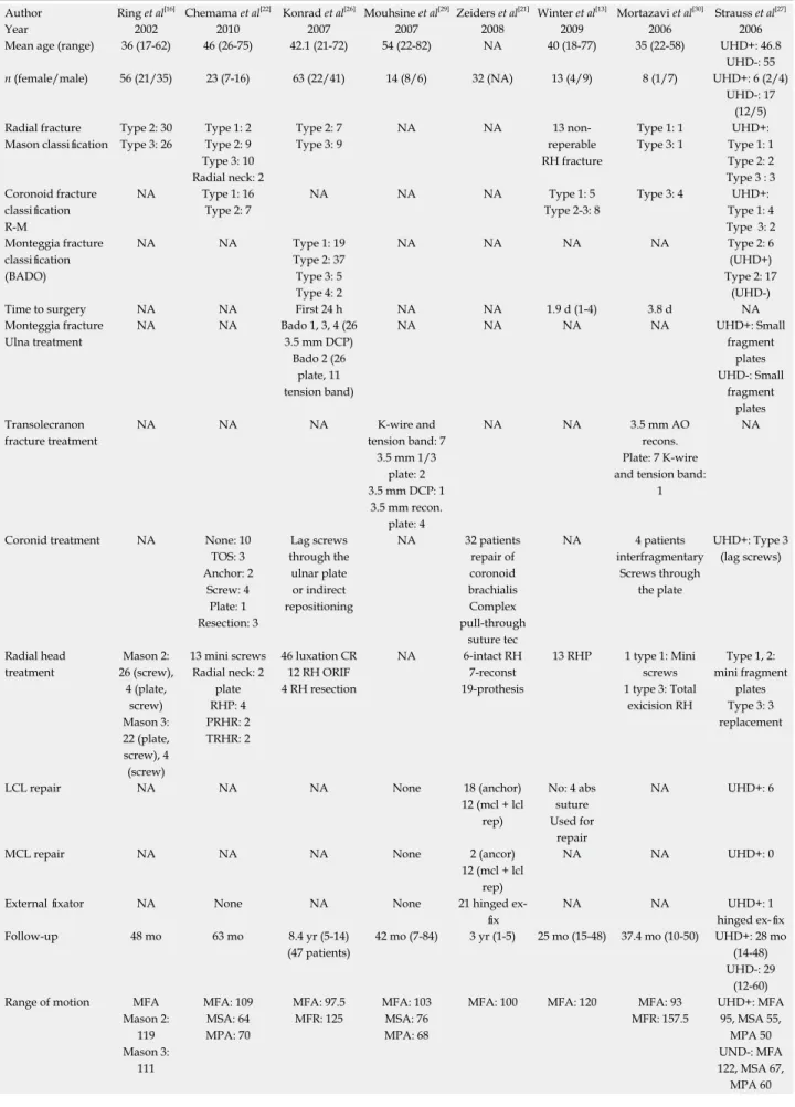
Author Ring et al[16] Chemama et al[22] Konrad et al[26] Mouhsine et al[29] Zeiders et al[21] Winter et al[13] Mortazavi et al[30] Strauss et al[27]
Year 2002 2010 2007 2007 2008 2009 2006 2006
Mean age (range) 36 (17-62) 46 (26-75) 42.1 (21-72) 54 (22-82) NA 40 (18-77) 35 (22-58) UHD+: 46.8
UHD-: 55
n (female/male) 56 (21/35) 23 (7-16) 63 (22/41) 14 (8/6) 32 (NA) 13 (4/9) 8 (1/7) UHD+: 6 (2/4) UHD-: 17 (12/5) Radial fracture Mason classification Type 2: 30 Type 3: 26 Type 1: 2 Type 2: 9 Type 3: 10 Radial neck: 2 Type 2: 7 Type 3: 9 NA NA 13 non-reperable RH fracture Type 1: 1 Type 3: 1 UHD+: Type 1: 1 Type 2: 2 Type 3 : 3 Coronoid fracture classification R-M NA Type 1: 16 Type 2: 7 NA NA NA Type 1: 5 Type 2-3: 8 Type 3: 4 UHD+: Type 1: 4 Type 3: 2 Monteggia fracture classification (BADO) NA NA Type 1: 19 Type 2: 37 Type 3: 5 Type 4: 2 NA NA NA NA Type 2: 6 (UHD+) Type 2: 17 (UHD-)
Time to surgery NA NA First 24 h NA NA 1.9 d (1-4) 3.8 d NA
Monteggia fracture Ulna treatment NA NA Bado 1, 3, 4 (26 3.5 mm DCP) Bado 2 (26 plate, 11 tension band) NA NA NA NA UHD+: Small fragment plates UHD-: Small fragment plates Transolecranon fracture treatment NA NA NA K-wire and tension band: 7 3.5 mm 1/3 plate: 2 3.5 mm DCP: 1 3.5 mm recon. plate: 4 NA NA 3.5 mm AO recons. Plate: 7 K-wire and tension band:
1
NA
Coronid treatment NA None: 10
TOS: 3 Anchor: 2 Screw: 4 Plate: 1 Resection: 3 Lag screws through the ulnar plate or indirect repositioning NA 32 patients repair of coronoid brachialis Complex pull-through suture tec NA 4 patients interfragmentary Screws through the plate UHD+: Type 3 (lag screws) Radial head treatment Mason 2: 26 (screw), 4 (plate, screw) Mason 3: 22 (plate, screw), 4 (screw) 13 mini screws Radial neck: 2 plate RHP: 4 PRHR: 2 TRHR: 2 46 luxation CR 12 RH ORIF 4 RH resection NA 6-intact RH 7-reconst 19-prothesis 13 RHP 1 type 1: Mini screws 1 type 3: Total exicision RH Type 1, 2: mini fragment plates Type 3: 3 replacement
LCL repair NA NA NA None 18 (anchor)
12 (mcl + lcl rep) No: 4 abs suture Used for repair NA UHD+: 6
MCL repair NA NA NA None 2 (ancor)
12 (mcl + lcl rep)
NA NA UHD+: 0
External fixator NA None NA None 21 hinged
ex-fix NA NA UHD+: 1 hinged ex-fix Follow-up 48 mo 63 mo 8.4 yr (5-14) (47 patients) 42 mo (7-84) 3 yr (1-5) 25 mo (15-48) 37.4 mo (10-50) UHD+: 28 mo (14-48) UHD-: 29 (12-60)
Range of motion MFA
Mason 2: 119 Mason 3: 111 MFA: 109 MSA: 64 MPA: 70 MFA: 97.5 MFR: 125 MFA: 103 MSA: 76 MPA: 68
MFA: 100 MFA: 120 MFA: 93
MFR: 157.5 UHD+: MFA 95, MSA 55, MPA 50 UND-: MFA 122, MSA 67, MPA 60
dislocation along with a coronoid fracture. Both medial and lateral compartments can be exposed through a posterior incision. The Kocher approach can be used for the radial head fracture. Hotchkiss type Ⅰ and type Ⅱ
radial head fractures can be fixed with headless screws or a plate[12]. Prosthetic replacement is mandatory for
comminuted radial head fractures (type Ⅲ) to avoid chronic instability. There is often a comminuted type 1 fracture in the coronoid, and it can usually only be fixed with a transosseous suture. If there is an isolated fragment that is sufficiently large, fixation with K wires or screws can be done[19]. The LCL is repaired last,
and elbow stability is assessed by fluoroscopy. In the presence of a residual instability, the MCL should also be repaired or a hinged external fixator should be applied[7,21,22] (Table 1).
MONTEGGIA-LIKE LESIONS
Monteggia injuries comprise a fracture of the ulnar shaft with an associated radial head dislocation. Monteggia originally described the lesions as a fracture of the proximal third of the ulna and an anterior dislocation of the proximal epiphysis of the radius[23]. Bado[23] classified
these injuries by primarily focusing on the radial component. Jupiter et al[24] modified this classification
by defining subtypes for the posterior Monteggia lesions (Bado type 2). Ulnohumeral dislocation, radial fracture, proximal and/or distal radioulnar dislocation, and interosseous membrane lesions may also accompany the ulnar fracture and radiohumeral dislocation. Each of these must be recognized and treated. The varying combinations of these injured structures explains the complexity and diversity of the management procedures.
Anatomic reduction and stabilization of the ulna and the ulnohumeral joint is the primary objective of surgical treatment for posterior elbow fracture-dislocations[25].
The radial head fracture is addressed initially. If the radial head cannot be salvaged satisfactorily, radial head arthroplasty is preferred. To size the radial head properly, the ulnar length should be restored by a provisional fixation[17]. The coronoid process is stabilized after the
ulnar shaft fracture has been addressed, and the olec-ranon is fixed with a dorsal plate. Finally, ligamentous
TRANSOLECRANON
FRACTURE-DISLOCATIONS (ANTERIOR OLECRANON
FRACTURE-DISLOCATIONS)
The radial head is dislocated anteriorly with an associated olecranon fracture in this injury pattern[28]. Two subtypes
have been described: One with a simple olecranon fracture, and one with a comminuted olecranon frac-ture[28]. The second subtype is more common and may
be associated with trochlear and coronoid fractures. This injury pattern is distinct from the anterior Monteggia (Bado type 1) lesion, because in transolecranon fracture-dislocation, the ulnohumeral stability is lost but the radioulnar relationship remains intact. Bony disruption is the main reason for the failure of the ulnohumeral joint rather than the ligamentous structures.
Anatomic reduction with particular attention to res-toring the ulnar length and greater sigmoid notch is ess-ential in the treatment[29] (Table 1). Restoration of the
ulnohumeral anatomy is crucial to prevent radiocapitellar instability or subluxation[25,30] (Table 1).
CONCLUSION
Surgical treatments of complex elbow fracture disloca-tions are among the most challenging procedures for orthopedic surgeons. Interpretation of the underlying mechanisms for elbow instability and accurate identifi-cation of the injured structures are crucial for surgical planning. Stable elbow fracture fixation is important for early elbow motion and avoiding joint stiffness. Recognition of the injury pattern and restoration of the joint stability are the prerequisites for any successful treatment of an unstable elbow injury.
In this review, we have examined the diagnosis, classification, and treatment of unstable elbow fractures. Future studies should be conducted to determine the optimal management strategies, the role of ligament reconstruction, and reductions in the complication rate.
REFERENCES
1 Parsons BO, Ramsey ML. Elbow instability and reconstruction.
In: Flynn JM. Orthopaedic knowledge update. Rosemont, IL:
Mean score Mason 2:
BM: 92 Mason 3: BM: 86 MEPS: 87 (75-100) 14 patients result BM: 87.2 (45-100) DASH: 17.4 (0-70) BM: 82 (78-100) DASH: 23 (19/28) BM: 86.5 (55-100) BM: 88 (71-100) ASES: 89 (69-100) UHD+: DASH 34 (0-80), BM: 73.8 UHD-: DASH 23 (0-70), BM: 83 Unsatisfactory Mason 2: 4 Mason 3: 14 BM: 0 BM: (9 fair) (4 poor) result BM: 2 fair 2 poor 2 (stiffness, infection) BM: 1 fair UHD+: 2 UHD-: 7 NA: Not applicable; UHD: Ulnohumeral dislocation; RH: Radial head; R-M: Roger Morrey; DCP: Dynamic compression plate; TOS: Transosseous suture; RHP: Radial head prosthesis; PRHR: Partial radial head resection; TRHR: Total radial head resection; MFA: Mean flexion arch; MSA: Mean supination arch; MPA: Mean pronation arch; MFR: Mean forearm rotation; BM: Broberg and Morrey index; MEPS: Mayo elbow performance score; DASH: Disabilities of the arm shoulder and hand; ASES: American Shoulder and Elbow Surgeons assessment system; LCL: Lateral collateral ligament; MCL: Medial collateral ligament.
2 Kuhn MA, Ross G. Acute elbow dislocations. Orthop Clin
North Am 2008; 39: 155-161, v [PMID: 18374806 DOI: 10.1016/
j.ocl.2007.12.004]
3 Josefsson PO, Nilsson BE. Incidence of elbow dislocation. Acta
Orthop Scand 1986; 57: 537-538 [PMID: 3577725 DOI: 10.3109/1
7453678609014788]
4 Glaser DL, Armstrong AD. Elbow and forearm trauma. In: Flynn
JM. Orthopaedic knowledge update. Rosemont, IL: American Academy of Orthopaedic Surgeons, 2011: 325-341
5 Nalbantoglu U, Gereli A, Kocaoglu B, Aktas S, Turkmen M.
Capitellar cartilage injuries concomitant with radial head fractures.
J Hand Surg Am 2008; 33: 1602-1607 [PMID: 18984344 DOI:
10.1016/j.jhsa.2008.05.016]
6 van Riet RP, Morrey BF, O’Driscoll SW, Van Glabbeek F.
Asso-ciated injuries complicating radial head fractures: a demographic study. Clin Orthop Relat Res 2005; 441: 351-355 [PMID: 16331026 DOI: 10.1097/01.blo.0000180606.30981.78]
7 Ring D, Jupiter JB, Zilberfarb J. Posterior dislocation of the elbow
with fractures of the radial head and coronoid. J Bone Joint Surg
Am 2002; 84-A: 547-551 [PMID: 11940613]
8 Kovar FM, Jaindl M, Thalhammer G, Rupert S, Platzer P, Endler
G, Vielgut I, Kutscha-Lissberg F. Incidence and analysis of radial head and neck fractures. World J Orthop 2013; 4: 80-84 [PMID: 23610756 DOI: 10.5312/wjo.v4.i2.80]
9 Søjbjerg JO, Ovesen J, Nielsen S. Experimental elbow instability
after transection of the medial collateral ligament. Clin Orthop
Relat Res 1987; (218): 186-190 [PMID: 3568478]
10 Morrey BF, Tanaka S, An KN. Valgus stability of the elbow. A definition of primary and secondary constraints. Clin Orthop Relat
Res 1991; (265): 187-195 [PMID: 2009657]
11 Mason ML. Some observations on fractures of the head of the radius with a review of one hundred cases. Br J Surg 1954; 42: 123-132 [PMID: 13209035 DOI: 10.1002/bjs.18004217203] 12 Hotchkiss RN. Displaced Fractures of the Radial Head: Internal
Fixation or Excision? J Am Acad Orthop Surg 1997; 5: 1-10 [PMID: 10797202]
13 Winter M, Chuinard C, Cikes A, Pelegri C, Bronsard N, de Peretti F. Surgical management of elbow dislocation associated with non-reparable fractures of the radial head. Chir Main 2009; 28: 158-167 [PMID: 19356963 DOI: 10.1016/j.main.2009.02.003]
14 Duckworth DG, Avakian Z, Chien C. Newly defined fracture pattern specific to Mason III radial head fractures: fracture description, management and outcomes using screw fixation.
ANZ J Surg 2012; 82: 434-438 [PMID: 22548755 DOI: 10.1111/
j.1445-2197.2012.06094.x]
15 Tan V, Daluiski A, Capo J, Hotchkiss R. Hinged elbow external fixators: indications and uses. J Am Acad Orthop Surg 2005; 13: 503-514 [PMID: 16330512]
16 Ring D, Quintero J, Jupiter JB. Open reduction and internal fixation of fractures of the radial head. J Bone Joint Surg Am 2002;
84-A: 1811-1815 [PMID: 12377912]
17 Giannicola G, Sacchetti FM, Greco A, Cinotti G, Postacchini F. Management of complex elbow instability. Musculoskelet Surg 2010; 94 Suppl 1: S25-S36 [PMID: 20383679 DOI: 10.1007/ s12306-010-0065-8]
18 Moon JG, Zobitz ME, An KN, O’Driscoll SW. Optimal screw orientation for fixation of coronoid fractures. J Orthop Trauma 2009;
23: 277-280 [PMID: 19318871 DOI: 10.1097/BOT.0b013e31819df8c9]
19 Budoff JE. Coronoid fractures. J Hand Surg Am 2012; 37: 2418-2423 [PMID: 23101539 DOI: 10.1016/j.jhsa.2012.09.002] 20 Garrigues GE, Wray WH, Lindenhovius AL, Ring DC, Ruch DS.
Fixation of the coronoid process in elbow fracture-dislocations. J
Bone Joint Surg Am 2011; 93: 1873-1881 [PMID: 22012524 DOI:
10.2106/JBJS.I.01673]
21 Zeiders GJ, Patel MK. Management of unstable elbows following complex fracture-dislocations--the “terrible triad” injury. J Bone
Joint Surg Am 2008; 90 Suppl 4: 75-84 [PMID: 18984720 DOI:
10.2106/JBJS.H.00893]
22 Chemama B, Bonnevialle N, Peter O, Mansat P, Bonnevialle P. Terrible triad injury of the elbow: how to improve outcomes?
Orthop Traumatol Surg Res 2010; 96: 147-154 [PMID: 20417913
DOI: 10.1016/j.otsr.2009.11.009]
23 Bado JL. The Monteggia lesion. Clin Orthop Relat Res 1967; 50: 71-86 [PMID: 6029027 DOI: 10.1097/00003086-196701000-00008] 24 Jupiter JB, Leibovic SJ, Ribbans W, Wilk RM. The posterior
Monteggia lesion. J Orthop Trauma 1991; 5: 395-402 [PMID: 1761999 DOI: 10.1097/00005131-199112000-00003]
25 Scolaro JA, Beingessner D. Treatment of monteggia and tran-solecranon fracture-dislocations of the elbow: A critical analysis review. JBJS Reviews 2014; 2: e3 [DOI: 10.2106/JBJS.RVW. M.00049]
26 Konrad GG, Kundel K, Kreuz PC, Oberst M, Sudkamp NP. Monteggia fractures in adults: long-term results and prognostic factors. J Bone Joint Surg Br 2007; 89: 354-360 [PMID: 17356149 DOI: 10.1302/0301-620X.89B3.18199]
27 Strauss EJ, Tejwani NC, Preston CF, Egol KA. The posterior Monteggia lesion with associated ulnohumeral instability. J Bone
Joint Surg Br 2006; 88: 84-89 [PMID: 16365126 DOI: 10.1302/03
01-620X.88B1.16704]
28 Biga N, Thomine JM. Trans-olecranal dislocations of the elbow.
Rev Chir Orthop Reparatrice Appar Mot 1974; 60: 557-567 [PMID:
4282168]
29 Mouhsine E, Akiki A, Castagna A, Cikes A, Wettstein M, Borens O, Garofalo R. Transolecranon anterior fracture dislocation. J
Shoulder Elbow Surg 2007; 16: 352-357 [PMID: 17188909 DOI:
10.1016/j.jse.2006.07.005]
30 Mortazavi SM, Asadollahi S, Tahririan MA. Functional outcome following treatment of transolecranon fracture-dislocation of the elbow. Injury 2006; 37: 284-288 [PMID: 16442109 DOI: 10.1016/ j.injury.2005.10.028]
P- Reviewer: Garg B, Malik H S- Editor: Gong XM L- Editor: A E- Editor: Li D