158
revealed a 45×14 mm sized mass lesion mixed with cystic and solid components, calcification foci extending from medulla oblongata to C2 level, with edema or infiltration areas extend-ing to C6 level, and the mass was heterogeneous on T1-weight-ed MR imaging, hyperintense on T2-weightT1-weight-ed MR imaging and solid components showed intense contrast-enhancing following injection of Gadolinium (Gd) (Fig. 1). Metastasis was consid-ered in the first place in preliminary diagnosis. The patient was scheduled for surgery. Suboccipital craniectomy and C1 lami-nectomy were performed following brainstem mapping with neurophysiological monitoring. The dura was opened in the midline. Cervicomedullary junction was found to be enlarged. After midline myelotomy, a stiff-structured gray tumor with unclear boundaries was encountered. Partial resection was per-formed with the help of ultrasonic aspirator (Fig. 2). Operation was terminated upon detection of increase in temporary so-matosensory evoked potential latencies and decrease in motor evoked potential amplitudes during the operation. Postopera-tive neurological examination was intact. On microscopic ex-amination, intense inflammation, lots of ganglion cells with focal calcification that stained positively with S100 and synaptophysin. No obvious glial component was detected with glial fibrillary
INTRODUCTION
Ganglion cell tumors (GCT) are divided into two sub-types : gangliocytoma and ganglioglioma. Although intramedullary gangliocytoma are extremely rare, intramedullary ganglioglio-mas are more frequent. Only ten cases of intramedullary gliocytoma have been reported in the literature. Only one gan-gliocytoma case located at cervicomedullary junction has been published15). Although association with scoliosis and Von
Reck-linghausen’s disease were previously reported in the literature, no gangliocytoma cases concomitant with neuroendocrine tu-mor of lung have been published. In this report, we aim to pres-ent a gangliocytoma case at cervicomedullary junction in a pa-tient with neuroendocrine tumor of lung and discuss diagnosis and treatment options with literature.
CASE REPORT
A 20-year-old male patient admitted with pain of neck and head. The patient underwent 2 surgeries previously 6 and 3 year ago due to neuroendocrine tumor of lung and had received oc-treotide therapy. Cervical magnetic resonance (MR) imaging
A Rare Case of Concomitant Intramedullary
Gangliocytoma at the Cervicomedullary Junction
in Patient with Neuroendocrine Tumor of Lung
Fatih Aydemir, M.D.,1 Melih Cekinmez, M.D.,1 Ozgur Kardes, M.D.,1 Fazilet Kayaselcuk, M.D.2Departments of Neurosurgery,1 Pathology,2 Baskent University Faculty of Medicine, Adana, Turkey
Ganglion cell tumors (GCT) are divided into two subtypes : gangliocytoma and ganglioglioma. Intramedullary gangliocytomas are extremely rare. A 20-year-old male patient with pain of neck, who also had a previously known neuroendocrine tumor of lung, was operated for mass found in the cervicomedullary junction with a presumptive diagnosis of metastases. Only partial resection could be performed. Pathological diagnosis had been reported as gangliocytoma. Only ten cases of intramedullary gangliocytoma have been reported in the literature. Although association with scoliosis and Von Recklinghausen’s disease were previously reported in the literature, no gangliocytoma case concomitant with endocrine tumor of lung have been published. Pathological study is the most important diagnostic method for gangliocytomas. Surgical excision is the primary treatment, but diffi-culty in total surgical tumor resection is the most important problem.
Key Words : Gangliocytoma · Intramedullary · Tumor. Case Report
•Received : October 21, 2015 •Revised : December 5, 2015 •Accepted : February 10, 2015 •Address for reprints : Fatih Aydemir, M.D.
Department of Neurosurgery, Baskent University Faculty of Medicine, Dadaloglu Street No. : 6, Adana 01250, Turkey Tel : +90-322-3272727, Fax : +90-322-3271273, E-mail : [email protected]
•This is an Open Access article distributed under the terms of the Creative Commons Attribution Non-Commercial License (http://creativecommons.org/licenses/by-nc/3.0) which permits unrestricted non-commercial use, distribution, and reproduction in any medium, provided the original work is properly cited.
J Korean Neurosurg Soc 59 (2) : 158-160, 2016
http://dx.doi.org/10.3340/jkns.2016.59.2.158
Copyright © 2016 The Korean Neurosurgical Society
PrintISSN 2005-3711 On-line ISSN 1598-7876
159
Intramedullary Gangliocytoma and Neuroendocrine Tumor of Lung |F Aydemir, et al.
with neurological symptoms ranging from pain, as in our case, radiculopathy, paraparesis and paraplegia. Although there are no significant radiological imaging features, they are usually hypointense on T1-weighted MR imaging and hyperintense on T2-weighted MR imaging and are enhancing lesions at different rates15). Astrocytoma, brain stem gliomas, ependymomas and
metastases should be considered in differential diagnosis. Ganglioglioma consist of dysplastic neurons and neoplastic glial cells, whereas gangliocytoma have dysplastic neural cells and normal glial cells2,12). Immunohistochemical study aids the
confirmation of diagnosis of ganglion cell tumor. The glial pop-ulations are reactive for GFAP, S-100 protein, and vimentin, but the neurons are for synaptophysin and Chromogranin A9,10,14).
Primary treatment of GCT is surgical excision and they are considered resistant to other adjuvant therapies, as they are well-differentiated slow-growing tumors. However, malignant transformations have been reported in the literature5).
Radio-logical follow-up is done for patients who underwent total exci-sion, but adjuvant therapy remains controversial in patients who underwent subtotal excision. Total resection is difficult in reported cervical-junction GCTs. As published in the literature, only four of 10 cases of intramedullary gangliocytoma under-went gross total resection6,7,13,15). Gangliocytoma cases published
in the literature do not have enough follow-up periods. Jacob et al5). reported no clinical and radiographic progression in a
par-tially resected case in 3-year follow-up. acidic protein (GFAP). Ki67 was 1%. In the light of these
find-ings, the case was reported as grade 1 gangliocytoma pathology (Fig. 3). No adjuvant therapy was given. The patient had no neurologic deficits at 3-month follow-up and radiographic pro-gression was not detected.
DISCUSSION
Ganglioglioma accounts for 0.4–7.6% of pediatric central nervous system (CNS) neoplasms and 1.3% of those in adults8).
They are slowly growing benign tumors and malignant trans-formation may be seen in less than 10%16). Generally, they are
located in the supratentorial region and most commonly in-volve temporal lobe. Brain stem and spinal location are rare. Spinal location has been reported to be associated with scolio-sis. Gangliocytoma are 0.1% to 0.5% of all CNS tumors and children and young adults constitute 60% of all patients5).
Intra-cranial location is usually seen in temporal lobe, frontoparietal region and floor of 3rd ventricle, gangliocytoma involving cere-bellum is described as Lhermitte-Duclos disease5). Association
with scoliosis and Von Recklinghausen’s disease was reported5).
Intramedullary spinal cord and cervicomedullary junction gan-gliocytomas are extremely rare. Up to date, nine cases for spinal cord and only one case cervicomedullary junction gangliocyto-mas were reported in the literature1-7,11,13,15).
Clinical symptoms usually vary depending on spinal cord compression or infiltration of located region. They may present
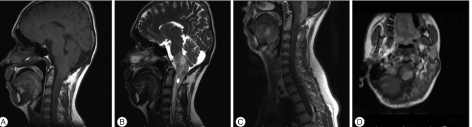
Fig. 1. Diffuse cord edema extending from medulla oblongata to C6 level and cystic component are seen on sagittal T1 (A) and T2 (B) weighted MR images. Intense contrast-enhancing lesion approximately 44×14 mm in size extending from medulla oblongata to C2 level on post-contrast sagittal T1 (C) and axial T1 (D) weighted MR images.
A B C D
Fig. 2. Expansion of the cord is observed at cervicomedullary junction fol-lowing dural opening (A), tumor cavity is seen after partial resection (B).
A B
Fig. 3. Neuroendocrine tumor of lung H&E ×100, inlet : closer view of tu-mor cells H&E ×400 (A), neoplastic development in ganglion cells H&E ×100, inlet : S-100 positive staining ganglion cells, original magnifica-tion H&E ×400 (B).
160 J Korean Neurosurg Soc 59 |March 2016
In our case, because of pre-existing neuroendocrine tumor of lung, metastases were considered in the first line and operation has been decided although there were no neurological deficits. Although association with scoliosis and Von Recklinghausen’s disease were previously reported in the literature, no GCT cases concomitant with neuroendocrine tumor of lung have been published.
With increasingly reported GCTs in recent years, they should be kept in mind in the differential diagnosis of intramedullary tumors especially in children and young adults. Pathological study is the most important diagnostic method for GCTs. Sur-gical excision is the primary treatment, but difficulty in total surgical tumor resection is the most important problem. References
1. Azzarelli B, Luerssen TG, Wolfe TM : Intramedullary secretory ganglio-cytoma. Acta Neuropathol 82 : 402-407, 1991
2. Baehring J, Ogle E, Sze G, Duncan C, Bannykh S : Ganglioneurocytoma of the spinal cord. J Neurooncol 71 : 149, 2005
3. Choi YH, Kim IO, Cheon JE, Kim WS, Yeon KM, Wang KC, et al : Gan-gliocytoma of the spinal cord : a case report. Pediatr Radiol 31 : 377-380, 2001
4. Coca S, Moreno M, Martos JA, Rodriguez J, Barcena A, Vaquero J : Neurocytoma of spinal cord. Acta Neuropathol 87 : 537-540, 1994 5. Jacob JT, Cohen-Gadol AA, Scheithauer BW, Krauss WE :
Intramedul-lary spinal cord gangliocytoma : case report and a review of the litera-ture. Neurosurg Rev 28 : 326-329, 2005
6. Kalyanaraman UP, Henderson JP : Intramedullary ganglioneuroma of spinal cord : a clinicopathologic study. Hum Pathol 13 : 952-955, 1982 7. Komotar RJ, O’Toole JE, Mocco J, Khandji AG, Keller CE, Connolly ES
Jr, et al. : Gangliocytoma of the spinal cord. Neurosurgery 60 : 895-900; discussion 895-900, 2007
8. Kwon JW, Kim IO, Cheon JE, Kim WS, Chi JG, Wang KC, et al. : Cere-bellopontine angle ganglioglioma : MR findings. AJNR Am J Neurora-diol 22 : 1377-1379, 2001
9. Lagares A, Gómez PA, Lobato RD, Ricoy JR, Ramos A, de la Lama A : Ganglioglioma of the brainstem : report of three cases and review of the literature. Surg Neurol 56 : 315-322; discussion 322-324, 2001 10. Milligan BD, Giannini C, Link MJ : Ganglioglioma in the
cerebellopon-tine angle in a child. Case report and review of the literature. J Neuro-surg 107 (4 Suppl) : 292-296, 2007
11. Ng TH, Fung CF, Goh W, Wong VC : Ganglioneuroma of the spinal cord. Surg Neurol 35 : 147-151, 1991
12. Rosenblum MK : Central nervous system, Rosai J (eds) : Rosai and Ack-erman’s Surgical Pathology, ed 9. Edinburgh : Mosby, 2004, pp2461-2622
13. Russo CP, Katz DS, Corona RJ Jr, Winfield JA : Gangliocytoma of the cervicothoracic spinal cord. AJNR Am J Neuroradiol 16 (4 Suppl) : 889-891, 1995
14. Shin JJ, Oh SH, Yoon DH, Kim TS : Cervical ganglioglioma. J Korean Neurosurg Soc 30 : 239-243, 2001
15. Wakao N, Imagama S : Intramedullary gangliocytoma with calcification and multiple intramedullary cysts. Neuroradiology 54 : 893-895, 2012 16. Yang C, Li G, Fang J, Wu L, Yang T, Deng X, et al. : Intramedullary
gan-gliogliomas : clinical features, surgical outcomes, and neuropathic scoli-osis. J Neurooncol 116 : 135-143, 2014