A. O. Vet. Fak. D"ı.
34 (3) : 562-569, 1987
SCANNING ELECTRON MICROSCOPIC EXAMINATION OF THE
TEGUMEN-TAL SURFACE OF TWO PARAMPHISTOMLD SPECIES (TREMATODA: PARAMP. HISTOMIDAE )
Şevki Z. Coşkun
*
Osman Sencer* *İki Paraınphistoınun Türünün Teguınental Yüzeylerinin Skaning Elektron Mik. roskopik Bakıııı.
Özet: Bu araştırmada taksonomik geçerlilikleri konusunda halen tartış-maların devam ettili iki Paramphistomun türüniin (Paramphistomum cervi Ze-der, 1790 ve P. leydeni Niismark, 1937) tepJimmtal )'üzC)'lerini kontrol etmek ve genital delik )'apılarındaki Izistomorfolojik ô'zellikleri karşılaştırmak amaç-lanmıştır.
lılu1)'eııe edilen 24 para:::.itten6 sının )'üzc)' .yapısl11dafarklılıklar göden-miştir. Ancak, bu para:::.itleringel/ital delik .yapılarında önemli bir fark bulu-llamamıştı)'. Bu sonuçlar, P. lC)'deni)i P. (avi' den ayırımda kullanılan kriter-lerin süreklilik i'C güvenilirliklerine şüphe düşürmüştür.
Summary: Tlze present investigation was carried out to investigate the tegumental sll1faces and to compaı'e the histomorphological pecularities-<Jfgenital opmings of two Paramphistomid species (Paramphistomum cerl'i Zeder, 1790 and P. leydeni Niismark, 1937) which thı~irtqxonomical validity are stil! under consideration.
The individual variations could be obsaved on the suıfiıce topography of 6 parasites out of 24 examined. Howeıoer, no signijicant diflerence were found in tlZe genital opening structures. These results cast doubts on the consisten~y and reliabili~'}'of criteria used in distinguishing P. lqdeni from P. cervi.
Introduction
The taxonomy of the family Paramphistomidac was based on the
histomorphological pccularities of certain muscular organs
(parti-• Dr. Department of Parasitology, Veterinal'Y Faeulıy, Ankara, Turkey . •• Doç. Dr. Electron Mieroseopy, Medicine Factılıy, Ankara, Turkey.
SCANNING ELECfRON MICROSCOPİC EXAMINATİoN... 563
cularly pharynx, genital opening and acetabulum) as seen in median
sagittal sections. Nasmark (3) had described Paramphistomum
ley-deni as a new species, and separated it from P. cervİ by having a well
developed genital opening (Leydeni type g.o., Sensu Nasmark). In
later years, Sey (5), after examining a large coııection of
Amphisto-mes from the different european sources and Nasmark's (3) original
preparations, has refused the validity of P. leydeni and proposed that
this species should be regarded as a synonym of P. cervİ. This synony-.
misation had already been accepted by Odening et aL. (4).
In recent years, sca,nning elec.t~on microscopy (SEM) has thrown
a new light upon the taxonomical studies. In the cas c of
Paramphis-tomid species which are sometimes morphologicaııy identical, the
usage of SEM took a special place indistinguishing these species.
Eduarda (1,2), for the first time, has announced that many
clo-sely related species could be separated from cach other by
theirsur-face stmctures. On this basis, he has separated P. leydeni from P.
cervi by the presence of dome to canical shaped tegumental papillae
which were conccntrated anteriorly around the oral opening and
ventraııy around the genital pore. P. ccrvi has been described as
completely without papillae. Eduarda (2) has also confirmed this
separation histomorphologically by deseribing new traits in the
ge-nitalopening of P. leydeni. In his descriptions, Leydeni type of
geni-tal opcning (P. leydeni) has been characterized by having thick and
large genital papillae and strongly developed radial fibres. However,
Gracile typc of genital opening (P. cervi) was characterized hy being
weakly developed and presence of only few radial musculature.
Rccendy, scanning electron microscopical examinations on the
surface of P. cervi revcaled that this species has also carried
tegu-mentalpapillac (6). However, the type of tegumental papillac of P.
ccrvi (5hort and stumpy papillae, sitting on a tegumental elevation.
Sensu Sey) \,'ere not identical with those of P. leydeni. Thus, the
ta-xonomical situation and identification of these species has become
confusing.
:The following possibilities were submitted by Sey (6) on this
subject.
r-
If any special importance is attributed to' the tegumentalpapillae, in that case P. cervi and P. leydeni are valid and has to be
564 ŞEVKİ Z. COŞKUN - OSMAN SENCER
2- If tegumental parjllae have considerahle indivudial
varıa-tions, in that case P. leydeni is a synonym of P. eervi.
\\'hile the ahove authors were indicating the taxonomic value of
tegumental pariHae, Tandon and Maitra (7) emphasized their
rhysi-ological (host-parasite interface relationship) importance.
With the present study we examincd the pattem and consisteney
of tegumental papiHae of both species and compared their genital
'opening structures.
Materiaı and Methods
The majority of the Paramphistomum species were ohtained
from the rumens of both sheep and eatde slaughtered in Ankara
abat-toir recently with the exception of one group of parasites which had
been obtained from sheep slaughtereel arounel iO years ago.
The samples were directly fixeel in 70
%
akohol except the 'oldgroup which had been fixed and stored in LO
%
forma1İne.Five Paramphistomid specimens were sectioned in median
sagit-tal plan from each group, carrying 8-12 LL thickness, and stained with
haematoxyline and eozin in usual microtechnical way.
A total of 8 group of parasites (6 from sheep and 2 from cattle)
comprisingo P. cervi and 101' P. leyeleni were separated for this
inves-tigation. From these groups, 24 parasites (3 from each group) were
dehydrated through an ascending series of akohol, transferred to
amylacetate-akohol mixtures, gradually up to absolute amylacetate.
Then they were critical point dried in liquid COı anel caated with
platinpalladium in a sputtering unit (Eiko IB-3 Ion caater).
Speci-mens were examİned in JEM 100-B-ASİD-I seanning electron
mic-roscape at 20 Kv.
Moreover, in order to examine the genital opening structure and
surface topography of the same specimen, three parasites showing
dif-ferent surface topography werc separated under stereo microseope
(x 80). Each of these parasites was sectioned with microtome until the
features seem to be sufficient for histomorphological diagnosis. Th('n
the remained parts of the same parasites wcre differentiated in xylol,
followed the above mentioned method, and then examined in SEl\1.
This method gaye us the oppurtunity\ to study each specimen his
SCA NNING ELECTRON MICROSCOPIC EXAMINA TION. . . 565
Results
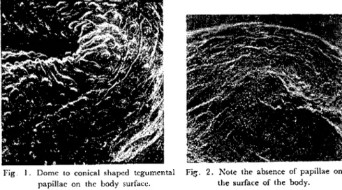
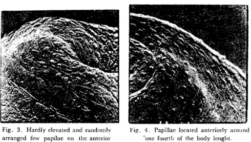
Sİxteen Oııt of 24 parasİtes examined in SEM showed from dome
to conical shaped tegumental papillae extending to the genital
ope-ning-Ievel (Fig. I). Only h.vo parasİtes from the old group were comp-letcly without papiııae (Fİg. 2). However, the rest 6 parasites showed
some variatİons on thcİr surface topography as listed bdow.
Fig. I. Domc to conical shaped tcgumental papiHac on thc body surfacc.
Fig. 2. Note the absencc of papillae on the surface of the body.
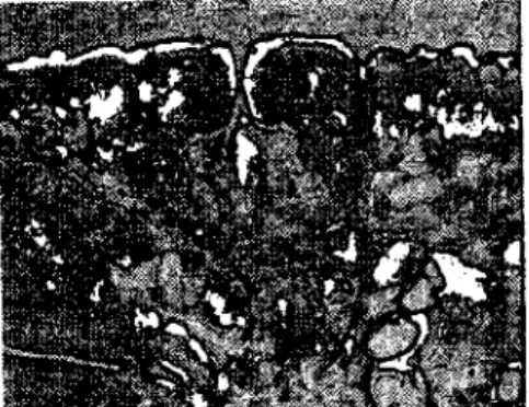
1- Three parasites slıowed hardly emevated and randomly
ar-ranged few papİııae on the anterior end around the oral openings
(Fİg. 3).
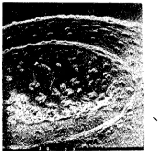
2- Two parasites were earrying papiııae loeated anterİorly around
one fourth of the body lenght. These papil1ae never extended ventraııy to the genital opening-Ievel (Fig. 4).
3- One parasite showed only a few papillae on anterior half of the body (Fig. 5).
Among the parasites examined, the rate of individual variations
of tegumental papillae seems to be worth attention.
In the histomorphologieal examinations of the genital openings
of these spceies it was obscrved that there was no a high eorrelation bctween thc thieknesscs of genital papillae and development rate of radial dibres.
566 ŞEVKI Z. COŞKUN - OSMAN SENCER
Fig. 3. Hardly clevated and randomly arranged few papilae on the anterior
end around the oral opening.
Fig. 4. Papiııae !ocated anteriorly around -one faurtlı of the body lenglıt.
As seen in Fig 6, in some genita! openings with tlıick genita!
pa-papillac radia! fibres were weakly deve!oped or completcly absent.
On the other hand, sametimes it was difficu!t to determine whether
genita! papillae were thick or thiıı in a genita! opening.
Fig. 5. Note the presence of only a few papiHae on anterior half of the body.
Fig. rı. Gracilc type of genital opening with thick genita! papillae.
In addition, not a good corrclation cou!d be observed between
the presence of tegumcnta! papillac on the surfaC(~ of the body and
l
SCANNING ELECTRON MICROSCorıc EXAMINATION... 567
in Figs. 7a and 7b, althoıİgh presence of tegumental papiliae on the
surface of the body, tlıere was only ;:l. few radial fibres in the genital
opening of the same specimen.
Fig. la. Surface topography of a speeimen ",iıh ıegurnental papiııac.
Fig. 7b. Genita! opcning slruclure of ıhe spccimcn iııustratcd in Fig 7a. Note the
presl'nce of only a few radial fibres.
Discussion and Condusian
Our observations on the consistency of tegumental papiliae
so-mewhat deviated from Eduardo's (1, 2) findings. The individual
vari-ability on the surface topography of these species could be observed at
a rate of that should not be overlookcd.
On the other hand, no parasites resembling to Sey's (6) P. cervı
could be observcd in this study.
it is of interest to point out that only the parasites from old group
were entirely without papiliae. A definite explanation for the waste
of these papiliae is far from being certain. It seems that the prefixative
treatment, starage time and condition for these parasites could be
the reason.
Two out of 6 parasites showing variations on their surface
topog-raphy ma)' give the impression that thesc. parasites are P. ichikawai
(Fig. 4). However, tlıe presence of long papiliae through the inner
surface of pharynx obviously reveals the fact that theyare not P.
~68 ŞEVKİ Z. COŞKUN - OSMAN SENCER
"
..Fig. 8. Long papiHae through the inner surface of pharynx of the specimen iHustrated in Fig. 4.
In histomorphologieal examinations, eontrary to Eduardo's
deseription on Leydeni type g.O., weakly developed radial fibres were
observed. On the other hand, absenee of a numerieal value for the
thiekneses of genital papillae made sametimes diffieult to determine
whether they were thiek or thin.
These results have east doubts on the eriteria used to separate
these speeies from eaeh other.
Comprehensive studies are necessary to elueidate the incidence
of variations on the surfaee topography of these speeies and to deteet
the preeise intel'mediate host (s) of these parasites to eonsider them
as different speeies.
References
1. Eduarda, S.L. (I 980a). TaxOllOmic value of legııınental stmclııre in the ideıılifiealioıı .of some spedes of Ihefamily Paramphislomidae Fischaeder, 1901 aeCllrring in mammals. Proc. EMOP,
3: 196.
:lo Eduarda, S.L. (I 980b). Tlıe laxonomy of the family Paramphistomidae Fisclıoeder, 1901 witlı special reference lo the morphalogy of speeies ocel/riııg in ruminanls. Thesis, Univ. London,
1-563.
3. Nasmark, K.E.(1937). Arevisian of ıhe Irematode family Paramphislomidae. Zool. Bidr. Upps., 16: 301-565.
SCANNING ELECTRON MICROSCOPIC EXAMINATION... 569
4. Odening, K., Bockhart, i. and Grafner, G. (I 978). Zur Frage der Pansenegelarten in der
DDR (Yreıııaloda: Paramplıistomidae) und ihrer Zwisclıenwirlsschnecken. Mb. Vet. Med., 33: i79-i8ı.
5. Sey, O. (1980). Revisiaıı of the Amphistomes of EUTopea,ı Rumiııaııts. Parasit. Hung.) 13: 13 25.
6. Sey, O. (1985). Persolid {oııımrmieatioıı.
7. Tandon, V. and Maitra, S.C. (ı 983). Surfaee ıııarphology of Gastrodiseoides haminis (Lcwis et "feCon/rel, 1876) Leiper, i9ı3 (Tremaloda: J)jgeırea) as Teı'ealedkvseanning eleetroıı mie-roseope..l. J-1clminth., 57: 339-342.