Department of Anatomy,
Faculty of Veterinary Medicine, University of Ankara Director: ProfDr.M.Cültekin
Morphological Aspect of Congenital Anophthalmia in the Ca1f fronı the Viewpoint of Conıparative Teratology
by
E. Deniz*
Özet: Araştırmada, yeni doğmuş bir erkek danada tesbit edilen bilateral .anophthal-mus congcnitus (Doğmalık göz yokluğu) olayı, 1976 yılı Dünyada Sağlık, Türkiye'de Körler Haftası münasebetile, morfolojik yönden incelenmiştir. Bu doğmalık körlük olayında ortaya çıkan anatomik bozukluklar ayrıntılarıyle tanıtılmış ve komparatifteratoloji açısından değer-lendirilmiştir. Olayda, özellikle N. opticus ve retina şekillenmemiş, her iki orbita adipes ve fibraz bir bağdoku ile dolu bulunmuştur. Literatürde bu malformasyonun sığırda en-dcr olmadığı verilerine dayanılarak, insandaki anophthalmus congenitus olayının nedenleri ve oluşum makanizmasının anlaşılabilmesi için, sığırın iyi bir deneme hayvanı olabileceği önerilmektedir.
Sununary: A true case of bilateral anophthalmus congenitus found in a newbom male calf was investigated morphologically on the occasion of World Health Week 1976 which was made the occasion in Turkey of BliT/dT/ess Week.
The main anatomical defects causing this blindness are described and discussed from the viewpoint of comparative teratology. The optic nerve and retina were abscnt, both or-bits were filled with a fatty and fibrous connective tissuc.
As a result of this study, and because the literature indicates that this malformation is not uneommon in cattle it is propose d that the cow wou!d be a good experimental animal for the invesrigation of congenital anophtha!mia in order to gain an understanding of the prineipa! causes and mechanism governing the development of congenita! anophthalmia in human beings.
Introduction
Congenİtal malformatİons are stmetural abnormalİtİes present at bİrth whİeh ean be expeeted to İnterfere partly or totalIy wİth the normal funetİons of an İndİvİduaL. Although mu ch researeh work has
• Professor for Comparative and Clinical Anaıomy, Faculty of Vetcrinary Medicine, University of Ankara. Ankara, Turkey
been done on this subject, there arc many such malformations, both in human beings and in animals, whose cause is still obscure. Therefore congenital anomalies are stili a challenge to medicine today. Many recent studies indicate that spontaously occuring congenital defects in animals can give life-saving clues to new cnvironmental teratogens
which are of importance to man, such as methyl-mercury which is
responsible for Minamata disease. Conditidns Well-known in man
which can be studied in domestic animals include the Klinefelter, Ehlers-Danlos and Chediak Higashi syndromcs, chimarism,
congeni-tal hearth disease (CHD), arthrogyriposis, cyclopia, intersexuaEty
and torticollis. it is bclieved that a systematic and comparative app-roach to the problem of congenital malformations would provide a
more effectivestrategy for their future prevention both in man and in
domestic animals.
Teratology is a very important subject in Vcterinary Medicine
today from the standpoint of economy, and for research into the actio-logy of cases of human malformation.
Mulvihill (1972) and Deniz (1975) have rcported that domestic animals providc good models for investigation of the causes of human congenital birth defects because of similarities in environmental fac-tors.
The aim of the study report ed in this paper was to investigate an
interesting case of bilateral congenital anophthalmia in a newbom
calf: this study was carried out on the occasion of World Health Week
1976, which was made the occasion in Turkey ofBlindness Week ..
Agenesis bulbi oculi and microphthalmia have been reported in
the literature both in man and in animals Wiedeking 1968, (Warkany
1971, Takano-Nishimura 1971). Complete absence of the eye is very
rare; many cases of microphthalmia have howevcr been erroneously
reported as anophthalmia, since clinical distinction is difficult withouf
microscopic examination. In a true casc. of anophthalmia both the
bulbus ocu1İ and the optic nerve are totally absent.
Anophthalmia may occur unilaterally or bilaterally. it is
rcpor-ted that the main causes of anophthalmia are either a unilateral or
bilateral nondevelopment of the eye-ball or a failure in normal deve-lopment of the optic nerve caused by abonormal amn{otic fluid
(Wies-ner). The hereditary morphogenesis of anophthalmia is still obscure
in animals, though simple recessive inheritance has ben reported in
the cow; in man anophthalmia normally shows autosorp.al recessiye
310 E. Deniz
has either been established or ean be assumed it can appear to show
autosomal dominant inheritanee.
Warkany and Kerse have also reported a trisomy Dı 13-15
cau-sing congenital anophthalmia and microphthalmia in man. The
gene-ral incidenee of anophthalmia in man has been reported as 0.1
%.
Takano and Nishimura (1957) report on an experiment in which in
rats treated with Alloxan, microphthalmia occured in a diabetic group,
and anophthalmia in a control group of Alloxan-treated non-diabetic
rats.
Beaudoin and Roberts (1966), testing thyrotropin as an
antithy-roid medication in rats, found 24.6
%
cases of anophthalmia withhydroeephalia (Nishimura 1968). Barbel' (1957), using Trypanblue,
was able to distinguish mice heterozygous for anophthalmia from
ho-mozygous normals. In an embryological investigation he found that
anophthalmic mice show an inhibition of the growth of the optic
ve-side on the tenth day of gestation.
Gilbert and Gilman (1954) deseribe the morphogenesis
ofTrypan-blue-indueed eye defects in the rat and tested the effeets of aza dyes.
Some authors have reported that the administration of excessive
Vi-tamin A (HyperviVi-taminos A) ean cause anophthalmia in mice, rats,
and rabbits. On the other hand Wiesner (1960) has reported that
Hypovitaminosis A can play a role in anophthalmia in pigs. Methlyl
salicylate, pantothenic aeid deficiencyand hypoglyeemia-inducing
compounds can also induce the development of anophthalmia
(War-kany).
Anophthalmia is relatively more frequent as abilateral condition.
Anophthalmia and microphthalmia can oecur in the same individual.
In the cow anophthalmia has been reported as assoeiated with
con-genital tailness (Kodı, Fischer, Schumann 1957). In the horse and
chicken anophthalmia is cOhsidered alethal factor named B7 in the
horse and E ı3in the chicken (Wiesner 1960).
Findings
The case reported in this paper is a native male calf which was
bom bIind, and showed bilateral anophthalmia (Fig i and 2). The
animal showed no other abnormalities. External examination
revc-aled that both eyes were smaller than normaL. The animal was unable
to maintain its cquilibrium. The animal was prepared for anatomical
investigation, and a detailed examination was made of both orbital
a .. Left rye (Fig 3 and 4). The superior and tertiary palpebrae
were absenL The lower eyelid (palpebra inferior) was present, but not
normaııy developed: its size was 2X
ı
cm, it was plicated, had fewci-Iia and was direeted within the orbital cavity. The form of the orbita
was rather quadrate (Fig 4). Incisurae were found at hoth sides of the
laerimal process; on the external surface of the lacrimal bone a smaıı
foramen was seen through whieh a nerve fibre and a branch of facial
artery passed into the orbita. The orbita was,mostly fiııed with a fatty
fibrous tissue mass, wIıich was the remnant oftIıe bulbus ocuıi. Within
this mass, tIıere were two rudimentary eye musdes (M.obliquus
superi-or et infcrisuperi-or) and small !acrimal gland. In the centre of the mass a
ru-elimentary eyebaıı some 5-7 mm in diamater was differentiated, with
a dark-pigmented tunica media and a fibrous tuniea externa. The lens
crystaııina and the retina were absenL At the apex of the orbita there
was a very smail optie foramen fiııeel with dura mater; there was no
optic nerve.
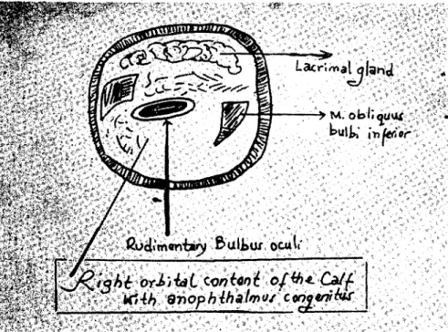
b- Right ,!ye (Fig. 5 and 6). The upper eyclid (palpebra superior)
had only a few eilia at its medial end. The inferior atretie palpebra formed a pliea on tIıe lateral anglc of the eye, direeted ventrolateraııy
to form arima. The margin of the inferior palpebra had normal eilia.
The orbita was again filled with fatty fibrous connective tissue, the
eye-musdes and rudimentary eyebaıı presented the same mall'ormed
ap-pearance as in the left eye. Again there was no optic nerve, no retina,
and no lens erystaııina.
c- Cranial cavi!)' : Investigation of the cranial cavity revelaed
that the brain and the meninges were normaııy devcloped, but the
volume of the total encephalon was relatively smaller than normaL.
The optic chiasma and optic nerves were not devcloped. In eontrast
to the optic structures, the olfaetory bulbus was larger than normaL.
Though searehecl for under the stcreomieroseope, the ophthalmie and
oeulomotor nerves eouJd not be identified.
Discussion
I n prenatal development, the aetivity of the albumen of the eell
is at its highest level at the beginning of organogenesis. As a result, at
this period the brail1, eyes, ears, palates and lips are very susceptible
to eraniofacial malformations; the eye is partieularly subjected to
malformation (Warkany, Ricck). According to Böhler (in Wiesner),
the main morphologicaJ results of anophthalmia and microphthalmia
312 E. Deniz
i. The retina, optic nerve and lcns crystallina may fail to develop,
2. The mesodermal parts of the bulbus may fail to form, or may
be malformed.
3. The ectodermal parts of the bulbus oculi which originate from
the ectoderm may develop abnormally.
The sensory part of the eye forms, together with the optic nerve,
an optic unİt which develop from a protrution of the forebrain
(pro-sencephalon). The lens crystallina cames from the ectoblast, the
midd-le and external tunica of the bulbus and the meninges are
mesenchy-matous. In the case of anophthalmia, the optic capsule fails to form,
and there is the n no optic nerve and retina.
In man, the eye is extremly sensitiye to teratogenetic factors:
exogenic factors, such as x-rays cause both anophthalmia and mi
c-rophthalmia.
The following lists cases reported in the literature in which
anopht-halmus congenitus or microphtanopht-halmus have been induced
experimen-tally in laboratory animals:
Author and Year Deseription of the ease
Anİmai speeies
Teratogen (used) Cohlan,S.Q. (1953) Mierophthalmia Rat H ypervi taminosis-A Cilbcrt, C. and
Cilman, J, (1954) Anophthalmia Rat Trypanblue Ciroud, A., Martinet,
M. (1956) Mieroph.halmia Rat H ypervi tamİnosis-A Barber, A.N. (1957) Anophthalmia Miee Maternal Hypoxia Ciroud, A. Martinet, M. (1959) Bcek, F., Lloyd, J.B. (1966) Takano, K., 1'iishimura, H. (1967) Mierophthalmia Rabbit Anophthalmia Mierophthalmia Rat Anophthamia Rat Mierophthalmia Miee H ypervi taminosis-A Azo Dyes Alloxan
According to the references citcd, congenital anophthalmia in
man has been said to have a recessive or an autosomal recessiye
inheri-tancc, that the mechanism is not understood in detai!. A Trisomy
Dt 13-15 has also been given as a possible cause for anophthalmia
in man. Toxoplasmosis and ru bella can be also cited as causes for
animals, anophthalmia is reported as a simple recessive character in the cow, and a Icthal trait in the horse and the ehieken.
In the Pig, Wiesner (ı960) has reported that Hypovitaminosis-A plays a role in the development of anoplıthalmia, while in the mouse, the rat and therabbit, Cohlan, Giroud and Martinet report that Hy-pervitaminosis-A is eoncerned in the development of congenital anop-hthalmia. Treatment with Trypanblue (Aza Dye), pantothenic acid deficiencyand hypopglycaemia inducing compounds can cause blind-ness in mice, rats and rabbits, eausing non-development of the optic capsulc alsa anophthalmia (Gilbert-Gilmann,
ı
954).I t is interesting to note that in some cases anophthalmia is associa-ted in the cow with the congenital absence of the taiL. In man anopht-halmia has been reported in combination with mental retardation, brain atrophy, hydrocephaly, mieroeephaly, CHD, ampholocel, uro-genital defects, celft palate, poly-and syndactyly (Warkany).
As summarised above, the ahnormal calf showed true bilateral anophthalmus congenitus. There was no retina, no optic nerve, no lens crystallina, no pupilla, no iris and no viterous body. As the blind-ness is caused by the nondevelopment of the optic parts of the eye this is a true case of anophthalmus congenitus. The orhita were filled with fatty fibrous tissue. In this cas e the exact cause could not be determi-nad, nor its mode of inheritance.
Anophthalmus congenitus has been reported IDore frequently in the cow than in other domestic animals, there is also a tendeney for it to occur more frequently in males. This is also suggested by of Sjög-ren and Larsson's findings that a partly sex-linked recessive mode of inheritance was responsible in eases of anophthalmia with oligophre-nia (Warkany).
In conclusion, i would like to propose that the cow is a good ex-perimental animal model for investigation of human congenital anop-hthalmia in order to obtain an understanding of the basic principal and mechanism govcming the development of this malf4:>rmation, which causes total blindness.
References:
Barher, A.N. (ı957): The efftct of maternal hypoxia on inheritance of recessive blindness in mice. Am..J .ophthalm. 44: 94-
ı
Oı.
Beck, F., Lloyd, j.B. (ı 966): The leratogenic efftcts of Azo Dyes. In Advances in Teralology, Vol.ı. ed.D.H.M.Wollam. Legos Press Book, London.
314 E. Deniz
Cohlan, S.Q. (19,):~): Exeeniı'e infake of Vitamin A as a eause eoııge-nital anumalies in the rat. Science, i17: 535.
Deniz, E. (i 975): Die Bedeuıuııg der Teratologie Jiir die Allalomie. An-kara Üniv.Vetcrin~r Fak.Derg. XXI, 3- 4 209-216.
Gilbert, C. and Gilman, J. (i951): The morplzogenesis oj Tıypaııb-lau indueed defeets ~fthe qe. Afr.J.~1ed.Sci. 19: 147-54.
Giroud, A., Martinent, M. (i956): l-ljpervitaminose el Izypervitami-nose A.elzez le jeune et elzez l'embriyon. Et.~eo-~atales, 5, 55.
Giroud, A., Martinent, M. (1950): Malformatione de la fec" et lzypeıvitamillOse A. Cit./ FattOli Metageneliei Esogeni Nelle }vfaljorma-zioni Congenite (Omfaciali) : Teratogenesi sperimentale Eriferimenti
clini-ci. ed. by Vichi, F., Masi, P., Orlondo, S., Pagni, L., Tollara, i. Pp.248-254. Ediziani Mincrva Medica.
Giroud, A., Martinent, M. (1959): Stades eüolufııfr de lapin soumıs a l'hyperüitaminose. BULL.Soc.Ophthalm. 3.
Kerse,
1.
(1968): Gelişme Bozukıııkları (Kongenital Ma(formasyonlar ve Anomaliler). Çocuk Sağlığı ve Hastalıkları Derg., 1I,4, 139-148.Koeh, P., Fiseher, H., Sehumann, H. (1957): Erbpatlzologie der Landu;iıtsclzajtlielzen Haustiere. Paul Parey, Berlin.
Muıvi~ill,
V.J.
(1972): Con,{f,enital and Genetic Diseases in DomesticAnimals. Science 176,132-137.
Murakami, U. (1966): Teratogenesis of eraniojacial maljormations in animals. Archives of Environmental Health, 13-695--698.
Rieek, W. (i 969): Giessener Beilıage zur Erhpatlzologie ıınd Zuclzt/ıygiene
2/ 3. Anhang 2.
Nishimura, H. (i 968): Manuel for tlZe Second lntemational Works/ıop in Teratolog)', Kyoto University, japan.
Tanako, K. (1968): Comparaliı'e teratological effects of mftabolic diseases of tlZe mothe.r. in lvlanudfor the Second International l;florkslzop in Teratolo.f!,}. Takano, K., Nisbimura, H. (1957): eongenital Ma(formations
indu-eedII),Alloxan diabetes in mice and rats. Anat.Ree. i58, 303-3 i2.
Warkany, J. (1971): eongenital lvfa(formations. Yearbook :Medical PubUne.Chicago.
Wiedeking, J.F. (1968): Terminologie der Embı)!onalen Entu;icklungs-stiiJ"llngen-Zlıgleiclz Bibliograplzie neııerer Literafıır aı!f dem Gebiet der
Teratulagie in der Vetemaermedizin.
Wiesner, E. (i 960): Die Erbsehiiden der Landwırfsclwftlic/ıen Nıııztiere,
Fig.ı.The head ofa ınale Calfwith bilateral Con~eııital anophthalınia. Right view.
Fig. 2.Thc lelfı "icw of the hcad of the Calf bornc biind. Afıer prcl'aration of ıhc orbital eontent.
316 E. Deniz : ~. <;li,~."~!'~,;:r~)ti!rar;,'t"'" jft<''!!'I:f '. ~'." ~'~ .' ~o:~I:~r.~ j./.ç:~~~~...{ ~t"~i•.i ~ ••.• ~~:~ "". '. 16wer
"J.liJ
~~;.,
d' .:••Lacriftl9~jtan.d."ti{:: ",~,~'f,f , ,;':' ı" " .. •~ ";!":" .:. ra~ilnd .fibro
ll$+i
SIIıQFig. 3. and 4. Drawings showİng the cxternal and İnternal orbital contents of the left eye. f'
't'
«'/