Journal of Clinical and Analytical Medicine |
O
h
r
c
i
r
g
a
in
e
a
s
l
R
e
1 Nezih Akkapulu, İlker Murat Arer, Abdirahman Sakulen Hargura, Murat Kuş, Hakan Yabanoğlu, Hüseyin Özgür AytaçDepartment of General Surgery, Baskent University Adana Research, and Teaching Hospital, Adana, Turkey Rectus sheath hematoma: A retrospective study
Factors affecting mortality in rectus sheath
hematoma: A retrospective study
DOI: 10.4328/JCAM.5453 Received: 26.10.2017 Accepted: 13.11.2017 Published Online: 13.11.2017 Printed: 01.01.2018 J Clin Anal Med 2018;9(1): 69-72 Corresponding Author: Nezih Akkapulu, Baskent University, Adana Hospital 01120 Yuregir, Adana, Turkey.
T.: + 90 3223272727 F.: +90 3223271274 E-Mail: [email protected]
Abstract
Aim: Rectus sheath hematoma is a rare clinical condition. Because it could mimic various intra-abdominal pathologies suspicious approach and attention is needed for proper diagnosis and management of rectus sheath hematoma. The aim of this study is a review of factors affecting mortality in patients with rectus sheath hematoma besides clinical features, diagnosis modalities, treatment periods and results of patients with rectus sheath hematoma. Material and Method: Twenty-three patients with rectus sheath hematoma from January 2012 to March 2017 in a tertiary care center were included in the study. Patients’ files were reviewed retrospectively. Reviewed variables were demographic and clinical features, symptom and findings, co-morbidities, medications, laboratory findings, diagnostic modalities, APACHE II scores, treatment approaches, transfused blood products and length of hospital stay. Results: Fifteen (65.2%) of the patients were women, and 8 (34.8%) of the patients were men. The mean age was 61.9 ±13.5. Seventeen (74%) of the patients had abdominal pain. Twenty (87%) of the patients had anticoagulant and antiplatelet therapy.The mean hematoma diameter was 6.3± 3.6 cm. The mean APACHE II score for the patients was 13.1±7.3. One patient had undergone surgery. There was mortality in 3 (13%) of the patients. The median length of stay in hospital was 5 (5) days. Discus-sion: The diagnosis of rectus sheath hematoma should be kept in mind while assessing old female patients and patients on anticoagulants with a complaint of abdominal pain. In our study, we identify risk factors such as higher APACHE –II scores, the presence of a transient ischemic attack, need for transfusion of more units of packed erythrocytes and more extended hospital stay as factors associated with mortality.
Keywords
Rectus Abdominis; Hematoma; Mortality
| Journal of Clinical and Analytical Medicine Rectus sheath hematoma: A retrospective study
2
Introduction
Rectus sheath hematoma (RSH) is a rare clinical condition that is caused by the occurrence of hematoma in rectus muscle or within muscle sheath following the injury to the branches of the superior epigastric artery. RSH could be observed after blunt or penetrating abdominal trauma, vigorous exercise, recent abdominal surgery, at trocar sites after laparoscopic surgery, abdominal wall injections, paracentesis or it could occur spon-taneously in the presence of predisposing factors such as an-ticoagulation therapy and intractable coughing [1]. RSH could also mimic appendicitis or sepsis [2].
There are well-defined predisposing factors, clinical features, diagnostic classification by abdominal computerized tomogra-phy (CT) and treatment options for RSH in the literature [3]. However; there is lacking consensus about RSH related mortal-ity and risk factors affecting mortalmortal-ity in patients with RSH. We aimed to investigate risk factors that influence mortality in patients with RSH primarily. The secondary purpose of this study is an overview of clinical characteristics, diagnosis mo-dalities, treatment options and results of the patients with RSH. Material and Method
Twenty-three patients with rectus sheath hematoma from January 2012 to March 2017 in a tertiary care center were included in the study. Patients’ files were reviewed retrospec-tively. Exclusion criterion was pediatric age group (0-16 years old) patients. We described 54 variables such as age, gender, symptoms, and clinical findings of the patient, co-morbidities, medications, laboratory findings, diagnostic modality, CT clas-sification, hematoma side, the diameter of hematoma, Acute Physiology and Chronic Health Evaluation (APACHE)- II score at the time of diagnosis, treatment options, transfused blood products, length of hospital stay (LOS) and presence of mortal-ity after reviewing the files.
We composed two independent groups due to the presence or absence of mortality to assess possible risk factors. IBM SPSS statistic software version 24.0 (IBM Corp., Armonk, NY, USA) was used for statistical analyses. Shapiro-Wilk test was applied to determine the distribution of normality. Normally distributed quantitative variables were expressed as the mean ± standard deviation (SD), quantitative variables not normally distributed were expressed as median with interquartile range and cate-gorical variables as frequency and percentage. Student-t-test was applied to normally distributed quantitative variables of independent groups. Mann-Whitney U test was used for quan-titative variables without normally distributed data of separate groups. Chi-square test was applied to categorical variables. Logistic regression model was carried out for multivariate analyses. A value of p< 0.05 was considered as statistically sig-nificant.
This study was approved by Baskent University Institutional Re-view Board (Project no: KA17/132)
Results
A total of the 23 patients, 65.2% were women, and 34.8% were men. The mean age was 61.9± 13.5 years (range 31 – 87 years). 87% of the patients had at least one co-morbid disease. 87% of the patients were receiving anticoagulant or antiplatelet
therapy, and 34.8% of patients were receiving both anticoagu-lant and antiplatelet therapy. RSH developed at trocar site after laparoscopy in three of the patients (13%), due to paracentesis in two of the patients (8.7%) and after blunt abdominal trauma in one of the patients (4.3%). One patient was pregnant. Con-servative treatment was chosen for 20 (87%) of the patients; transcatheter embolization was performed for two (8.7%) of the patients, and one (4.3%) patient was operated on. Patients’ clinical features are shown in Table 1 and Table 2.
Mortality rate was 13%. The mean age was 53.3±7.5 (range 48- 62), the mean Co-morbid disease numbers were 3.3± 1.1 (range: 2- 4), the mean APACHE-II score was 27 ±4.5 (range: 23- 32), the median transfused packed erythrocytes units were 9(0) (Range: 6- 16 units), the median LOS days were 11(0) (range: 8- 21 days) in mortality group. Conservative management was utilized for two of the patients and surgery was selected for one patient in the group.
We detected a significant difference between mortality and APACHE –II scores, the presence of a transient ischemic attack, LOS, and transfused units of packed erythrocytes.
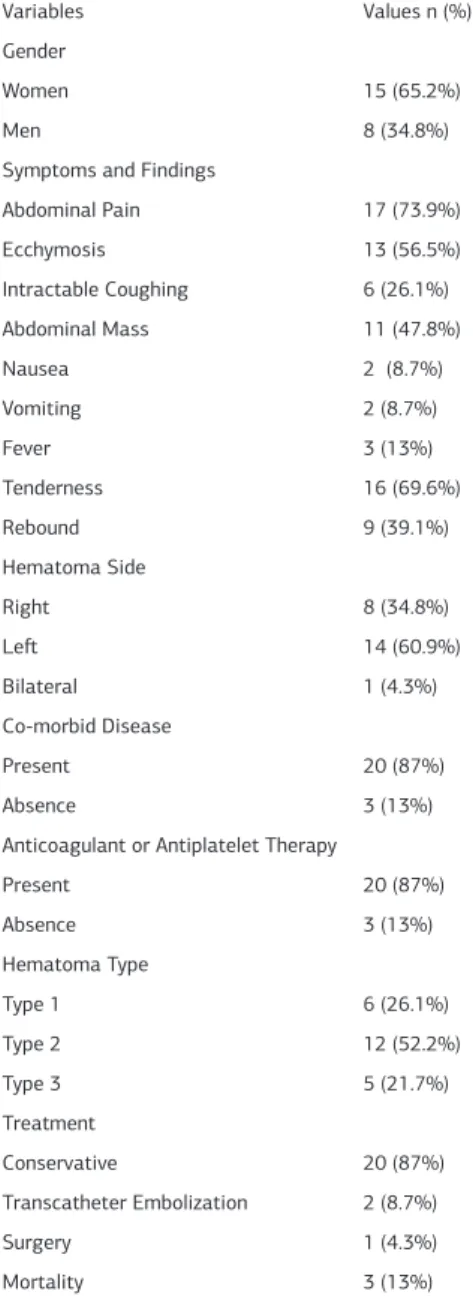
Table 1. Categorical variables of the Patients
Variables Values n (%)
Gender
Women 15 (65.2%)
Men 8 (34.8%)
Symptoms and Findings
Abdominal Pain 17 (73.9%) Ecchymosis 13 (56.5%) Intractable Coughing 6 (26.1%) Abdominal Mass 11 (47.8%) Nausea 2 (8.7%) Vomiting 2 (8.7%) Fever 3 (13%) Tenderness 16 (69.6%) Rebound 9 (39.1%) Hematoma Side Right 8 (34.8%) Left 14 (60.9%) Bilateral 1 (4.3%) Co-morbid Disease Present 20 (87%) Absence 3 (13%)
Anticoagulant or Antiplatelet Therapy
Present 20 (87%) Absence 3 (13%) Hematoma Type Type 1 6 (26.1%) Type 2 12 (52.2%) Type 3 5 (21.7%) Treatment Conservative 20 (87%) Transcatheter Embolization 2 (8.7%) Surgery 1 (4.3%) Mortality 3 (13%)
| Journal of Clinical and Analytical Medicine
70
| Journal of Clinical and Analytical Medicine Rectus sheath hematoma: A retrospective study
3
Discussion
We investigated factors affecting mortality and overviewed clinical features in the patients with RSH in this retrospective study. We found that mortality rate is 13% and this ratio is compatible with the literature. The mortality rate of the largest case series of 126 patients from Mayo Clinic was 1.6% [4]. The mortality rate in patients with RSH ranges from 0% to 14.7 % in various studies [5]. Villa et al. [6]reported that mortality rate was 17% among the patients who had to undergo sur-gery because of the hemodynamic instability of the patients. Carkman et al. [7]stated that the mortality rate could reach 20% in elderly and patients receiving anticoagulant therapy presenting with RSH. In this univariate analysis, we found that higher APACHE- II scores, the presence of TIA, need for more erythrocytes transfusion, and longer LOS are associated with the occurrence of mortality. We could not reveal significant dif-ferences between mortality and any variable in the multivari-ant analysis. While the median units of transfused erythrocytes were 3.5 units in the previous study [4], our patients needed more erythrocytes transfusion (the median is nine units). The length of stay in hospital ranges from 4 to 10 days in previous studies [8]. In our study, the median LOS was 11 days in mortal-ity group and 4.5 days in surviving group. We think that patients had to limit physiologic conditions beforehand resulting in more
transfusions and longer stay in hospital in mortality group. Higara et al. [9] reported that mortality rate was 0.28% in isch-emic stroke patients with RSH and they indicated that patients were receiving both antiplatelet and anticoagulant therapy. In our study, mortality occurred in two of the three TIA patients. While one of the mortality cases was receiving both antiplatelet and anticoagulant therapy, the other patient was receiving only anticoagulant therapy. However, we could not derive a relation between mortality and anticoagulant therapy.
We noted that women to men ratio was 1.8:1 and is compatible with previous studies, nevertheless, we could not reveal any ad-ditional risk factor for female patients with RSH.
The range of age was 46 – 69 years in previous research [10] and the mean age was 61.9 ±13.5 (range: 31-87) in our study. Patients developing RSH are mostly elderly, and this could be explained with these elderly patients being more liable to be on anticoagulant therapy. We also found lower INR levels than the previous study [4].
CT scan was performed for all patients except the pregnant one. CT scan is widely accepted as first-line diagnostic modal-ity because of its superiormodal-ity to ultrasound. Because it reveals hematoma size, location, and relation with peritoneum, CT scan could aid in shortening time for surgical decision-making pe-riod.
Most of the RSH patients were treated conservatively, that includes nil per os, intravascular hydration, bed rest and ad-ministering analgesics. Transcatheter embolization was per-formed for two patients, and surgery for hematoma drainage with bleeding control was conducted for one patient. Surgical decision-making depends on the hemodynamic instability of the patient with RSH [6]. Scoring systems such as APACHE- II could be used to decide which patient to take to surgery before devel-oping hemodynamic instability.
The study has some limitations: its retrospective in nature, a tertiary center analysis and a small number of patients that could have resulted in findings of higher mortality rate. In conclusion, there are various predisposing factors for RSH. RSH should be kept in mind while evaluating geriatric female patients and patients on anticoagulant with a complaint of ab-dominal pain. Furthermore, we note that risk factors such as higher APACHE –II scores, the presence of TIA, transfusion of more units of packed erythrocytes and longer hospital stays are associated with an increase in mortality.
Scientific Responsibility Statement
The authors declare that they are responsible for the article’s scientific content including study design, data collection, analy-sis and interpretation, writing, some of the main line, or all of the preparation and scientific review of the contents and ap-proval of the final version of the article.
Animal and human rights statement
All procedures performed in this study were in accordance with the ethical standards of the institutional and/or national re-search committee and with the 1964 Helsinki declaration and its later amendments or comparable ethical standards. No ani-mal or human studies were carried out by the authors for this article.
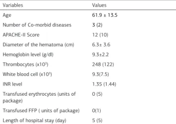
Table 2 Quantitative variables of the patients
Variables Values
Age 61.9 ± 13.5
Number of Co-morbid diseases 3 (2)
APACHE-II Score 12 (10)
Diameter of the hematoma (cm) 6.3± 3.6 Hemoglobin level (g/dl) 9.3±2.2 Thrombocytes (x103) 248 (122)
White blood cell (x103) 9.3(7.5)
INR level 1.35 (1.44)
Transfused erythrocytes (units of
package) 0 (5)
Transfused FFP ( units of package) 0(1) Length of hospital stay (day) 5 (5)
INR: International normalized ratio; FFP: Fresh Frozen Plasma Table 3. Comparison of variables in patients with or without mortality
Variables Mortality P value
Present Absence
Age 53.3±7.5 63.2±13.9 0.2
Co-morbid disease number 4(0) 3(3) 0.8
Hematoma diameter (cm) 8.7±4 6.1±3.5 0.3
Hemoglobin (g/dl) 8.2±2.9 9.5±2.2 0.3
WBC (x103) 9.3(0) 9.1(8.2) 1
Thrombocytes (x103) 266(0) 241(132) 0.4
INR 1.6(0) 1.2(1.5) 0.4
Erythrocytes (Unit of packages) 9(0) 0(2) 0.001*
FFP (Unit of packages) 26(0) 0(1) 0.1
APACHE II score 26(0) 11(9) 0.001*
Length of hospital stay (days) 11(0) 4(3) 0.012*
WBC: White blood cell; INR: International normalized ratio; FFP: Fresh frozen plasma
*Statistically significance
Journal of Clinical and Analytical Medicine | 71
| Journal of Clinical and Analytical Medicine Rectus sheath hematoma: A retrospective study
4
Funding None
Conflict of interest
None of the authors received any type of financial support that could be considered potential conflict of interest regarding the manuscript or its submission.
References
1. Hatjipetrou A, Anyfantakis D, Kastanakis M. Rectus sheath hematoma: a review of the literature. Int J Surg. 2015;13:267-71.
2. Kasotakis G. Retroperitoneal and rectus sheath hematomas. Surg Clin North Am. 2014;94(1):71-6.
3. Oh JH, Kim TH, Cha SJ, Kim SH. Rectus sheath hematoma caused by non-contact strenuous exercise mimicking acute appendicitis. J Emerg Med. 2010;39(3):e117-9.
4. Hamid NS, Spadafora PF, Khalife ME, Cunha BA. Pseudosepsis: rectus sheath hematoma mimicking septic shock. Heart Lung. 2006;35(6):434-7.
5. Berna JD, Garcia-Medina V, Guirao J, Garcia-Medina J. Rectus sheath hematoma: diagnostic classification by CT. Abdom Imaging. 1996;21(1):62-4.
6. Cherry WB, Mueller PS. Rectus sheath hematoma: review of 126 cases at a single institution. Medicine (Baltimore). 2006;85(2):105-10.
7. Anyfantakis D, Kastanakis M, Petrakis G, Bobolakis E. Rectus sheath he-matoma in a single secondary care institution: a retrospective study. Hernia. 2015;19(3):509-12.
8. Akturk OM, Kayilioglu SI, Aydogan I, Dinc T, Yildiz B, Cete M et al. Spontane-ous Rectus Sheath Hematoma: an Overview of 4-Year Single Center Experience. Indian J Surg. 2015;77(Suppl 3):1219-21.
9. Villa M, Grande M, Rulli F, Konda D, Perretta T, Amabile D et al. Rectus sheath haematoma: are there prognostic risk factors of haemodynamic instability moti-vating an early operative treatment? Eur J Trauma Emerg Surg. 2012;38(5):537-41.
10. Sheth HS, Kumar R, DiNella J, Janov C, Kaldas H, Smith RE. Evaluation of Risk Factors for Rectus Sheath Hematoma. Clin Appl Thromb Hemost. 2016;22(3):292-6.
11. Carkman S, Ozben V, Zengin K, Somuncu E, Karatas A. Spontaneous rec-tus sheath hematoma: an analysis of 15 cases. Ulus Travma Acil Cerrahi Derg. 2010;16(6):532-6.
12. Hiraga A, Nakagawa Y, Kamitsukasa I, Suzuki T, Kuwabara S. Muscle hae-matoma due to antithrombotic treatment for ischaemic stroke. J Clin Neurosci. 2015;22(7):1160-3.
How to cite this article:
Akkapulu N, Arer İM, Hargura AS, Kuş M, Yabanoğlu H, Aytaç HÖ. Factors affect-ing mortality in rectus sheath hematoma: A retrospective study. J Clin Anal Med 2018;9(1): 69-72.
| Journal of Clinical and Analytical Medicine
72