Ultrasonographic Evaluation of Flap
Thickness In Patients with Pilonidal
Sinus-Original Article
Pilonidal Sinüs Nedeni İle Opere Edilen Hastalarda Flep
Kalınlığının Ultrasonografik Değerlendirmesi
Enis Dikicier
1, Fatih Altıntoprak
1, Mustafa Yener Uzunoğlu
2,
Yusuf Arslan
2, Kıyasettin Asil
3, İsmail Zengin
21 Department of General Surgery, İstinye University Faculty of Medicine, İstanbul, Turkey 2 Department of General Surgery, Sakarya University Research and Educational Hospital,
Sakarya, Turkey
3 Department of Radiology, Sakarya University Research and Educational Hospital, Sakarya,
Turkey
Yazışma Adresi / Correspondence:
Enis Dikicier
İstinye University School of Medicine, İstanbul, Turkey T: +90 505 252 08 02 E-mail: e-mail:[email protected] Geliş Tarihi / Received : 05.05.2018 Kabul Tarihi / Accepted : 07.06.2018
RESEARCH ARTICLE / Araştırma Makalesi
Abstract
Aim Various risk factors have been defi ned for recurrence in pilonidal disease. This study was designed to investigate the local subcutaneous tissue thickness in patients who were operated on for sacrococcygeal pilonidal disease. ( Sakarya Med J 2018, 8(3):505-510) Methods Ninety-eight patients who had been operated in General Surgery Clinics of Sakarya University Research and Educational Hospital on with
the diagnosis of pilonidal sinus were retrospectively evaluated between January 2008 and January 2014. In evaluation of the fl ap thickness, the nearest and farthest margins were measured by ultrasonography between the skin and sacrum.This is a cross-sectional and descriptive study and local ethics approval was obtained from Sakarya University Medical School
Results Resection and primary closure and Limberg fl ap transposition were performed in 46 (46.9%) and 52 (53.1%) patients, respectively. Mean fl ap thickness was found to be 17.2 mm (range: 3.9 - 36.4) and 20.5 mm (range: 4.0 - 42.3) in the primary repair group and the Limberg group, respectively. The recurrence rate was 10.8% in the primary repair group, 3.8% in the Limberg group at a mean of 27.1 months (range: 12-54 months) of follow-up.
Conclusion Subcutaneous tissue thickness in the place of surgery was greater in patients with the Limberg fl ap procedure compared to the patients with primary closure in pilonidal sinus disease. Flap thickness may be a parameter that can be used to predict the possibilty of recurrence Keywords Pilonidal sinus; Surgery; Limberg
Öz
Amaç Pilonidal sünis nedeni ile opere edilen hastalarda nüksün pek çok nedeni vardır. Bu çalışmanın amacı pilonidal sinüs nedeni ile limberg fl ep uygulanan hastalarda, fl ep kalınlığının hastalık tekrarına etkisini araştırmaktır ( Sakarya Tıp Dergisi 2018, 8(3):505-510 ).
Yöntem Ocak 2008 ve Ocak 2014 yılları arasında, Sakarya Üniversitesi Eğitim ve Araştırma Hastanesi Genel Cerrahi Kliniği’nde pilonidal sinüs ne-deni ile opere edilen 98 hastanın verileri retrospektif olarak incelendi. Flep kalınlığı deri ile sakrum arasındaki en yakın ve en uzak mesafeler ultrasonografi k olarak ölçülerek değerlendirildi. Bu araştırma kesitsel ve tanımlayıcı bir çalışmadır ve Sakarya Üniversitesi Tıp Fakültesi Etik Kurulu’ndan etik kurul onayı alınmıştır
Bulgular Limberg fl ep uygulanan 46 hasta (%46.9) ve rezeksiyon ve primer kapama uygulanan 52 hasta (%53.1) sonuçları geriye dönük değerlen-dirildi. Ortalama fl ep kalınlığı primer onarım grubu ve limberg fl ep grubunda sırası ile,17.2 mm (3.9 - 36.4 mm) ve 20.5 mm (4 -42.3 mm) olarak bulundu. Nüks oranı primer kapama grubunda %10.8 limberg fl ep grubunda % 3.8 olarak saptandı. Olguların ortalama takip süresi 27.1 ay (12-54 ay) olarak bulundu.
Sonuç Subkutan doku kalınlığı Limberg fl ep uygulanan hastalarda primer kapama grubuna göre belirgin olarak fazlaydı. Flep kalınlığı pilonidal sinüs nedenli opere edilen hastalarda nüksü azaltmada önemli bir parametre olarak kullanılabilir.
Anahtar
Introduction
Pilonidal sinus (PS) is a chronic benign disorder, commonly localized to sacrococcygeal region. It is most frequently seen in young adults and in men. Although it is a benign disease, it might affect the entire society since it causes an important amount of loss of labor and interruption of the edu-cational processes when the affected gender and age group are taken into account.
Numerous operative and non-operative treatment methods have been described for treatment of pilonidal sinus; however, recurrence is still a serious problem whose defi nitive solution is not yet known. More than 20 techniques have been described for the treatment of pilonidal disease (1), and signifi cant recurrence rates have been reported with each one. This suggests that the factors effective on recurrence are still not entirely known.
In this study, subcutaneous tissue thickness, which was measured postoperatively in cases that underwent two different types of treatment for sacrococcygeal PS was evaluated to defi ne whether it could be used as an effective parameter to determine the success of the surgery.
Materİals And Methods
Patients who were either operated on with the indication of recurrent or complicated PS, who had Diabetes Mellitus, who were using steroids on a regular basis for various reasons, with a body mass index of <18.5 and >29.9, with age >65 years or whose follow-up were not adequate were excluded from the study.
The records of 98 patients who were operated with the diagnosis of simple sacrococcygeal pi-lonidal sinus between January 2008 and January 2014 in General Surgery Clinics of Sakarya University Research and Educational Hospital, were evaluated in detail in terms of demographics, the technique of operation, recur-rence and fl ap thickness.
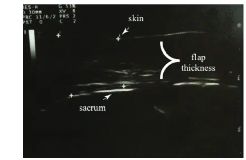
The cyst was excised through an elliptical incision and the subcutaneous tissue was fi xed to the presacral fascia using absorbable sutures in the primary repair technique (PR). In the other case, the cyst was excised through a rhomboid incision and the fasciocutaneous fl ap prepared from the right gluteus maximus muscle was fi xed to the presacral fascia using absorbable sutures in the Lim-berg fl ap transposition technique (LF). The patients received a single dose of a prophylactic antibi-otic (Cefazolin sodium, 1gr) and an aspirative drain was placed in the surgical fi eld in both cases. Follow-up of the patients were performed at the outpatient clinics. The diagnosis of recurrent disease was determined based on the physical examination fi ndings (the presence of recurrent sinus openings at the localization of previous operation). Ultrasonographic measurements were applied between postoperative 3th and 7th days. The term “fl ap thickness” was used to defi ne the thickness of the tissue between the skin and sacrum at the midline of the previous surgical fi eld and was calculated as the mean of the shortest and the farthest of the ultrasonographic me-asurements (Aplio MX, Toshiba, Tokyo, Japan) (Figure 1). All the meas-urements were performed by the same radiologist.
Statistical analyses were performed using the Statistical Package for the Social Sciences (SPSS) version 10.0 (SPSS, Inc., Chicago, IL, USA). The differences between the groups were analyzed DİKİCİER et al.
Ultrasonographic Evaluation of Flap Thickness In Patients with Pilonidal Sinus - Original Article Sakarya Medical Journal 2018;8(3):505-510
DİKİCİER et al.
Ultrasonographic Evaluation of Flap Thickness In Patients with Pilonidal Sinus Sakarya Medical Journal 2018;8(3):505-510
507
by the Mann-Whitney U-test and Fisher’s exact test. A value of p < 0.05 was considered to be statistically signifi cant.
Figure 1. Ultrasonographic appearance of sacrococcygeal subcutaneous tissue in the postopera-tive period.
Results
Thirteen of the patients were females (13.2%) while 85 were males (86.8%) with a mean age of 29.7 years (range:18-63). Patients were divided into two groups according to the procedure per-formed after the excision of the sinus. There were 46 patients in the primary repair group (46.9%) and 52 patients in the Limberg fl ap group (53.0%). The demographics of the patients, fl ap thick-nesses, and the results of follow-up are shown in Table 1.
Table. 1: Demographic fi ndings, type of surgery, fl ap thickness and follow-up results in all patients
Variables Primary closure (n=46, %100) (n=52, %100)Limberg fl ap p value Age (mean) 30.1 (18-55) 29.1 (18-63) 0.119 Sex 0.112 Male 39, 84.8 46, 88.5 Female 7, 15.2 6, 11.5 BMI 0.987 Minimum 23.0 20.0 Maximum 29.8 29.6 Avarage 26.6 25.1 Flap thickness (mm) 0.004 Minimum 3.9 4.0 Maximum 36.4 42.3 Avarage 17.2 20.5 Recurrence 5, 10.8 2, 3.8 0.145 Follow-up (months) 33.1 (12-54) 21.1 (12-49) 0.06 Abbrevations: BMI; Body Mass Index, mm; milimeter
DİKİCİER et al.
Ultrasonographic Evaluation of Flap Thickness In Patients with Pilonidal Sinus Sakarya Medical Journal 2018;8(3):505-510
The recurrence rate was 10.8% (n=5) in the primary repair group, 3.8% (n=2) in the Limberg gro-up at a mean of 27.1 months (range: 12-54 months) of follow-gro-up. None of these patients had any early postoperative complication (wound infection, hematoma etc.) in the course of ultrasonog-raphic examination. Recurrence time were determined as postoperative 18th and 27th months in Limberg fl ap group; 9th, 14th, 17th, 19th and 34th months in primary repair group, respectively. No statistically signifi cant differences in age (p=0.119), gender (p=0.112), and in the rate of recur-rence (p=0.145) between the two groups were determined. Mean fl ap thicknesses in all patients was greater in the LF group compared to the PC group (p=0.004).
Dicussion
Although it was thought to be mostly congenital when the pilonidal sinus disease was fi rst defi ned, currently it is accepted to be mostly an acquired disease. After the understanding of its acquired etiology, some indi-vidual, local and mechanical risk factors facilitating the development of piloni-dal sinus disease, such as poor personal hygiene, hairy body, stiffness of body hair, sweat, deep intergluteal groove, and the rolling move-ments of the buttock surface and prolonged sitting have been defi ned2,3. The most signifi cant factors were suggested to be the presence of a deep natal cleft that collects hair that has fallen from other parts of the body4,5.
After better understanding of the risk factors, numerous types of treatment have been introduced with a baseline reference point to remove the sinus and/or facilitating factors. The ideal method of treatment for pilonidal sinus is one with minimal tissue loss, minimal postoperative morbidity, ex-cellent cosmetic results, rapid resumption of daily activities, low cost, and a low recurrence rate6. Nevertheless, none of the present treatment options fulfi ll all these requirements.
Primary repair is the most frequently used surgical treatment in uncomplicated pilonidal sinus. Although the durations of wound healing, hospital stay and return to normal activity are shorter, the post-operative rate of complications and recurrences are high7-9. The reasons for failure have been suggested as the midline incision, tissue tension, and unfl attening of the natal cleft. To over-come the negative results of primary clo-sure, asymmetrical or oblique incision options have been developed10-14; however, currently, fl ap closure techniques have been selected predominantly15. The most frequently used fl ap technique is the Limberg fl ap. Limberg fl ap has lower postoperative complica-tion and recurrence rates, a shorter hospital stay, and better cosmetic results, when compared to primary repair7,8,16. These advantageous results are obtained by using the Limberg fl ap repair through the re-moval of the midline scar, providing a wound healing without tension, and by fl attening and lateralizing the natal cleft17. In addition, Topgul et al.16 recommended applying some technical points when preparing the fl ap, such as leaving the gluteal fascia under the fl ap and the suture fi xation of the fl ap to the presacral fascia to decrease the rate of recurrence.
High body mass index was associated with the development of complications in pilonidal sinus and obesity was suggested as a risk factor for the development of recurrence16,18,19, Balik et al20, on the other hand, demonstrated a greater local subcutaneous fat tissue thickness in the sacrococc-ygeal region in pa-tients with pilonidal sinus compared to controls, and reported that the thickness of the local subcutaneous fat tissue, rather than the whole body fat might be associated with the development of pilonidal sinus.
DİKİCİER et al.
Ultrasonographic Evaluation of Flap Thickness In Patients with Pilonidal Sinus Sakarya Medical Journal 2018;8(3):505-510
509
In this study, the subcutaneous fat tissue thickness was measured in the long term after surgery and this measurement was found to be greater in all patients who underwent Limberg fl ap applica-tion compared to patients with primary repair (p=0.004). Although this result is one that would be expected after fl ap recon-struction, there is no data about postoperative local tissue thickness in patients with pilonidal sinus. The thicker postoperative local tissue after Limberg fl ap reconstructi-on might be reconstructi-one of the facts explaining the better postoperative results of Limberg fl ap technique compared to primary repair.
The limiting factors in this study were the small number of patients which prohibited us from analy-zing the factors affecting the development of recurrence, and the presence of only two groups of PC and LF. To fur-ther evaluate the effects of fl ap thickness on the development of recurrent disease, large series including more numbers of recurrences and analyzing other techniques as well in the treatment of pilonidal sinus.
In conclusion, subcutaneous tissue thickness in the place of surgery was greater in patients with the Lim-berg fl ap procedure compared to the patients with primary closure in pilonidal sinus di-sease. Flap thickness may be a parameter that can be used to predict the possibilty of recurrence.
DİKİCİER et al.
Ultrasonographic Evaluation of Flap Thickness In Patients with Pilonidal Sinus Sakarya Medical Journal 2018;8(3):505-510
REFERENCES
1. Sharma PP. Multiple Z-plasty in pilonidal sinus--a new technique under local anesthesia. World J Surg 2006;30(12):2261-5.
2. Harlak A, Mentes O, Kilic S, Coskun K, Duman K, Yilmaz F. Sacrococc-ygeal pilonidal dis-ease: analysis of previously proposed risk factors. Clinics (Sao Paulo) 2010;65(2):125-31.
3. Akinci OF, Bozer M, Uzunkoy A, Duzgun SA, Coskun A. Incidence and aetiological factors in pilonidal sinus among Turkish soldiers. Eur J Surg 1999;165(4):339-42.
4. Karydakis GE. Easy and successful treatment of pilonidal sinus after explanation of its caus-ative process. Aust N Z J Surg 1992;62(5):385-9.
5. Page BH. The entry of hair into a pilonidal sinus. Br J Surg 1969;56(1):32.
6. Akinci OF, Coskun A, Uzunköy A. Simple and effective surgical tre-atment of pilonidal sinus: asymmetric excision and primary closure using suction drain and subcuticular skin closure. Dis Co-lon Rectum 2000;43(5):701-6.
7. Ertan T, Koc M, Gocmen E, Aslar AK, Keskek M, Kilic M. Does tech-nique alter quality of life after pilonidal sinus surgery? Am J Surg 2005;190(3):388-92.
8. Urhan M, Kücükel F, Topgül K, Ozer I, Sari S. Rhomboid excision and Limberg fl ap for man-aging pilonidal sinus: results of 102 cases. Dis Colon Rectum 2002;45(5):656-9.
9. Holm J, Hulten L. Simple primary closure for pilonidal disease. Acta Chir Scand 1970;136(6):537-40.
10. Karydakis GE. New approach to the problem of pilonidal sinus. Lancet 1973; 2(7843):1414-5.
11. Bascom J. Pilonidal disease: origin from follicles of hairs and results of follicle removal as treatment. Surgery 1980;87(5):567-72.
12. Doll D, Petersen S. Multiple Z-plasty in pilonidal sinus--a new technique under local anes-thesia. World J Surg 2006;30(12):2261-5. 13. Toubanakis G. Treatment of pilonidal sinus disease with the Z-plasty
procedure (modifi ed). Am Surg 1986;52(11):611-2.
14. Roth RF, Moorman WL. Treatment of pilonidal sinus and cyst by conservative excision and W-plasty closure. Plast Reconstr Surg 1977;60(3):412-5.
15. el-Khadrawy O, Hashish M, Ismail K, Shalaby H. Outcome of the rhomboid fl ap for recurrent pilonidal disease. World J Surg 2009;33(5):1064-8.
16. Topgul K, Ozdemir E, Kilic K, Gokbayir H, Ferahkose Z. Long-term re-sults of limberg fl ap procedure for treatment of pilonidal sinus: a report of 200 cases. Dis Colon Rectum 2003;46(11):1545-8.
17. Petersen S, Koch R, Stelzner S, Wendlandt TP, Ludwig K. Primary clo-sure techniques in chronic pilonidal sinus: a survey of the results of dif-ferent surgical approaches. Dis Colon Rectum 2002;45(11):1458-67. 18. Arda IS, Güney LH, Sevmiş S, Hiçsönmez A. High body mass index as
a possible risk factor for pilonidal sinus disease in adolescents. World J Surg 2005;29(4):469-71.
19. Cubukçu A, Gönüllü NN, Paksoy M, Alponat A, Kuru M, Ozbay O. The role of obesity on the recurrence of pilonidal sinus disease in patients, who were treated by excision and Limberg fl ap transposition. Int J Co-lorectal Dis 2000;15(3):173-5.
20. Balik O, Balik AA, Polat KY, Aydinli B, Kantarci M, Aliagaoglu C, et al. The importance of local subcutaneous fat thickness in pilonidal disea-se. Dis Colon Rectum 2006;49(11):1755-7.