65
Turk J Hematol 2020;37:57-76
Address for Correspondence/Yazışma Adresi: Evrim Çifçi Sunamak, M.D., Dr. Lütfi Kırdar Kartal Training and Research Hospital, Child Health and Diseases, İstanbul, Turkey
Phone : +90 216 458 30 00
E-mail : [email protected] ORCID: orcid.org/0000-0003-2952-3094
Received/Geliş tarihi: July 25, 2019 Accepted/Kabul tarihi: September 16, 2019 DOI: 10.4274/tjh.galenos.2019.2019.0283 ©Copyright 2020 by Turkish Society of Hematology
Turkish Journal of Hematology, Published by Galenos Publishing House
CMV-specific T-Cells for Treatment of CMV Infection after
Hematopoietic Stem Cell Transplantation in a Pediatric Case: First
Application in Turkey
Pediatrik Bir Olguda HKHN Sonrası CMV Spesifik T Hücre Kullanımı: Türkiye’deki İlk
Uygulama
Sevil Celilova1, Ersin Toret1, Başak Aksoy Adaklı1, Ercüment Ovalı2, Ceyhun Bozkurt3
1Altınbaş University Faculty of Medicine, Medicalpark Bahçelievler Hospital, Department of Pediatric Hematology-Oncology & Bone Marrow
Transplantation Unit, İstanbul, Turkey
2Acıbadem University Faculty of Medicine, Altunizade Hospital, Department of Hematology, İstanbul, Turkey
3İstinye University Faculty of Medicine, Medicalpark Bahcelievler Hospital, Department of Pediatric Hematology-Oncology & Bone Marrow
Transplantation Unit, İstanbul, Turkey
To the Editor,
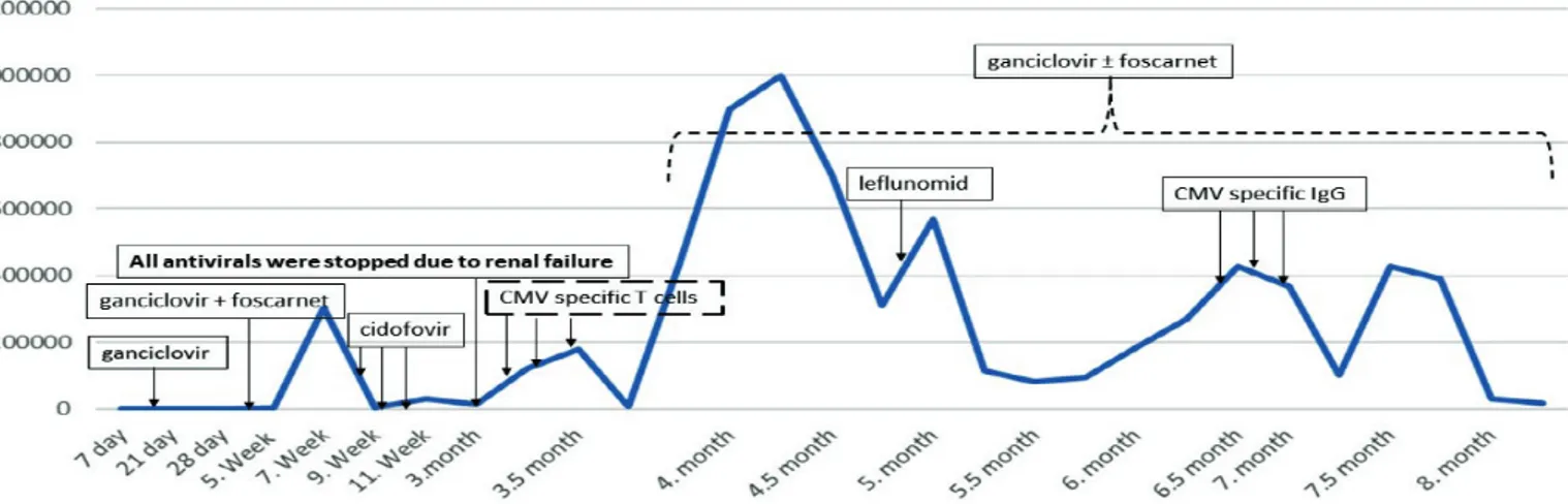
Cytomegalovirus (CMV) infection is still a major complication after allogeneic hematopoietic stem cell transplantation (HSCT) [1,2]. Unfortunately, prolonged antiviral treatment of CMV infection causes a delayed CMV-specific immune reconstitution. At this point, adoptive immunotherapy by CMV-specific T-cells can control CMV infection or provide immune reconstruction [3,4,5]. A 17-year-old boy with high-risk T-cell acute lymphoblastic leukemia underwent HSCT from one antigen-mismatched unrelated donor. He was conditioned with treosulfan, fludarabine, thiotepa, and rabbit anti-thymocyte globulin at 15 g/m2 for 3 consecutive days (days -2 to 0). The patient also received cyclosporine A (CsA) divided into two doses: 3 mg/kg daily from day -1 to post-transplant days +20 and +30 intravenously then switched to approximately 6 mg/kg peroral daily (targeted blood concentration: 200-250 ng/mL with monitoring). CsA was tapered quickly and stopped in the third month of transplant due to renal failure. Methotrexate was administered on days +1 (10 mg/m2), +3 (8 mg/m2), and +6 (8 mg/m2). He achieved neutrophil engraftment on day +17 and thrombocyte engraftment on day +32. Full donor chimerism was observed in the first and third months. Lymphoid engraftment was achieved on day +75 but generally the absolute lymphocyte
count was under 1500/mm3. He was CMV immunoglobulin G (IgG)-seropositive and CMV-DNA polymerase chain reaction (PCR) was negative before transplantation. Unfortunately, his donor was CMV IgG-seronegative. CMV infection (reactivation) occurred on day +19. Ganciclovir was started at 10 mg/kg/day and no response was obtained in 14 days. CMV drug resistance mutation was detected in the UL54 polymerase gene. Foscarnet was administered at 180 mg/kg/day on day +34. First, an increase of CD3+ lymphocytes was seen in the lymphocyte subtype analyses around the third month after the transplant. As a comorbidity, in spite of the fact that fluoroquinolone was administered until +30 day, BK virus infection developed in the patient and cidofovir was used at 5 mg/kg/week on days +52, +67, and +79. No response was achieved with the antiviral treatment and renal failure developed in the patient on day +82. All antivirals were stopped. According to the recent literature, the transplant council decided to use CMV-specific T-cells for the patient’s ongoing CMV infection. Informed consent was received from his family and the application was approved by the Ministry of Health’s Scientific Advisory Commission on Stem Cell Transplantation. In accordance with cGMP standards, peptide-specific T lymphocytes were isolated and amplified by a interferon-γ cytokine capture system using the fully automated CliniMACS Prodigy device at Acıbadem Labcell, İstanbul. The
8. Bradley AM, Buie LW, Kuykendal A, Vorhees PM. Successful use of intrathecal carboxypeptidase G2 for intrathecal methotrexate overdose: a case study and review of the literature. Clin Lymphoma Myeloma Leuk 2013;13:166-170.
9. Kazancı E, Gülen H, Erbay A, Vergin C. Treatment of intrathecal methotrexate overdose with folinic acid rescue and lumbar cerebrospinal fluid exchange: a report of two cases. Turk J Hematol 2011;28:63-67.
66
infusion doses of third-party CMV-specific T-cells were 2x10⁴ cell/kg and 1x10⁴ cell/kg in the 20th and 22nd weeks after transplantation, respectively. While the recommended dose of T-cells was 2x10⁶/m2 [6], we reduced the dose due to the risk of graft-versus-host disease (GvHD). The CMV-DNA PCR level was higher than 1x10⁵ copies/mL before infusion and had decreased to 8x10⁴ copies/mL on the 15th day after infusion. The patient had no immunosuppression at the time of T-cell infusion and did not develop GvHD after the infusion. In follow-up, CMV-DNA PCR increased to more than 3.5x10⁵ copies/mL in the first month of the cell infusion and the sixth month after transplantation. In this period, CD3-CD16+56+ (natural killer) and CD3+CD8+ (T cytotoxic) lymphocyte subtypes were increased. Nevertheless, the patient developed respiratory distress and CMV infection was detected from the bronchoalveolar lavage sample, and the CMV DNA titer was 152,000 copies/mL. After losing partial response to CMV-specific T-cells, CMV pneumonia was proved and then leflunomide was tested, but there was no response. Finally, CMV-specific IgG was administered once weekly three times. This treatment managed to decrease the CMV DNA copies to under 20,000 copies/mL. The treatment process according to the course of CMV DNA titer is shown in Figure 1.
CMV reactivations/infections are life-threatening complications in the transplant setting, especially if the recipient and donor are CMV mismatches. From our experience with this case, CMV-specific T-cells can control viral replication to a certain extent, but not enough for permanent results. The answer may be CMV-specific IgG, which controlled CMV reactivation best in our case, and antivirals may be used in combination.
Keywords: Childhood, Hematopoietic stem cell transplant, CMV,
Specific T-cell, Therapy
Anahtar Sözcükler: Çocukluk çağı, Hematopoetik kök hücre
nakli, CMV, Spesifik T hücre, Tedavi
Informed Consent: Informed consent was received from the family. Authorship Contributions
Surgical and Medical Practices: S.C., E.T, B.A.A., E.O., C.B.; Design: E.T., C.B.; Data collection or Processing: S.C., E.T, B.A.A.; Analyses or Interpretration: E.O., C.B.; Literature Search: E.T., B.A.A. ; Writing: E.T.
Conflict of Interest: No conflict of interest was declared by the
authors.
Financial Disclosure: The authors declared that this study
received no financial support.
References
1. Einsele H, Roosnek E, Rufer N, Sinzger C, Riegler S, Löffler J, Grigoleit U, Moris A, Rammensee HG, Kanz L, Kleihauer A, Frank F, Jahn G, Hebart H. Infusion of cytomegalovirus (CMV)-specific T cells for the treatment of CMV infection not responding to antiviral chemotherapy. Blood 2002;99:3916-3922. 2. Espigado I, de la Cruz-Vicente F, BenMarzouk-Hidalgo OJ, Gracia-Ahufinger
I, Garcia-Lozano JR, Aguilar-Guisado M, Cisneros JM, Urbano-Ispizua A, Perez-Romero P. Timing of CMV-specific effector memory T cells predicts viral replication and survival after allogeneic hematopoietic stem cell transplantation. Transpl Int 2014;27:1253-1262.
3. Boeckh M, Nichols WG, Papanicolaou G, Rubin R, Wingard JR, Zaia J. Cytomegalovirus in hematopoietic stem cell transplant recipients: current status, known challenges, and future strategies. Biol Blood Marrow Transplant 2003;9:543-558.
4. Scheinberg P, Melenhorst JJ, Brenchley JM, Hill BJ, Hensel NF, Chattopadhyay PK, Roederer M, Picker LJ, Price DA, Barrett AJ, Douek DC. The transfer of adaptive immunity to CMV during hematopoietic stem cell transplantation is dependent on the specificity and phenotype of CMV-specific T cells in the donor. Blood 2009;114:5071-5080.
Turk J Hematol 2020;37:57-76
Figure 1. Treatment process according to the course of CMV DNA titer (copies/mL).
CMV: Cytomegalovirus. LETTERS TO THE EDITOR
67
Turk J Hematol 2020;37:57-76
Address for Correspondence/Yazışma Adresi: Ersin Töret, M.D., Altınbaş University Faculty of Medicine, Medicalpark Bahçelievler Hospital, Department of Pediatric Hematology-Oncology & Bone Marrow Transplantation Unit, İstanbul, Turkey
Phone : +90 505 799 42 34
E-mail : [email protected] ORCID: orcid.org/0000-0002-6379-8326
Received/Geliş tarihi: August 6, 2019 Accepted/Kabul tarihi: November 12, 2019 DOI: 10.4274/tjh.galenos.2019.2019.0293 ©Copyright 2020 by Turkish Society of Hematology
Turkish Journal of Hematology, Published by Galenos Publishing House
Comparison of Different Culture Conditions for Mesenchymal
Stem Cells from Human Umbilical Cord Wharton’s Jelly for Stem
Cell Therapy
Kök Hücre Tedavisi için İnsan Kordon Kanı Wharton Jel’inden Üretilen Mezenkimal Kök
Hücreler için Farklı Kültür Ortamlarının Karşılaştırılması
Yu Bao1, Shumin Huang2, Zhengyan Zhao2
1Zhejiang University Faculty of Medicine, Children’s Hospital, Department of Nephrology, Zhejiang, China
2Zhejiang University Faculty of Medicine, Children’s Hospital, Clinic of Division of Child Health Care, Zhejiang, China
To the Editor,
Many recent studies have demonstrated that the umbilical cord is an excellent source of mesenchymal stem cells (MSCs) [1,2,3]. However, in order to use human umbilical cord Wharton’s jelly-derived mesenchymal stem cells (hUC-MSCs) in clinical therapy, a suitable culture procedure for good manufacturing practice-compliant production is mandatory. Nutritional deficiency is the major pathophysiological situation in an ischemic microenvironment in the clinic [4]. Thus, the development of serum-free culture systems is needed [5]. Furthermore, hypoxia is common in vivo in mammals [6]. The average oxygen tension falls to 1% in some cases of pathological ischemia, including fracture hematoma, and in cases of myocardial ischemia [7]. Hence, the investigation of biological characteristics of hUC-MSCs exposed to hypoxic and/or serum-free conditions is of great interest.
In our study, we conducted parallel assays by using four cell groups. For the hypoxic controls, cells from group A (n=10) and group B (n=10) were exposed to 5% CO2 and 94% N2 in an airtight modular incubator chamber (Billups-Rothenberg Inc., Del Mar, CA, USA). The final oxygen tension was 1%-3% as
measured by an oximeter (Oxybaby M+, Witt Technology, Solza, Italy). For the normoxic controls, cells from group C (n=10) and group D (n=10) were placed in an incubator at 37 °C, 5% CO2, and 21% O2. Cells from group A and group C were expanded in a mixture of Dulbecco’s modified Eagle’s medium and nutrient mixture F-12 (GIBCO, USA) supplemented with 10% fetal bovine serum (GIBCO, USA). Cells from group B and group D were expanded in StemPRO MSC serum-free medium (StemRD, USA). Flow cytometric analysis, differentiation potential, proliferative activities, cell cycle analysis, and apoptosis analysis of these four cell populations were evaluated. We repeated all these experiments 3 times.
Flow cytometry analysis of MSC-specific surface marker expression showed that hUC-MSCs cultured under four experimental conditions for six passages were positive for CD44, CD73, CD90, CD105, CD29, and HLA-ABC (BD Pharmingen, USA) and negative for CD34, CD45, CD14, and HLA-DR (BD Pharmingen, USA); no significant differences were detected between the four cell populations (Figure 1). This finding indicates that culturing cells under hypoxic and/or serum-free conditions did not induce significant variations in the typical MSC marker expression profile.
hUC-5. Poiret T, Axelsson-Robertson R, Remberger M, Luo XH, Rao M, Nagchowdhury A, Von Landenberg A, Ernberg I, Ringden O, Maeurer M. Cytomegalovirus-specific CD8+ T-cells with different T-cell receptor affinities segregate T-cell phenotypes and correlate with chronic graft-versus-host disease in patients post-hematopoietic stem cell transplantation. Front Immunol 2018;9:760. 6. Tzannou I, Papadopoulou A, Naik S, Leung K, Martinez CA, Ramos CA,
Carrum G, Sasa G, Lulla P, Watanabe A, Kuvalekar M, Gee AP, Wu MF, Liu H, Grilley BJ, Krance RA, Gottschalk S, Brenner MK, Rooney CM, Heslop
HE, Leen AM, Omer B. Off-the-shelf virus-specific T cells to treat BK virus, human herpesvirus 6, cytomegalovirus, Epstein-Barr virus, and adenovirus infections after allogeneic hematopoietic stem-cell transplantation. J Clin Oncol 2017;35:3547-3557.