http://journals.tubitak.gov.tr/veterinary/ © TÜBİTAK
doi:10.3906/vet-1411-45
The effect of macromolecule and growth factor combinations on in vitro
development of bovine embryos
Alper KOÇYİĞİT1, Mesut ÇEVİK2,*, Uğur ŞEN3, Mehmet KURAN4
1Department of Reproduction and Artificial Insemination, Faculty of Veterinary Medicine, Cumhuriyet University, Sivas, Turkey
2Department of Reproduction and Artificial Insemination, Faculty of Veterinary Medicine, Ondokuz Mayıs University, Samsun, Turkey
3Department of Agricultural Biotechnology, Faculty of Agriculture, Ahi Evran University, Kırşehir, Turkey
4Department of Agricultural Biotechnology, Faculty of Agriculture, Ondokuz Mayıs University, Samsun, Turkey
1. Introduction
In vitro fertilization (IVF) and development of bovine embryos up to the blastocyst stage has been successfully achieved with a variety of culture systems. Numerous studies have been performed to improve developmental competence of mammalian embryos by supplementation of culture media with gonadotropins, steroid hormones, serum, and growth factors. Therefore, current culture media usually are supplemented with various proteins (1,2). The protein supplements with the addition of embryo culture are thought to play several important roles in the culture media: 1) reducing embryo toxicity, 2) as a source for basic nutritive requirements of early embryos, and 3) as a source of growth factors promoting embryonic development directly or indirectly via cumulus-cell proliferation (3–5).
It is well defined that serum and bovine serum albumin (BSA) are complex and undefined mixtures of different proteins containing various energy substrates, small peptides or growth factors that play an important role in embryonic development (6). In several studies it has been
shown that serum or BSA has been replaced with synthetic macromolecules such as polyvinyl-alcohol (PVA) or polyvinylpyrrolidone (PVP) to prevent the stickiness of the embryos that occurs when protein-free medium is used (7,8). These synthetic polymers are used widely as a serum/BSA substitute in chemically defined embryo culture media, especially for bovine embryos. Among these polymers, PVA has been preferred as an additive, particularly having surfactant activity similar to that of albumin. PVA is more frequently used in culture media (1,9) than PVP (10) because its surface-active properties are greater, while the use of PVP in embryo freezing media (6,11) is attributed with colloidal rather than surface-active properties (12). More recently, this list has been extended to include glycosaminoglycans, in particular hyaluronic acid, already known to stimulate cattle embryo development in vitro (13,14).
Growth factors like epidermal growth factor (EGF) and insulin like growth factor-I (IGF-I) are known to have effects on preimplantation development by stimulating the metabolism and growth of embryos (5,15). In bovine
Abstract: This study was conducted to determine the effects of different macromolecule sources added to synthetic oviduct fluid (SOF)
culture medium supplemented with growth factors on the development of bovine embryos and blastocyst morphology. Zygotes were distributed into 5 treatment groups. Cleavage, morula, and blastocyst rates were evaluated under a stereomicroscope. Trophectoderm (TE) and inner cell mass (ICM) cells were determined by differential staining method. It was found that bovine serum albumin (BSA), either alone or in combination with growth factors, as compared to the control or polyvinyl-alcohol (PVA) resulted in higher embryo yield and faster development during early bovine embryo culture. The quality of bovine embryos, based on the number of blastocyst cells and the ratio of ICM to total blastomeres, was affected by the sources of macromolecules and their combinations with growth factors. Growth factors supplemented to SOFaa media with BSA and PVA significantly increased the number of ICM cells and the ratio of ICM cells to total number of cells. In conclusion, replacing BSA with PVA depressed the blastocyst rate and cell numbers, and the number of blastomeres and ICM and TE cell numbers were affected by both the type of macromolecule and the growth factor supplements.
Key words: Bovine, embryo culture, blastocyst quality, macromolecules, growth factors
Received: 24.11.2014 Accepted/Published Online: 24.02.2015 Printed: 10.06.2015 Research Article
embryos, poor morphology is generally associated with hatched blastocysts with low inner cell mass (ICM) cell numbers. The number of blastomeres and the ratio of ICM cells to total cells are considered to be potential indicators of embryo quality (16). The culture conditions influence the cell allocation in different species (7,17). The objective of the present study was to replace serum with BSA as a less complex protein source or with a synthetic macromolecule (PVA) and growth factors and to observe the effects of additional culture supplements on morphologic quality and cell allocation of bovine embryos.
2. Materials and methods
All chemicals and media used in this study were purchased from Sigma-Aldrich (St. Louis, MO, USA), except where otherwise indicated.
2.1. Collection and in vitro maturation of oocytes Bovine ovaries were collected from a local slaughterhouse and transported to the laboratory at approximately 36 ± 2.0 °C in physiological saline solution supplemented with gentamycin sulfate (0.1 µL/mL). Cumulus oocyte complexes (COCs) were recovered from follicles 2–8 mm in diameter by aspiration. The COCs were collected in 3–4 mL of HEPES-buffered Medium-199 containing Earle’s salts and supplemented with 1% v/v antibiotic-antimycotic solution. Before in vitro maturation, the COCs were assessed morphologically and only oocytes with compact, nonatretic cumulus investment and evenly granulated cytoplasm were selected for maturation. Maturation medium was sodium bicarbonate-buffered Medium-199 with sodium pyruvate (5.5 µg/mL), antibiotic-antimycotic solution (1% v/v), and heat-inactivated fetal calf serum (FCS, 10% v/v). The COCs were matured for 22 h in a humidified atmosphere of 5% CO2 in air at 38.5 °C. 2.2. Spermatozoa preparation and in vitro fertilization After a 22-h maturation period, oocytes were transferred into 44-µL fertilization drops. The fertilization medium was glucose-free modified TALP supplemented with bicarbonate (25 mM), Na-lactate (22 mM), Na-pyruvate (1 mM), fatty acid-free BSA (6 mg/mL), heparin-sodium salt (184 U/mg heparin, 10 mg/mL), and antibiotic-antimycotic solution (0.5 µL/mL) (pH 7.4 and 280–300 mOsm/kg). Frozen-thawed semen was used for the fertilization of oocytes. A Percoll density gradient system was used for the separation of the motile fraction of the frozen-thawed semen (18). Sperm were then diluted to 50 × 106 spermatozoa/mL in TL-HEPES, including
2 × 10⁶ spermatozoa/mL as the final concentration. The
fertilization procedure was completed by adding 2 µL of diluted sperm, 2 µL of heparin (5 µg/mL), and 2 µL of PHE solution (20 µM penicillamine, 10 µM hypotaurine, and 1 µM epinephrine in final concentration) into the fertilization drops containing oocytes. The oocytes were
fertilized with 2 µL of diluted semen per fertilization drop for 22 h in a humidified atmosphere of 5% CO2 in air at 38.5 °C.
2.3. In vitro culture
Cumulus cells surrounding the oocytes were removed from presumptive zygotes at approximately 22 h after insemination by vortexing for 3 min. The zygotes were transferred in groups of 20–30 for the culture droplets. The mSOF medium was supplemented with pyruvate (0.4 mM), BSA-FAF (4 mg/mL), 100X MEM (20 µL/mL), 50X BME (10 µL/mL), penicillin (100 U/mL), and streptomycin (100 µg/mL) on the day of use. Macromolecules and growth factors added to the culture medium were as follows: 1) essentially fatty-acid free BSA (4 mg/mL), 2) BSA + IGF-I (100 ng/mL) + EGF (10 ng/mL), 3) PVA (1 mg/mL), 4) PVA + IGF-I + EGF, and 5) FCS (10% , v/v), for 9 days under low oxygen tension (5% CO2, 5% O2, 90% N2 atmosphere) at 38.5 °C. Cleavage, morula, and blastocyst development rates were evaluated from the zygotes on days 3, 5, and 8. 2.4. Determining of inner cell mass and trophectoderm Expanding blastocysts characterized by zona pellucidae showing signs of thinning and with slightly increased embryo diameters by day 7 or 8 were stained as described by Van Soom et al. (16). Embryos were examined under a fluorescence microscope (Nikon Invert Microscope Eclipse Ti-FL). ICM nuclei labeled with bisbenzimide appeared blue and trophectoderm (TE) nuclei labeled with both bisbenzimide and propidium iodide appeared pink to red. The numbers of ICM and TE nuclei were counted directly under the fluorescence microscope.
2.5. Statistical analysis
Embryos were randomly allocated to each treatment group and all experiments were replicated six or seven times. Statistical data analysis was performed using the GLM procedure of SAS (19). One-way analysis of variance (ANOVA) followed by Duncan’s multiple comparison test was used for statistical comparison of the groups. All values are reported as least-squares means ± SEM, and statistical differences were considered as significant when P-values were less than 0.05.
3. Results
3.1. Embryonic development
The effects of different macromolecule sources alone or combined with growth factors on in vitro bovine embryo development potential were evaluated. As shown in Table 1, there were significant differences among culture groups in terms of cleavage and morula rates and development to the blastocyst stage (P < 0.001). BSA alone or combined with growth factors and PVA supplemented with EGF and IGF-I resulted in the highest cleavage rate in contrast to the FCS- and PVA-alone treatments. Furthermore, the
results of our study demonstrated that bovine embryos in the culture medium containing BSA developed faster and had increased blastocyst cell counts when compared to bovine embryos cultured in 1 mg/mL PVA-supplemented media (105.15 and 88.57, P < 0.05). However, BSA alone or supplemented with IGF-I and EGF yielded similar results (23.1% and 28.9 %, respectively) in terms of development to the blastocyst stage. Additionally, FCS, PVA alone, or PVA supplemented with IGF-I and EGF combination resulted in similar blastocyst rates (16.5%, 11.4%, and 15.1%, respectively). FCS had the lowest percentage of embryos that cleaved, and it resulted in 24.8% morula development, suggesting that FCS delays embryonic cleavage compared to the BSA and growth factor-supplemented culture media. Similarly, there was a difference in the overall blastocyst yield at days 7–9 (range: 11%–28%) among the groups.
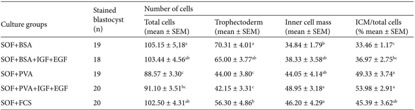
3.2. Effects of culture supplements on cell allocation The data on cell allocation of ICM and TE cells are presented in Table 2. There were significant differences between treatment groups in terms of the number of
blastomeres and ICM cells (P < 0.05), and in the ratio of ICM cells to total cells (P < 0.001) harvested on days 7, 8, and 9 of culture after fertilization. Numbers of ICM and TE cells were influenced by both the type of macromolecule and the growth factor supplements. As shown in Table 2, successive use of BSA alone or in combination with IGF-I and EGF, and also FCS in the SOF medium, significantly increased the number of blastomeres in blastocysts (103.4, 105.1, and 102.5 cells/blastocyst, respectively) (Figure 1). In general, embryos produced in PVA alone or supplemented with growth factors had a lower number of cells in blastocysts (P < 0.05) (Figures 2). However, significant differences were found in the numbers of blastomeres and ICM cells (P < 0.05) and in TE cells and the ratio of ICM cells to total cells among the treatment groups (P < 0.001). A higher ratio of ICM cells to total cells was observed when embryos were cultured in the media supplemented with FCS, PVA alone, or growth factors compared to embryos cultured in the BSA alone and the BSA plus growth factors combination (45.3 and 49.3 and 53.9 versus 33.4 and 36.9) (P < 0.001).
Table 1. Effect of growth factors and macromolecules on in vitro developmental competence of bovine embryos.
Culture
groups Presumptive zygotes(n)
Percentage of embryos developed to Cleavage
(% mean ± SEM) Morula (% mean ± SEM) Blastocyst (% mean ± SEM)
SOF+BSA 281 79.71 ± 2.40a 37.72 ± 2.89a 23.13 ± 2.51ab
SOF+BSA+IGF+EGF 308 69.48 ± 2.62b 37.01 ± 2.75a 28.89 ± 2.58a
SOF+PVA 245 54.69 ± 3.18c 19.59 ± 2.54b 11.42 ± 2.03c
SOF+PVA+IGF+EGF 283 71.73 ± 2.68b 24.38 ± 2.55b 15.19 ± 2.13c
SOF+FCS 230 54.78 ± 3.28c 24.78 ± 2.85b 16.52 ± 2.45bc
abc: Rates and values with different letters in the same column are statistically significant in bovine IVF embryos (P < 0.001).
Table 2. Differential cell counts of blastocysts cultured in medium containing different macromolecules and growth factors.
Culture groups Stained blastocyst (n) Number of cells Total cells
(mean ± SEM) Trophectoderm(mean ± SEM) Inner cell mass(mean ± SEM) ICM/total cells(% mean ± SEM)
SOF+BSA 19 105.15 ± 5,18a 70.31 ± 4.01a 34.84 ± 1.79b 33.46 ± 1.17c
SOF+BSA+IGF+EGF 18 103.44 ± 4.56ab 65.00 ± 3.77ab 38.33 ± 3.58ab 36.97 ± 2.75bc
SOF+PVA 19 88.57 ± 3.30c 44.00 ± 3.80c 44.05 ± 4.14ab 49.33 ± 3.74a
SOF+PVA+IGF+EGF 20 91.10 ± 3.51bc 42.15 ± 3.31c 48.95 ± 3.18a 53.98 ± 2.91a
SOF+FCS 20 102.50 ± 4.31ab 56.30 ± 4.86b 46.20 ± 4.29a 45.39 ± 3.62ab
4. Discussion
The purpose of the present study was to replace serum with BSA as a less complex protein source or with PVA as a synthetic macromolecule and to observe the effects on early embryonic development and morphologic quality in preimplantation bovine embryos. The present study showed that different macromolecule sources (especially BSA) alone or combined with growth factors were significantly effective on in vitro bovine embryo development, but no effect of PVA alone or in combination with growth factors on cleavage and blastocyst rates and blastocyst cell number was observed compared other culture groups.
Recent studies focused on amino acid or growth factor supplementation to in vitro culture media of mammalian embryos. Therefore, a wide variety of epigenetic factors, including ions, energy substrates, amino acids, vitamins, growth factors, cytokines, and hormones, play an important role in early embryonic development (7,11). Defined media with PVP and PVA, semidefined media with BSA, and undefined media with FCS have all been added to mammalian embryo media as energy sources. In a modified SOF culture system, the successive use of BSA and FBS during early and late preimplantation development was the most effective regime. BSA may also provide as of yet undefined embryotrophic compounds, function as a heavy metal ion chelator/free radical scavengers, protect cellular constituents against the effect of toxins, and regulate redox potential, pH, and osmolarity. The serum contains not only a variety of energy sources (3,6) but also growth factors (8,13) needed for in vitro embryonic development. The beneficial effect of more complex protein supplements is evident after the
activation of the embryonic genome and probably due to the presence of growth factors (10). One of the most commonly used supplements, BSA is an embryotrophic macromolecule used in embryo culture media, which is commonly replaced with synthetic compounds, such as PVA. However, the polyvinyl polymers PVP and PVA have been commonly used as substitutes for BSA in media for the culture of mammalian preimplantation embryos. The more frequent use of PVA in culture media (1,9,20) than PVP (10) could be related to its greater surface-active properties or may be related to colloidal rather than surface-active properties (3,12).
The present study showed that exposure of bovine embryos to BSA alone or in combination with growth factors accelerated progression of blastocyst in bovine embryos. These data are in agreement with several researchers, such as as Lonergan et al. (4) and Rieger et al. (21). Some of the beneficial effects of BSA may be due to its action as a chelating agent, regulator of oxidation reduction potential, cell surface protector, or enzyme protector. Furthermore, our results demonstrated that there was no effect of PVA alone or combined with growth factors on cleavage and blastocyst rates and blastocyst cell number in comparison with other groups. These results are in agreement with the findings of Kuran et al. (20) and Duque et al. (7), but in contrast to those of Wrenzycki et al. (8), Krisher et al. (11), and Mingoti et al. (13). Eckert and Niemann (2) demonstrated that embryos cultured in SOFaaBSA consumed more oxygen than their SOFaaPVA counterparts, despite having significantly lower pyruvate uptake. This suggests that embryos grown in PVA may have reduced viability. In this study, the reduction in
Figures 1 and 2. Differentially stained (Hoechst 33342 and propidium iodide) bovine blastocysts with blue nuclei
representing the inner cell mass (ICM) and pink to red nuclei representing outer cells (TE). 1- Cell allocation of blastocyst in BSA group, 2- cell allocation of blastocyst in PVA group.
blastocyst quality in medium supplemented with PVA was associated with reduced blastocyst cell numbers.
EGF and IGF-I have been reported to be mitogens inducing a positive effect on preimplantation development and stimulating metabolism and development of embryos (5,22,23). Moreover, these factors are involved in the compaction and formation of the blastocyst, activation of transport systems responsible for the uptake of glucose, enhancement of endocytosis, and probably protein turnover (24,25).
Neira et al. (26) reported that IGF-I accelerated development of bovine blastocysts to hatched blastocysts and a combination with growth factors and cytokines added to SOF medium produced greater percentages of blastocysts and hatched blastocysts than the others. According to several researchers (5,21,24), IGF-I and EGF had mitogenic effects and stimulated the growth and metabolism of embryos. These findings are consistent with our results. In our study, the data observed suggest that when culture media are supplemented with growth factors, the culture system produces more blastocysts and embryos produced have a higher number of cells. Furthermore, the addition of serum to embryo culture medium was similar in its effect in terms of embryonic development with only BSA or BSA supplemented with growth factors, while it was similar to PVA alone or PVA supplemented with growth factors in terms of blastocyst cell allocation.
In assessing the viability of the embryos, morphological observations are most widely used as a gross indicator of embryo viability, but the cell number of the blastocysts and the ICM/total cells ratio are valid indicators of the viability of preimplantation embryos, while morphological criteria alone are poor indicators (27,28). Van Soom et al. (16) reported that blastocyst formation is the first differentiation process during early embryonic development in mammals, yielding the ICM and TE cells. The ICM cells will differentiate into all tissues of the developing fetus. It is well known that the ICM cells contribute to all embryonic tissues and to a part of the extraembryonic membranes, whereas the TE cells mainly form the outer layer of the placenta. Both cell lineages are vital for embryonic and fetal survival (17). In bovine embryos, poor morphology is associated with blastocysts with low numbers of ICM cells (28,29).
According to Koo et al. (17), some factors in the culture media, such as growth factors present in the serum, can modify the distribution of the embryonic cells in favor of the TE cells, which can lead to pregnancy abnormalities encountered after transfer of in vitro cultured bovine and ovine embryos. However, IVF-derived embryos also showed a moderate increase in the ICM/TE cell ratio as compared with in vivo derived embryos. Diaz-Cueto and Gerton (22) reported that IGF-I and IGF-II receptors are only localized in the ICM. Stimulation of cell proliferation is specific to the ICM and the TE cells are not affected. The present study indicates that the addition of BSA and/or PVA supplemented with EGF and IGF-I into the embryo culture medium led to increased ICM cell differentiation. The ICM/total cell ratios of the bovine blastocysts obtained in the present study were similar to those of the previous findings of Choi et al. (3) and Orsi and Leese (30), but lower than in the studies of Duque et al. (7) and higher than those of Krisher et al. (11) and Shirazi et al. (14). Differences between the present study and others may be related to differences in culture and laboratory conditions. Van Soom et al. (16) reported that especially ICM cell numbers were sensitive to environmental influences; precisely for this reason, significantly lower ICM cell numbers were detected in in vitro produced bovine embryos. A difference in allocation of inner cells was noted for in vitro produced bovine embryos, depending on the medium combinations.
In conclusion, the present study demonstrated that defined medium supplemented with PVA can be used in in vitro culture of bovine embryos, but both blastocyst rates and the numbers of cells were significantly lower than those of undefined medium. Bovine embryos cultured in SOF medium supplemented with BSA alone or together with growth factors cleaved faster and reached blastocyst stage earlier than culture medium supplemented with PVA alone or with growth factors. The addition of BSA alone or with growth factors to culture medium also resulted in better blastocyst quality than in the other groups.
Acknowledgments
We wish to thank Dr Arslan for statistical analysis. This work was supported by the Research Foundation of Ondokuz Mayıs University (PYO.VET.1901.09.002). References
1. Ali A, Sirard MA. Effect of the absence or presence of various protein supplements on further development of bovine oocytes during in vitro maturation. Biol Reprod 2002; 66: 901–905. 2. Eckert J, Niemann H. In vitro maturation, fertilization and
culture to blastocysts of bovine oocytes in protein-free media. Theriogenology 1995; 43: 1211–1225.
3. Choi YH, Lee BC, Lim JM, Kang SK, Hwang WS. Optimization of culture medium for cloned bovine embryos and its influence on pregnancy and delivery outcome. Theriogenology 2002; 58: 1187–1197.
4. Lonergan P, Carolan C, Van LA, Donnay I, Khatir H, Mermillod M. Role of epidermal growth factor in bovine oocyte maturation and preimplantation embryo development in vitro. Biol Reprod 1996; 54: 1420–1429.
5. Srisathien S, Hernandez-Fonseca HJ, Brackett BG. Influences of epidermal growth and insulin-like growth factor-I on bovine blastocyst development in vitro. Anim Reprod Sci 2003; 77: 21–32.
6. Rizos D, Gutierrez-Adan A, Perez-Garnelo S, de la Fuente J, Boland MP, Lonergan P. Bovine embryo culture in the presence or absence of serum: Implications for blastocyst development, cryotolerance, and messenger RNA expression. Biol Reprod 2003; 68: 236–243.
7. Duque P, Hidalgo CO, Gómez E, Pintado B, Facal N, Díez C. Macromolecular source as dependent on osmotic pressure and water source: effects on bovine in vitro embryo development and quality. Reprod Nutr Dev 2003; 43: 487–496.
8. Wrenzycki C, Herrmann D, Carnwath JW, Niemann H. Alterations in the relative abundance of gene transcripts in preimplantation bovine embryos cultured in medium supplemented with either serum or PVA. Mol Reprod Dev 1999; 53: 8–18.
9. Lee ES, Fukui Y. Effect of various growth factors in a defined culture medium on in vitro development of bovine embryos matured and fertilized in vitro. Theriogenology 1995; 44: 71–83. 10. Kim JY, Kim SB, Park MC, Park H, Park YS, Park HD, Lee JH, Kim JM. Addition of macromolecules to PZM-3 culture medium on the development and hatching of in vitro porcine embryos. Asian-Aust J Anim Sci 2007; 20: 1820–1826. 11. Krisher RL, Lane M, Bavister BD. Developmental competence
and metabolism of bovine embryos cultured in semi-defined and defined culture media. Biol Reprod 1999; 60: 1345–1352. 12. Palasz AT, Thundathil J, Verrall RE, Mapletoft RJ. The effect
of macromolecular supplementation on the surface tension of TCM-199 and the utilization of growth factors by bovine oocytes and embryos in culture. Anim Reprod Sci 2000; 58: 229–240.
13. Mingoti GZ, Castro VS, Méo SC, Sá Barretto LS, Garcia JM. The effects of macromolecular and serum supplements and oxygen tension during bovine in vitro procedures on kinetics of oocyte maturation and embryo development. In Vitro Cell Dev Biol Anim 2001; 47: 361–367.
14. Shirazi A, Ardali MA, Ahmadi E, Nazari H, Mamuee M, Heidari B. The effect of macromolecule source and type of media during in vitro maturation of sheep oocytes on subsequent embryo development. J Reprod Infertil 2012; 13: 13–19.
15. Quetglas MD, Coelho LA, Garcia JM, Oliveira FEB, Esper CR. Effect of insulin-like growth factor-I during in vitro oocyte maturation and in vitro culture of bovine embryos. Arq Bras Med Vet Zootec 2001; 53: 1–5.
16. Van Soom A, Boerjan M, Ysebaert MT, Kruif A. Cell allocation to the inner cell mass and the trophectoderm in bovine embryos cultured in two different media. Mol Reprod Dev 1996; 45: 171–182.
17. Koo DB, Kang YK, Choi YH, Park JS, Kim HN, Oh KB, Son DS, Park H, Lee KK, Han YM. Aberrant allocations of inner cell mass and trophectoderm cells in bovine nuclear transfer blastocysts. Biol Reprod 2002; 67: 487–492.
18. Parrish JJ, Krogenaes A, Susko-Parrish JL. Effects of bovine sperm by either swim-up or Percoll method on success of in vitro fertilization and early embryonic development. Theriogenology 1995; 44: 859–869.
19. SAS. SAS User Guide Ver. 9.01. Cary, NC, USA: SAS; 2007. 20. Kuran M, Robinson JJ, Staines ME, McEvoy TG. Development
and de novo protein synthetic activity of bovine embryos produced in vitro in different culture systems. Theriogenology 2001; 55: 593–606.
21. Rieger D, Luciano AM, Modina S, Pocar P, Lauria A, Gandolfi F. The effect of epidermal growth factor and insulin-like growth factor I on the metabolic activity, nuclear maturation and subsequent development of cattle oocytes in vitro. J Reprod Fertil 1998; 112: 123–130.
22. Diaz-Cueto L, Gerton GL. The influence of growth factors on the development of preimplantation mammalian embryos. Arch Med Res 2001; 32: 619–626.
23. Makarevich AV, Markkula M. Apoptosis and cell proliferation potential of bovine embryos stimulated with insulin-like growth factor I during in vitro maturation and culture. Biol Reprod 2002; 66: 386–392.
24. Herrler A, Lucas-Hahn A, Niemann H. Effects of insulin-like growth factor-I on in vitro production of bovine embryos. Theriogenology 1992; 37: 1213–1224.
25. Kurzawa R, Glabowski W, Baczkowski T, Wiszniewska B, Marchlewicz M. Growth factors protect in vitro cultured embryos from the consequences of oxidative stress. Zygote 2004; 12: 231–240.
26. Neira JA, Tainturier D, Pena MA, Martal J. Effect of the association of IGF-I, IGF-II, bFGF, TGF-β1, GM-CSF, and LIF on the development of bovine embryos produced in vitro. Theriogenology 2010; 73: 594–604.
27. Stojkovic M, Büttner M, Zakhartchenko V, Brem G, Wolf E. A reliable procedure for differential staining of in vitro produced bovine blastocysts: comparison of tissue culture medium 199 and Menezo’s B2 medium. Anim Reprod Sci 1998; 50: 1–9. 28. Van Soom A, Ysebaert MT, Kruif AD. Relationship between
timing of development, morula morphology, and cell allocation to inner cell mass and trophectoderm in in vitro produced bovine embryos. Mol Reprod Dev 1997; 47: 47–56.
29. Kuran M, Robinson JJ, Brown DS, McEvoy TG. Development, amino acid utilization and cell allocation in bovine embryos after in vitro production in contrasting culture systems. Reproduction 2002; 124: 155–165.
30. Orsi NM, Leese HJ. Amino acid metabolism of preimplantation bovine embryos cultured with bovine serum albumin or polyvinyl alcohol. Theriogenology 2004; 61: 561–572.